Advertisement

Main menu

  • Home
  • Content
    • Current Issue
    • Accepted Manuscripts
    • Article Preview
    • Past Issue Archive
    • Video Articles
    • AJNR Case Collection
    • Case of the Week Archive
    • Case of the Month Archive
    • Classic Case Archive
  • Special Collections
    • AJNR Awards
    • Low-Field MRI
    • Alzheimer Disease
    • ASNR Foundation Special Collection
    • Photon-Counting CT
    • View All
  • Multimedia
    • AJNR Podcasts
    • AJNR SCANtastic
    • Trainee Corner
    • MRI Safety Corner
    • Imaging Protocols
  • For Authors
    • Submit a Manuscript
    • Submit a Video Article
    • Submit an eLetter to the Editor/Response
    • Manuscript Submission Guidelines
    • Statistical Tips
    • Fast Publishing of Accepted Manuscripts
    • Graphical Abstract Preparation
    • Imaging Protocol Submission
    • Author Policies
  • About Us
    • About AJNR
    • Editorial Board
    • Editorial Board Alumni
  • More
    • Become a Reviewer/Academy of Reviewers
    • Subscribers
    • Permissions
    • Alerts
    • Feedback
    • Advertisers
    • ASNR Home

User menu

  • Alerts
  • Log in

Search

  • Advanced search
American Journal of Neuroradiology

American Journal of Neuroradiology

ASHNR American Society of Functional Neuroradiology ASHNR American Society of Pediatric Neuroradiology ASSR
  • Alerts
  • Log in

Advanced Search

  • Home
  • Content
    • Current Issue
    • Accepted Manuscripts
    • Article Preview
    • Past Issue Archive
    • Video Articles
    • AJNR Case Collection
    • Case of the Week Archive
    • Case of the Month Archive
    • Classic Case Archive
  • Special Collections
    • AJNR Awards
    • Low-Field MRI
    • Alzheimer Disease
    • ASNR Foundation Special Collection
    • Photon-Counting CT
    • View All
  • Multimedia
    • AJNR Podcasts
    • AJNR SCANtastic
    • Trainee Corner
    • MRI Safety Corner
    • Imaging Protocols
  • For Authors
    • Submit a Manuscript
    • Submit a Video Article
    • Submit an eLetter to the Editor/Response
    • Manuscript Submission Guidelines
    • Statistical Tips
    • Fast Publishing of Accepted Manuscripts
    • Graphical Abstract Preparation
    • Imaging Protocol Submission
    • Author Policies
  • About Us
    • About AJNR
    • Editorial Board
    • Editorial Board Alumni
  • More
    • Become a Reviewer/Academy of Reviewers
    • Subscribers
    • Permissions
    • Alerts
    • Feedback
    • Advertisers
    • ASNR Home
  • Follow AJNR on Twitter
  • Visit AJNR on Facebook
  • Follow AJNR on Instagram
  • Join AJNR on LinkedIn
  • RSS Feeds

AJNR Awards, New Junior Editors, and more. Read the latest AJNR updates

Research ArticleBrain
Open Access

Neurovascular Manifestations in Hereditary Hemorrhagic Telangiectasia: Imaging Features and Genotype-Phenotype Correlations

T. Krings, H. Kim, S. Power, J. Nelson, M.E. Faughnan, W.L. Young, K.G. terBrugge and the Brain Vascular Malformation Consortium HHT Investigator Group
American Journal of Neuroradiology May 2015, 36 (5) 863-870; DOI: https://doi.org/10.3174/ajnr.A4210
T. Krings
aFrom the Division of Neuroradiology (T.K., S.P., K.G.t.B.), Department of Medical Imaging
bDivision of Neurosurgery (T.K.), Department of Surgery, Toronto Western Hospital, University of Toronto, Toronto, Ontario, Canada
  • Find this author on Google Scholar
  • Find this author on PubMed
  • Search for this author on this site
H. Kim
cDepartment of Anesthesia and Perioperative Care (H.K., J.N., W.L.Y.), Center for Cerebrovascular Research
dDepartment of Epidemiology and Biostatistics (H.K.), University of California San Francisco, San Francisco, California
  • Find this author on Google Scholar
  • Find this author on PubMed
  • Search for this author on this site
S. Power
aFrom the Division of Neuroradiology (T.K., S.P., K.G.t.B.), Department of Medical Imaging
  • Find this author on Google Scholar
  • Find this author on PubMed
  • Search for this author on this site
J. Nelson
cDepartment of Anesthesia and Perioperative Care (H.K., J.N., W.L.Y.), Center for Cerebrovascular Research
  • Find this author on Google Scholar
  • Find this author on PubMed
  • Search for this author on this site
M.E. Faughnan
eDivision of Respirology (M.E.F.), Department of Medicine and Li Ka Shing Knowledge Institute, St. Michael's Hospital, Toronto, Ontario, Canada
fDivision of Respirology (M.E.F.), Department of Medicine, University of Toronto, Toronto, Ontario, Canada.
  • Find this author on Google Scholar
  • Find this author on PubMed
  • Search for this author on this site
W.L. Young
cDepartment of Anesthesia and Perioperative Care (H.K., J.N., W.L.Y.), Center for Cerebrovascular Research
  • Find this author on Google Scholar
  • Find this author on PubMed
  • Search for this author on this site
K.G. terBrugge
aFrom the Division of Neuroradiology (T.K., S.P., K.G.t.B.), Department of Medical Imaging
  • Find this author on Google Scholar
  • Find this author on PubMed
  • Search for this author on this site
  • Article
  • Figures & Data
  • Supplemental
  • Info & Metrics
  • Responses
  • References
  • PDF
Loading

Article Figures & Data

Figures

  • Tables
  • Fig 1.
    • Download figure
    • Open in new tab
    • Download powerpoint
    Fig 1.

    Left ICA angiogram, lateral view, depicts typical features of a capillary vascular malformation in the left frontal opercular region, with a subcentimeter vascular blush identified in the arterial (A) persisting into early venous phase (B, arrow), without evidence of arteriovenous shunting. On MR imaging, this appears as a focal region of hyperintensity on axial T2 FLAIR imaging (C, arrow), with fluffy enhancement on coronal gadolinium-enhanced T1-weighted imaging (D, arrow). No dilated arterial feeder or dilated draining vein is seen.

  • Fig 2.
    • Download figure
    • Open in new tab
    • Download powerpoint
    Fig 2.

    Lateral projection of right ICA angiogram (A–C) and axial T1-weighted gadolinium-enhanced MR imaging (D) demonstrate the typical imaging features of a capillary vascular malformation in a 17-year-old male patient with HHT. A focal subcentimeter blush (arrows) is identified on the arterial phase of a right carotid angiogram, in the frontal, temporal, and parietal regions, in keeping with multiple capillary telangiectasia. Magnified view of the right parietal lesion shows typical angiographic features, with vascular blush identified in the arterial (B) persisting into early venous phase (C); no arteriovenous shunting, dilated arterial feeder, or dilated draining vein are seen. MR imaging appearance of the right parietal lesion demonstrates the typical features of fluffy contrast enhancement in a superficial cortical/subcortical location, without abnormal vascular dilation (D, arrow).

  • Fig 3.
    • Download figure
    • Open in new tab
    • Download powerpoint
    Fig 3.

    Lateral view of a right ICA angiogram in a 43-year-old male patient with HHT demonstrates a capillary vascular malformation in the right anterior parietal region. A subcentimeter blush is seen in the arterial phase (A, arrow), which persists in the late arterial phase (B) without evidence of arteriovenous shunting.
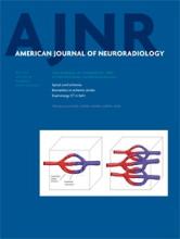
  • Fig 4.
    • Download figure
    • Open in new tab
    • Download powerpoint
    Fig 4.

    Left ICA catheter angiogram (A and B), microcatheter injection (C and D), axial T2-weighted (E), and contrast-enhanced T1-weighted MR imaging (F) demonstrate imaging findings of a typical arteriovenous malformation. Anteroposterior projection of a left ICA injection demonstrates filling of the AVM nidus through an enlarged anterior temporal branch of the left MCA in the early arterial phase (A, arrow). There is arteriovenous shunting with early venous drainage through an enlarged left inferior temporal vein (B, arrows). Microcatheter injection in frontal (C) and lateral (D) projections demonstrates a typical glomerular well-defined compact nidus supplied by a single terminal arterial feeder with shunting into a dilated vein. MR imaging shows the superficial location of the AVM, with vascular flow voids seen on T2-weighted imaging (E, arrow) and enhancement on postgadolinium T1-weighted imaging (F, arrow). Evidence of previous hemorrhage related to AVM rupture, with a slit-like hematoma cavity in the left inferior frontal lobe, and surrounding hemosiderin staining (E, black arrow).

  • Fig 5.
    • Download figure
    • Open in new tab
    • Download powerpoint
    Fig 5.

    Typical MR imaging features of an arteriovenous malformation in a 50-year-old male patient with HHT. Axial T2-weighted imaging demonstrates a cluster of flow voids in the left occipital lobe, in keeping with a vascular nidus (A). A linear flow void of the dilated draining vein is seen inferior to the nidus on axial T2 (B) and coronal postcontrast T1-weighted imaging (C). Axial MRA MIP demonstrates a compact glomerular-type nidus in the left occipital lobe (D).

  • Fig 6.
    • Download figure
    • Open in new tab
    • Download powerpoint
    Fig 6.

    Right ICA catheter angiogram in lateral (A) and frontal (B) projections demonstrates 2 separate high-flow single-hole pial arteriovenous fistulas, 1 in the right frontal lobe and 1 in the right temporal lobe. Arterial supply to each fistula is through a single enlarged right MCA branch, which empties directly into a large venous pouch. Venous drainage is superficial, with both fistulas draining through enlarged cortical veins to the superior sagittal sinus. On axial T2-weighted MR imaging (C and D) venous pouches appear as flow voids and exert mass effect on the adjacent right temporal lobe. Evidence of venous congestion with venous reflux through dilated transdmedullary veins to the deep venous system is demonstrated on a coronal reconstruction of CTA (arrows).

  • Fig 7.
    • Download figure
    • Open in new tab
    • Download powerpoint
    Fig 7.

    Lateral view of a left ICA angiogram in an 8-year-old male patient with HHT. Angiography in early arterial (A) and venous (B) phases demonstrates a high-flow single-hole pial arteriovenous fistula in the left frontal lobe. Arterial supply is through a single enlarged prefrontal branch of the left MCA, which empties directly into a large venous pouch. Venous drainage is superficial through an enlarged cortical vein to the superior sagittal sinus.

Tables

  • Figures
    • View popup
    Table 1:

    Overview of 75 subjects with HHT and brain vascular malformations

    CharacteristicSummarya
    Demographics
        Female sex41/75 (55%)
        Age at enrollment (yr) (n = 75)36.6 ± 19.9
        Age at brain malformation diagnosis (yr) (n = 68)30.1 ± 19.7
    HHT-related symptoms
        Epistaxis66/73 (90%)
        Anemia20/72 (28%)
        GI bleeding5/67 (7%)
        Pulmonary AVM45/69 (65%)
        Liver VM4/66 (6%)
    HHT-causing mutation
        ALK113/45 (29%)
        Endoglin27/45 (60%)
        SMAD41/45 (2%)
        All test findings negative4/45 (9%)
    • Note:—GI indicates gastrointestinal; VM, vascular malformations.

    • ↵a Summary is the No. of observations with specified value over the total No. (and the percentage) or the mean ± SD.

    • View popup
    Table 2:

    Brain vascular malformation phenotypes of 75 subjects with HHT

    Capillary Vascular Malformation
    012+
    0 AVMs/AVFs1 (1%)a17 (23%)18 (24%)b
    1 AVM20 (27%)5 (7%)3 (4%)
    2 AVMs1 (1%)0 (0%)1 (1%)
    1 AVF4 (5%)1 (1%)0 (0%)
    2 AVFs1 (1%)1 (1%)0 (0%)
    1 AVM and 1 AVF2 (3%)0 (0%)0 (0%)
    • ↵a This individual had 1 dural arteriovenous fistula only.

    • ↵b One individual in this group had a developmental venous anomaly.

    • View popup
    Table 3:

    Characteristics of 80 capillary vascular malformations

    CharacteristicSummarya
    Size of maximal dimension (mm) (n = 80)4.8 ± 2.1
    Lobe
        Frontal30/80 (38%)
        Parietal16/80 (20%)
        Occipital14/80 (18%)
        Temporal10/80 (13%)
        Cerebellum7/80 (9%)
        Brain stem2/80 (3%)
        Thalamus/basal ganglia1/80 (1%)
    Location
        Deep3/80 (4%)
        Subcortical14/80 (18%)
        Superficial63/80 (79%)
    MRI visible66/71 (93%)
    • ↵a Summary is the No. of observations with specified value over the total No. (and the percentage) or the mean ± SD.

    • View popup
    Table 4:

    Characteristics of 34 AVMs

    CharacteristicSummarya
    Size of maximal dimension (mm) (n = 34)18.3 ± 5.3
    Eloquent13/34 (38%)
    Deep venous drainage5/31 (16%)
    Spetzler-Martin score
        116/31 (52%)
        212/31 (39%)
        32/31 (6%)
        41/31 (3%)
        50/31 (0%)
    Lobe
        Frontal14/34 (41%)
        Parietal5/34 (15%)
        Occipital4/34 (12%)
        Temporal5/34 (15%)
        Cerebellum2/34 (6%)
        Brain stem2/34 (6%)
        Thalamus/basal ganglia2/34 (6%)
    Location
        Deep4/34 (12%)
        Subcortical5/34 (15%)
        Superficial25/34 (74%)
    Nidus en passage (vs terminal)2/20 (10%)
    Diffuse nidus (vs sharp)6/34 (18%)
    Pial-to-pial collateralization5/31 (16%)
    Moyamoya-type changes0/33 (0%)
    Stenoses on arteries0/31 (0%)
    Type, pial (vs choroidal)33/34 (97%)
    Associated aneurysms0/30 (0%)
    Multiple draining veins3/29 (10%)
    Venous ectasia8/31 (26%)
    Venous reflux1/29 (3%)
    Pseudophlebitic pattern0/29 (0%)
    • ↵a Summary is the No. of observations with specified value over the total No. (and the percentage) or the mean ± SD.

    • View popup
    Table 5:

    Characteristics of 11 AVFs

    CharacteristicSummarya
    Single hole with pouch10/11 (91%)
    Lobe
        Frontal4/11 (36%)
        Parietal0/11 (0%)
        Occipital3/11 (27%)
        Temporal3/11 (27%)
        Cerebellum0/11 (0%)
        Brain stem0/11 (0%)
        Thalamus/basal ganglia1/11 (9%)
    Location
        Deep1/11 (9%)
        Subcortical0/11 (0%)
        Superficial10/11 (91%)
    Pial-to-pial collateralization3/11 (27%)
    Moyamoya-type changes1/11 (9%)
    Stenoses on arteries1/11 (9%)
    Type, pial (vs choroidal)2/11 (18%)
    Associated aneurysms2/11 (18%)
    Multiple draining veins5/11 (45%)
    Venous ectasia11/11 (100%)
    Venous reflux0/11 (0%)
    Pseudophlebitic pattern3/11 (27%)
    • ↵a Summary is the No. of observations with specified value over the total No. (and the percentage).

PreviousNext
Back to top

In this issue

American Journal of Neuroradiology: 36 (5)
American Journal of Neuroradiology
Vol. 36, Issue 5
1 May 2015
  • Table of Contents
  • Index by author
  • Complete Issue (PDF)
Advertisement
Print
Download PDF
Email Article

Thank you for your interest in spreading the word on American Journal of Neuroradiology.

NOTE: We only request your email address so that the person you are recommending the page to knows that you wanted them to see it, and that it is not junk mail. We do not capture any email address.

Enter multiple addresses on separate lines or separate them with commas.
Neurovascular Manifestations in Hereditary Hemorrhagic Telangiectasia: Imaging Features and Genotype-Phenotype Correlations
(Your Name) has sent you a message from American Journal of Neuroradiology
(Your Name) thought you would like to see the American Journal of Neuroradiology web site.
CAPTCHA
This question is for testing whether or not you are a human visitor and to prevent automated spam submissions.
Cite this article
T. Krings, H. Kim, S. Power, J. Nelson, M.E. Faughnan, W.L. Young, K.G. terBrugge, the Brain Vascular Malformation Consortium HHT Investigator Group
Neurovascular Manifestations in Hereditary Hemorrhagic Telangiectasia: Imaging Features and Genotype-Phenotype Correlations
American Journal of Neuroradiology May 2015, 36 (5) 863-870; DOI: 10.3174/ajnr.A4210

Citation Manager Formats

  • BibTeX
  • Bookends
  • EasyBib
  • EndNote (tagged)
  • EndNote 8 (xml)
  • Medlars
  • Mendeley
  • Papers
  • RefWorks Tagged
  • Ref Manager
  • RIS
  • Zotero
0 Responses
Respond to this article
Share
Bookmark this article
Neurovascular Manifestations in Hereditary Hemorrhagic Telangiectasia: Imaging Features and Genotype-Phenotype Correlations
T. Krings, H. Kim, S. Power, J. Nelson, M.E. Faughnan, W.L. Young, K.G. terBrugge, the Brain Vascular Malformation Consortium HHT Investigator Group
American Journal of Neuroradiology May 2015, 36 (5) 863-870; DOI: 10.3174/ajnr.A4210
del.icio.us logo Twitter logo Facebook logo Mendeley logo
  • Tweet Widget
  • Facebook Like
  • Google Plus One
Purchase

Jump to section

  • Article
    • Abstract
    • ABBREVIATIONS:
    • Materials and Methods
    • Results
    • Discussion
    • Conclusions
    • Appendix
    • Footnotes
    • REFERENCES
  • Figures & Data
  • Supplemental
  • Info & Metrics
  • Responses
  • References
  • PDF

Related Articles

  • PubMed
  • Google Scholar

Cited By...

  • Current Practice: Rationale for Screening Children with Hereditary Hemorrhagic Telangiectasia for Brain Vascular Malformations
  • Prevalence and Characteristics of Intracranial Aneurysms in Hereditary Hemorrhagic Telangiectasia
  • Endovascular treatment in the multimodality management of brain arteriovenous malformations: report of the Society of NeuroInterventional Surgery Standards and Guidelines Committee
  • Comparison of MRI, MRA, and DSA for Detection of Cerebral Arteriovenous Malformations in Hereditary Hemorrhagic Telangiectasia
  • A theory for polymicrogyria and brain arteriovenous malformations in HHT
  • Management of Brain Arteriovenous Malformations: A Scientific Statement for Healthcare Professionals From the American Heart Association/American Stroke Association
  • Natural history of brain capillary vascular malformations in hereditary hemorrhagic telangiectasia patients
  • Neurovascular Manifestations of Hereditary Hemorrhagic Telangiectasia: A Consecutive Series of 376 Patients during 15 Years
  • Cerebrovascular Manifestations of Hereditary Hemorrhagic Telangiectasia
  • Crossref
  • Google Scholar

This article has not yet been cited by articles in journals that are participating in Crossref Cited-by Linking.

More in this TOC Section

  • Usefulness of Quantitative Susceptibility Mapping for the Diagnosis of Parkinson Disease
  • White Matter Alterations in the Brains of Patients with Active, Remitted, and Cured Cushing Syndrome: A DTI Study
  • Qualitative and Quantitative Analysis of MR Imaging Findings in Patients with Middle Cerebral Artery Stroke Implanted with Mesenchymal Stem Cells
Show more Brain

Similar Articles

Advertisement

Indexed Content

  • Current Issue
  • Accepted Manuscripts
  • Article Preview
  • Past Issues
  • Editorials
  • Editor's Choice
  • Fellows' Journal Club
  • Letters to the Editor
  • Video Articles

Cases

  • Case Collection
  • Archive - Case of the Week
  • Archive - Case of the Month
  • Archive - Classic Case

More from AJNR

  • Trainee Corner
  • Imaging Protocols
  • MRI Safety Corner
  • Book Reviews

Multimedia

  • AJNR Podcasts
  • AJNR Scantastics

Resources

  • Turnaround Time
  • Submit a Manuscript
  • Submit a Video Article
  • Submit an eLetter to the Editor/Response
  • Manuscript Submission Guidelines
  • Statistical Tips
  • Fast Publishing of Accepted Manuscripts
  • Graphical Abstract Preparation
  • Imaging Protocol Submission
  • Evidence-Based Medicine Level Guide
  • Publishing Checklists
  • Author Policies
  • Become a Reviewer/Academy of Reviewers
  • News and Updates

About Us

  • About AJNR
  • Editorial Board
  • Editorial Board Alumni
  • Alerts
  • Permissions
  • Not an AJNR Subscriber? Join Now
  • Advertise with Us
  • Librarian Resources
  • Feedback
  • Terms and Conditions
  • AJNR Editorial Board Alumni

American Society of Neuroradiology

  • Not an ASNR Member? Join Now

© 2025 by the American Society of Neuroradiology All rights, including for text and data mining, AI training, and similar technologies, are reserved.
Print ISSN: 0195-6108 Online ISSN: 1936-959X

Powered by HighWire