
Article Figures & Data
Figures
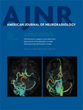
- Fig 1.
3D blood flow visualization in a left medial occipital AVM (AVM-17) with time-integrated pathlines (A) and quantification of the peak velocity and blood flow in the major feeding arteries (B, example for LPCA) and a perfusion model (C), including 3 pairs of ROIs (Pi/Pc, Hi/Hc, and Ri/Rc) used for perfusion analysis in CBF (D), CBV (E), and MTT (F) images (example for AVM-4). LMCA indicates left MCA; LPCA, left PCA; RMCA, right MCA; RPCA, right PCA; Contra, contralateral artery; Pc, Hc, and Rc, corresponding ROIs in the contralateral hemisphere.
- Fig 2.
Phase-contrast MRA (PC-MRA), time-integrated 3D pathlines, and perfusion (CBF) for 4 patients with AVMs (SMG = 1–4). AVM arterial feeding (solid white arrows) and venous draining patterns (open white arrows) can be clearly appreciated in the PC-MRA and 4D flow pathlines. The draining veins of AVM-17 and AVM-14 are obscured by pathlines and are not shown. 4D flow pathlines also illustrate the distribution of blood flow velocities in the entire brain. Note the flow voids (thin white arrows) within the nidus for AVM-11, AVM-17, and AVM-14 due to high transnidal shunt flow. LPICA indicates left posterior inferior cerebellar artery; LACA, left anterior cerebral artery; DV, draining vein; RTS, right transverse sinus; StrS, straight sinus; SSS, superior sagittal sinus.
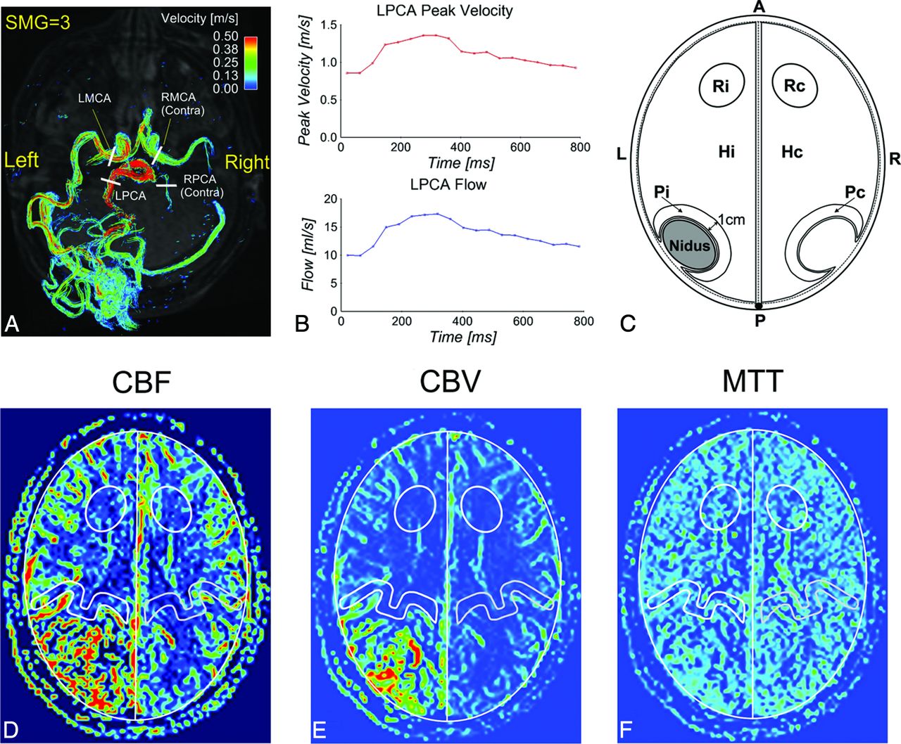
- Fig 3.
Distribution of peak velocities and blood flow in the AVM feeding (Feeders) and corresponding normal contralateral arteries (Contra) (A and E), as well as in the major feeding arteries (B and F), draining veins (C and G), and the straight sinus (D and H) for the low (SMG-A) and high (SMG-B) grade AVM groups. The feeding arteries had significantly higher peak velocity and blood flow compared with the normal contralateral arteries. The high-grade AVM group (SMG-B) had significantly higher peak velocity and blood flow in the major feeding arteries and the straight sinus and significantly higher blood flow in the draining veins compared with the low-grade group (SMG-A). The asterisk indicates significant difference with P < .05; NS, not significant.
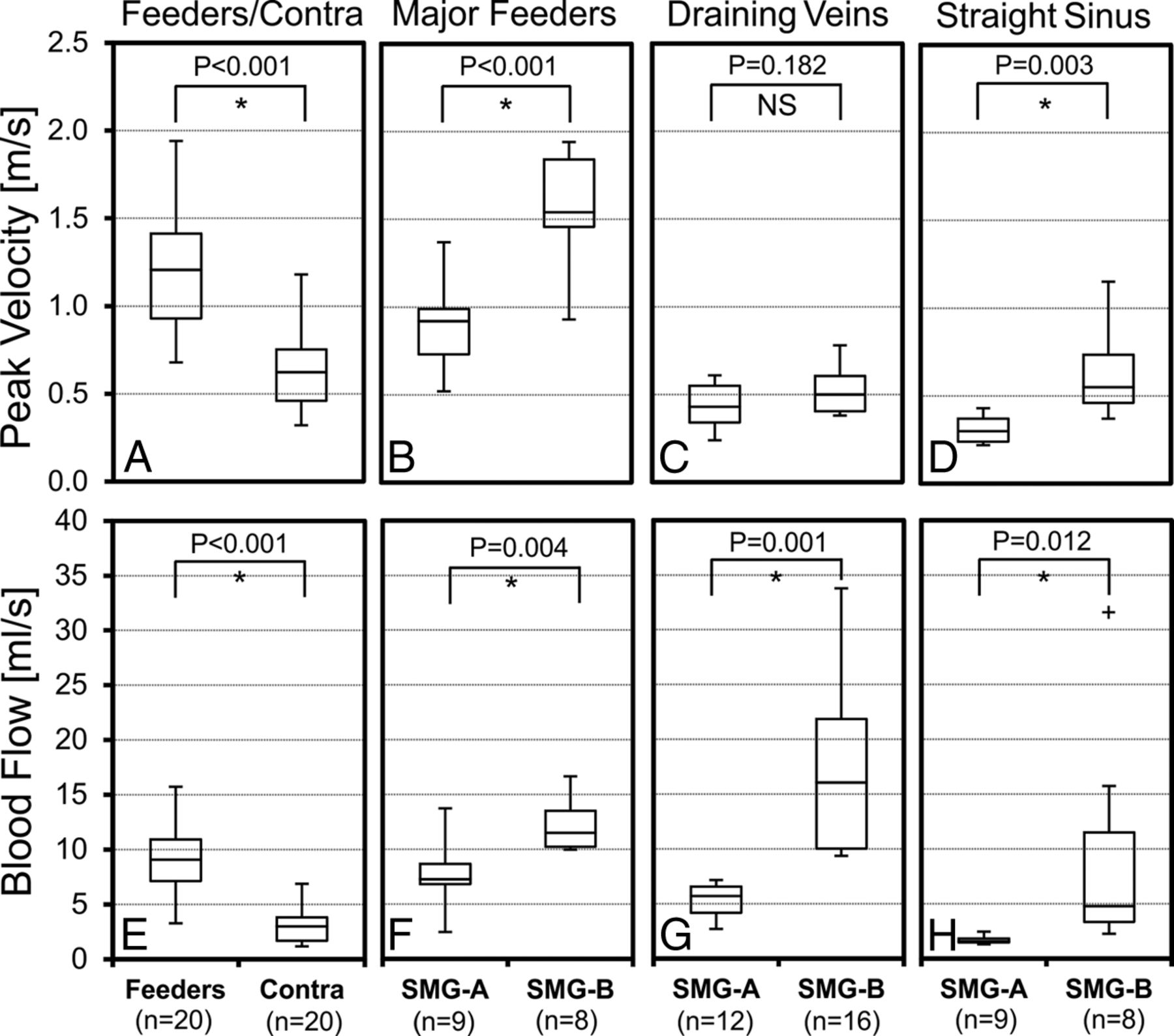
- Fig 4.
Ipsilateral-to-contralateral perfusion ratios (CBF, CBV, and MTT ratios) in the 3 pairs of ROIs (Pi/Pc, Hi/Hc, and Ri/Rc) for 16 AVMs (A) and perfusion ratios in the low (SMG-A, n = 9) and high (SMG-B, n = 7) grade AVM groups for CBF (B), CBV (C), and MTT (D). The asterisk indicates a significant difference between perinidal (Pi/Pc) and remote (Ri/Rc) perfusion ratios.


Tables
Demographics and AVM features of the 17 patients with AVMs included in the study
AVM (No.) Age (yr) Sex Nidal Size (cm3) Drainage Eloquence SMG (1–5) Major Feeding Arteries 1 43 M 5.6 × 4.1 × 5.1 D+S N 3 LMCA, LACA, RACA 2 29 M 4.0 × 3.5 × 3.4 D+S N 3 RPcomA, RPCA 3 43 M 2.7 × 3.1 × 3.1 D+S Y 3 RPcomA, RPCA 4 21 F 4.0 × 3.4 × 3.8 D+S Y 4 LMCA, LPCA, LMMA 5 22 M 2.5 × 2.3 × 2.4 D+S N 2 RMCA 6 68 F 4.0 × 3.0 × 2.5 D+S Y 4 LACA, RACA, RMCA 7a 34 M 3.3 × 2.3 × 2.5 S N 1 RACA 8 40 F 3.0 × 2.0 × 4.0 D+S N 2 LMCA, LPCA 9 25 F 2.3 × 2.3 × 1.7 D+S N 2 RMCA 10 52 F 2.4 × 2.1 × 2.6 S N 1 LPICA 11a 16 F 2.3 × 2.8 × 2.8 S Y 2 LACA, LMCA 12 55 M 2.1 × 3.6 × 2.6 S N 2 LMCA, LPCA 13 49 M 1.9 × 2.4 × 2.4 S N 1 RMCA 14 41 F 6.6 × 3.7 × 5.2 D+S Y 4 LMCA, LPCA, LACA 15 29 M 5.4 × 3.7 × 5.5 D+S Y 4 RMCA, RPCA 16a 36 M 3.6 × 3.4 × 3.2 S N 2 RACA, RMCA 17 66 M 2.6 × 2.3 × 1.7 D+S Y 3 LMCA, LPCA Note:—D indicates deep; S, superficial; L, left; R, right; Y, yes; N, no; LPICA, left posterior inferior cerebellar artery; LACA, left anterior cerebral artery; RACA, right anterior cerebral artery; RPcomA, right posterior communicating artery; LMMA, left middle meningeal artery.
↵a Contralateral arteries not visible in the 4D flow data.


Jump to section
Related Articles
Cited By...
- Hemodynamic Analysis of Cerebral AVMs with 3D Phase-Contrast MR Imaging
- How Flow Reduction Influences the Intracranial Aneurysm Occlusion: A Prospective 4D Phase-Contrast MRI Study
- In-room assessment of intravascular velocity from time-resolved rotational angiography in patients with arteriovenous malformation: a pilot study
- In Vivo Assessment of the Impact of Regional Intracranial Atherosclerotic Lesions on Brain Arterial 3D Hemodynamics
- Age-Related Changes of Normal Cerebral and Cardiac Blood Flow in Children and Adults Aged 7 Months to 61 Years