
Article Figures & Data
Figures
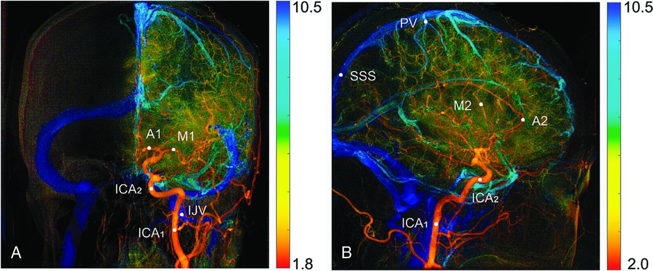
- Fig 1.
Color-coded DSA images in the PA (A) and lateral (B) views. Eleven ROIs are manually selected for quantification of perfusion parameters. ICA1 on the PA view and ICA1 on lateral view are planned to be at the same place.
- Fig 2.
The measured (blue open circle) and fitted (orange line) time-concentration curves. Hemodynamic parameters such as maximum enhancement, time to peak, bolus arrival time, wash-in slope, washout slope, and full width at half maximum are all derived from the fitted curve.
- Fig 3.
Measured (blue circle) and fitted (orange line) time-concentration curves of representative ROIs before the stent-placement treatment: ICA2 (A), M2 (B), PV (C), SSS (D). Note that the time-concentration curve of the PV (C) lacks the middle and lower descending portion. The ME in the time-concentration curve of the SSS (D) is at the last temporal point. AU indicates arbitrary unit.
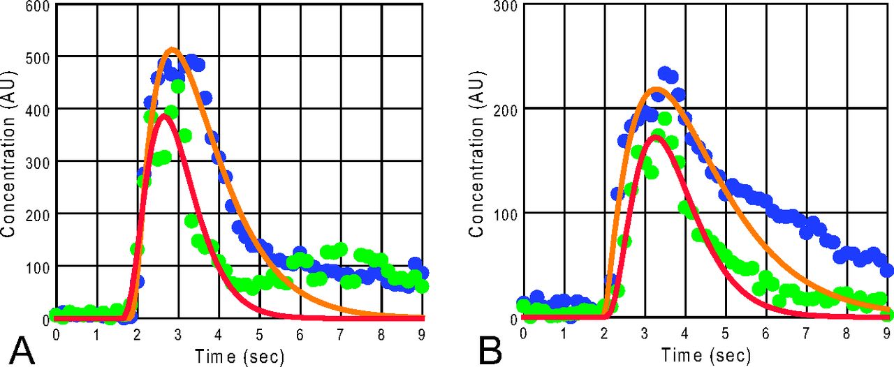
- Fig 4.
A comparison of the time-concentration curves before and after the stent placement treatment for the ICA2 (A) and M2 (B) in the lateral view. The blue solid circles are measured data, and orange lines are the fitted curves before the treatment. The green solid circles and red lines are measured data and fitted curves, respectively, after treatment. AU indicates arbitrary unit.
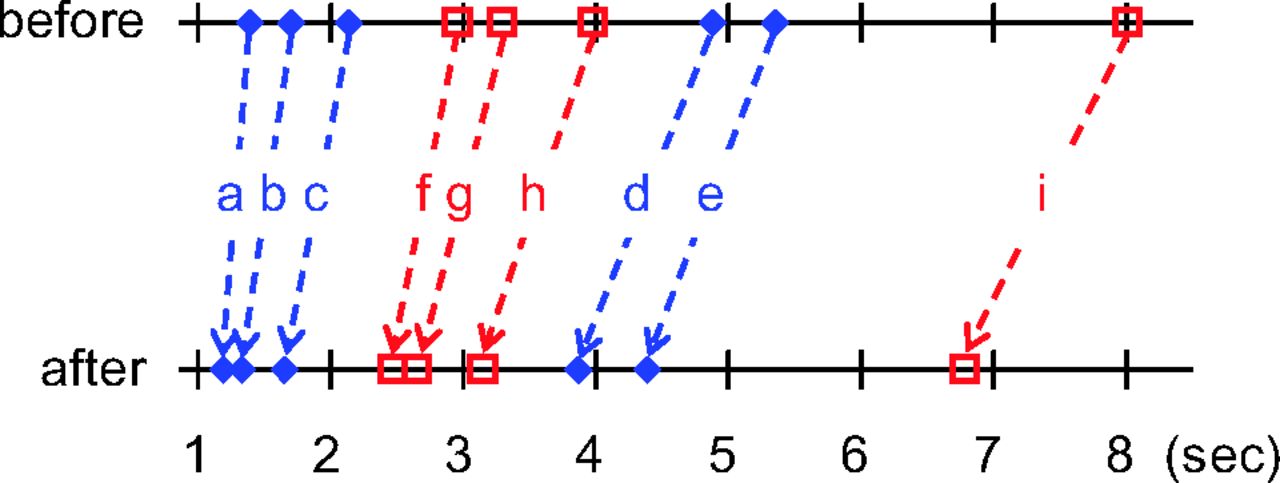
- Fig 5.
Comparison of the mean BAT (blue, a–e) and mean TTP (red, f–i) before (upper line) and after (lower line) treatment for different vascular ROIs in the lateral view: ICA1 (a and f), ICA2 (b and g), M2 (c and h), PV (d and i), and SSS (e). The TTP and rTTP are reduced after treatment in the extracranial (a and f) and intracranial segments, being more obvious in segments between the ICA1 and the PV (a–d and f–i) and less obvious from PV to SSS (d and e).


Tables
ROI Locations PA View Lateral View ICA1 ICA2 M1 A1 IJV ICA1 ICA2 M2 A2 PV SSS ME .27 .11 .15 .79 NA .68 .99 .20 * .55 NA TTP * * * * NA * * * * * NA rTTP – *b *b *b NA – *c *d *d *d NA BAT * * * * .32 * * * * * * FWHM * * * .08 NA * * * * NA NA WI .97 .88 .09 .64 .15 .37 .20 .12 .06 .20 NA WO * * * * NA * * * * NA NA AUC * * .06 .39 NA * * * .83 NA NA Note:—NA indicates not available because of incomplete data; –, not calculated; *, significant difference before and after stenting (P < .05); IJV, internal jugular vein.
↵a Details for significant results are shown in Tables 2 and 3.
↵b TTP relative to ICA1 in the PA view.
↵c TTP relative to ICA1 in the lateral view.
↵d TTP relative to ICA2 in the lateral view.
- Table 2:
Results for the hemodynamic parameters with significant changes (P < .05) after the stenting procedure in the PA view
ROI Before Treatment (Mean ± SD) After Treatment (Mean ± SD) No. TTP (sec) ICA1 2.89 ± 0.62 2.46 ± 0.27 34 ICA2 3.16 ± 0.70 2.60 ± 0.25 34 M1 3.70 ± 0.92 2.95 ± 0.31 34 A1 3.42 ± 0.67 2.95 ± 0.37 18 rTTP (sec) ICA1–ICA2 0.27 ± 0.22 0.14 ± 0.13 34 ICA1–M1 0.81 ± 0.43 0.49 ± 0.18 34 ICA1–A1 0.74 ± 0.25 0.54 ± 0.23 18 BAT (sec) ICA1 1.37 ± 0.29 1.15 ± 0.20 34 ICA2 1.63 ± 0.37 1.27 ± 0.29 34 M1 1.91 ± 0.48 1.49 ± 0.31 34 A1 1.85 ± 0.45 1.51 ± 0.31 18 FWHM (sec) ICA1 2.54 ± 0.81 1.95 ± 0.46 34 ICA2 2.53 ± 0.89 1.96 ± 0.50 34 M1 3.27 ± 0.91 2.62 ± 0.63 34 WO (AU/s) ICA1 −209 ± 79 −264 ± 80 34 ICA2 −202 ± 80 −247 ± 91 34 M1 −86 ± 44 −112 ± 44 34 A1 −73 ± 29 −90 ± 30 18 AUC (AU/sec) ICA1 1268 ± 426 942 ± 324 34 ICA2 1188 ± 476 866 ± 269 34 Note:—No. indicates number of cases measured; AU, arbitrary unit.
- Table 3:
Results for the hemodynamic parameters with significant changes (P < .05) after the stenting procedure in the lateral view
ROI Before Treatment (Mean ± SD) After Treatment (Mean ± SD) No. TTP (sec) ICA1 2.96 ± 0.69 2.47 ± 0.25 34 ICA2 3.30 ± 0.81 2.66 ± 0.27 34 M2 3.98 ± 0.98 3.15 ± 0.30 34 A2 3.70 ± 0.88 2.97 ± 0.41 17 PV 8.01 ± 1.28 6.79 ± 0.68 30 rTTP (sec) ICA1–ICA2 0.34 ± 0.24 0.19 ± 0.13 34 ICA2–M2 0.68 ± 0.25 0.50 ± 0.16 34 ICA2–A2 0.66 ± 0.38 0.34 ± 0.17 17 ICA2–PV 4.73 ± 1.00 4.14 ± 0.69 30 M2–PV 4.06 ± 0.93 3.67 ± 0.65 30 BAT (sec) ICA1 1.39 ± 0.37 1.19 ± 0.26 34 ICA2 1.70 ± 0.37 1.33 ± 0.29 34 M2 2.14 ± 0.54 1.65 ± 0.34 34 A2 2.17 ± 0.69 1.74 ± 0.37 17 PV 4.88 ± 0.94 3.87 ± 0.69 30 SSS 5.35 ± 1.53 4.39 ± 1.09 34 FWHM (sec) ICA1 2.66 ± 1.01 1.93 ± 0.41 34 ICA2 2.66 ± 0.93 2.01 ± 0.50 34 M2 3.22 ± 0.88 2.54 ± 0.65 34 A2 2.90 ± 0.84 2.27 ± 0.83 17 WO (AU/s) ICA1 −217 ± 110 −281 ± 91 34 ICA2 −229 ± 91 −305 ± 107 34 M2 −90 ± 49 −120 ± 46 34 A2 −79 ± 37 −116 ± 34 17 AUC (AU/sec) ICA1 1413 ± 692 1002 ± 340 34 ICA2 1561 ± 753 1160 ± 385 34 M2 879 ± 377 750 ± 299 34 ME (AU) A2 203 ± 74 234 ± 72 17 Note:—No. indicates number of cases measured; AU, arbitrary unit.


Jump to section
Related Articles
Cited By...
- Combined collaterals and hemodynamic features to predict the prognosis in acute ischemic stroke patients undergoing mechanical thrombectomy
- Quantitative evaluation of hemodynamics after partial embolization of brain arteriovenous malformations
- Carotid webs produce greater hemodynamic disturbances than atherosclerotic disease: a DSA time-density curve study