
Article Figures & Data
Figures
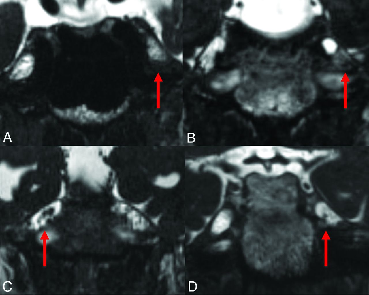
- Fig 1.
Coronal CISS precontrast images at the level of Meckel's cave. A, Decreased T2 signal intensity and poorly delineated nerve rootlets in the left Meckel's cave after rhizotomy. Note a normal-appearing right Meckel's cave. B, A different patient with clumping of the nerve rootlets inferiorly within the left Meckel's cave post-rhizotomy. C, Central clumping of nerve rootlets in the right Meckel's cave post-rhizotomy. D, A different patient with more subtle clumping of the nerve rootlets in the left Meckel's cave and subtle decreased CISS SI post-rhizotomy.
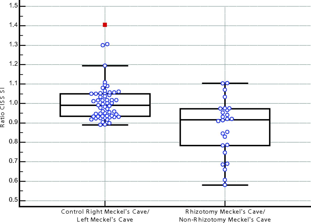
- Fig 2.
Graph showing the ratio of CISS SI of the right and left Meckel's caves in control patients and rhizotomy/nonrhizotomy in Meckel's caves in post-rhizotomy patients. The control group ratio was 0.99 compared with 0.87 for patients who underwent rhizotomy (P < .001).
- Fig 3.
Coronal CISS precontrast at the level of Meckel's caves. Encephalomalacia of the medial left temporal lobe (straight arrow) adjacent to Meckel's cave. Also note clumping of the nerve rootlets in the left Meckel's cave (curved arrow) status post rhizotomy.


Tables
Post-Rhizotomy Treatment-Naïve P Value No. of Subjects 26 54 Age (mean) (range) (yr) 60 (27–85) 55 (29–74) .09 Sex 6 Men, 20 women 20 Men, 34 women .31 - Table 2:
Frequency of findings within Meckel's cave in treatment-naïve versus post-rhizotomy patientsa
Post-Rhizotomy Treatment-Naïve P Value Subjective clumping 16/26 (62%) 3/54 (6%) <.001 Decreased CISS 13/26 (50%) 3/54 (6%) <.001 Subjective clumping without decreased CISS SI 4/26 (15%) 0/54 (0%) .01 ↓ CISS SI without clumping 1/26 (4%) 0/54 (0%) .33 Subjective nerve clumping and ↓ CISS SI 12/26 (46%) 3/54 (6%) <.001 Subjective nerve clumping and/or ↓ CISS SI 17/26 (65%) 3/54 (6%) <.001 Note:—↓ indicates decrease.
↵a Interobserver agreement = 90%, κ = 0.69.
Control Right MC/Left MC (mean ratio ± SD) Rhizotomy MC/Contralateral MC (mean ratio ± SD) P Value Ratio of CISS SI 0.99 (±0.09) 0.87 (±0.15) <.001 Note:—MC indicates Meckel cave.
- Table 4:
Frequency of findings in the adjacent structures in treatment-naïve versus post-rhizotomy patients
Post-Rhizotomy Treatment-Naïve P Value Interobserver Agreement κ Encephalomalacia 8/26 (31%) 0/54 (0%) <.001 98.8% 0.93 Hematoma 1/26 (4%) 0/54 (0%) .33 100% 1 Atrophy of muscles of mastication 3/26 (10%) 3/54 (4%) .38 96.3% 0.64 CN V enhancement 0/26 (0%) 0/54 (0%) 1.00 100% 1 Vascular injury 0/26 (0%) 0/54 (0%) 1.00 100% 1 Note:—CN indicates cranial nerve.


Jump to section
Related Articles
Cited By...
- No citing articles found.