
Article Figures & Data
Figures
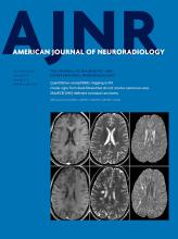
- Fig 1.
Three-month-old patient. A, Axial CT demonstrates interhemispheric subdural hemorrhage (arrowhead) and symmetric edema of the bilateral occipital lobes (arrows). There is abnormal low attenuation in the basal ganglia. Superior frontal parietal edema is present as well (not shown). B, Sagittal midline STIR image shows interspinous ligamentous injury at all cervical levels (arrows), paraspinous muscular injury, nuchal ligament injury (arrowhead), and marrow edema involving the lower cervical and upper thoracic vertebral bodies, most prominent at T1 (long arrow).
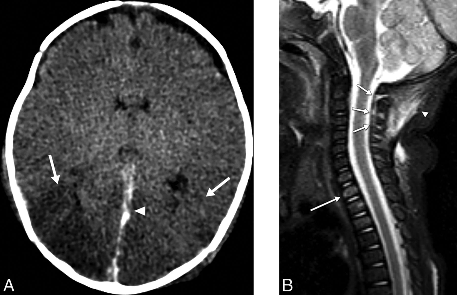
- Fig 2.
Five-month-old patient. Sagittal midline STIR image shows a dens fracture (arrowhead) and disruption of the inferior tectorial ligament anterior longitudinal ligament junction (short arrow). Extensive injury to the C1–2 interspinous ligamentous structures (long arrow) and edema in the posterior paraspinal musculature are present. Diffuse parenchymal injury was present on CT and MR imaging (not shown).
- Fig 3.
Two-month-old patient. A, Axial trace DWI shows diffuse restricted diffusion involving the bilateral cerebral hemispheres. B, Sagittal paramidline STIR image shows abnormal fluid within the atlanto-occipital joint space with mild distension (arrow), consistent with capsular injury.
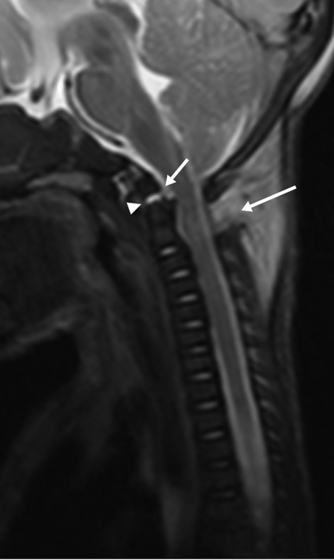
- Fig 4.
Four-month-old patient. Sagittal midline T2WI shows abnormal posterior extradural fluid within the cervical and thoracic region (arrows). The fluid was isointense to CSF on T1-weighted images. Interspinous ligamentous injury is present (not shown).
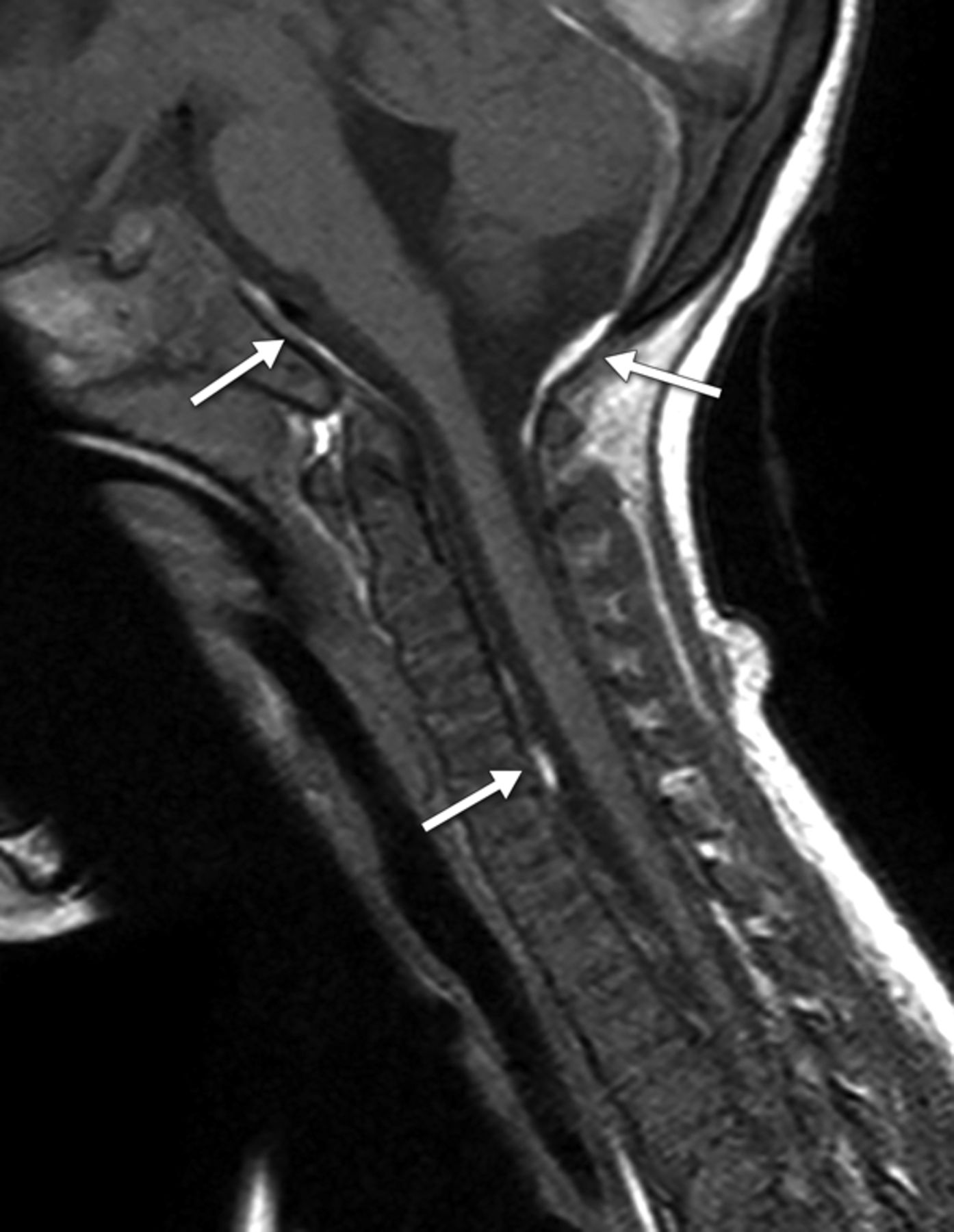
- Fig 5.
Three-month-old patient. Sagittal midline T1WI shows intracranial and intraspinal T1 hyperintense subdural hemorrhage (arrows).
- Fig 6.
Seven-month-old patient. A, Axial DWI shows focal restricted diffusion in the left parietal lobe (arrow). B, Sagittal midline STIR image demonstrates interspinous ligamentous injury (arrows) and injury to the nuchal ligament (arrowhead).
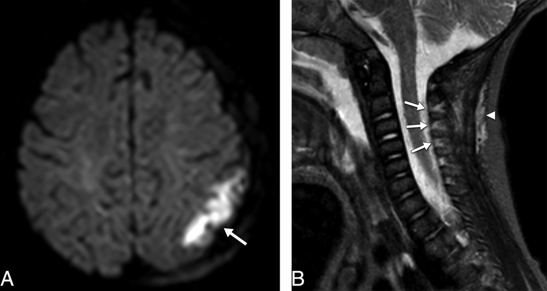
- Fig 7.
Seven-month-old patient. A, Axial DWI shows diffuse cerebral restricted diffusion, worse on the left. There is mild midline shift, left to right, caused by a left hemispheric convexity subdural hemorrhage (not shown). B, Sagittal midline STIR image demonstrates interspinous (long arrows) and nuchal ligament injury (short arrow). There is also prevertebral edema (arrowhead).


