
Article Figures & Data
Figures
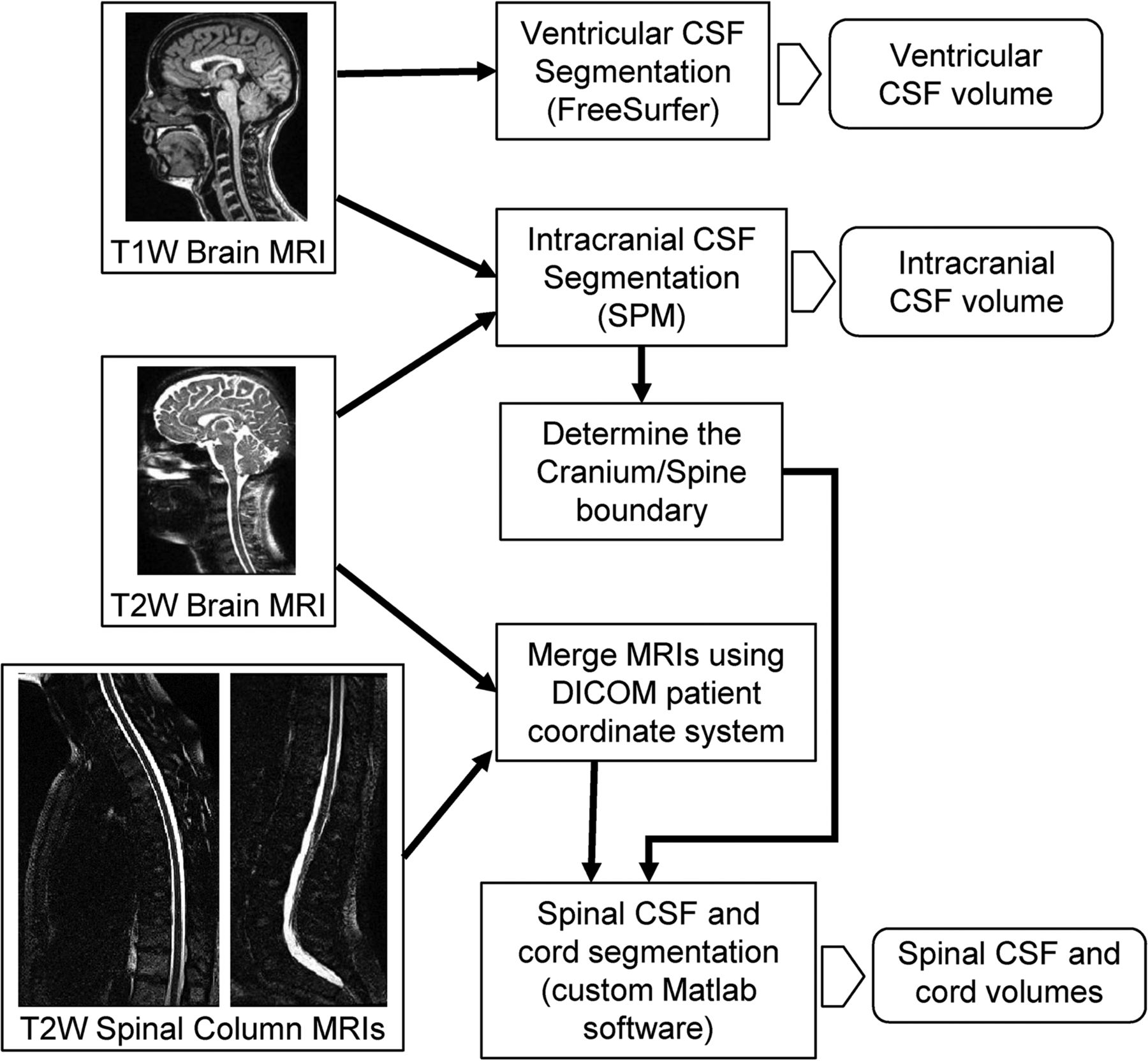
- Fig 1.
Flow chart of the CSF segmentation method. T1- and T2-weighted brain images are used to obtain the ventricular and intracranial CSF volumes by using publicly available software packages. Spinal CSF and cord volumes are obtained using the 3 T2-weighted scans with a custom-developed software.
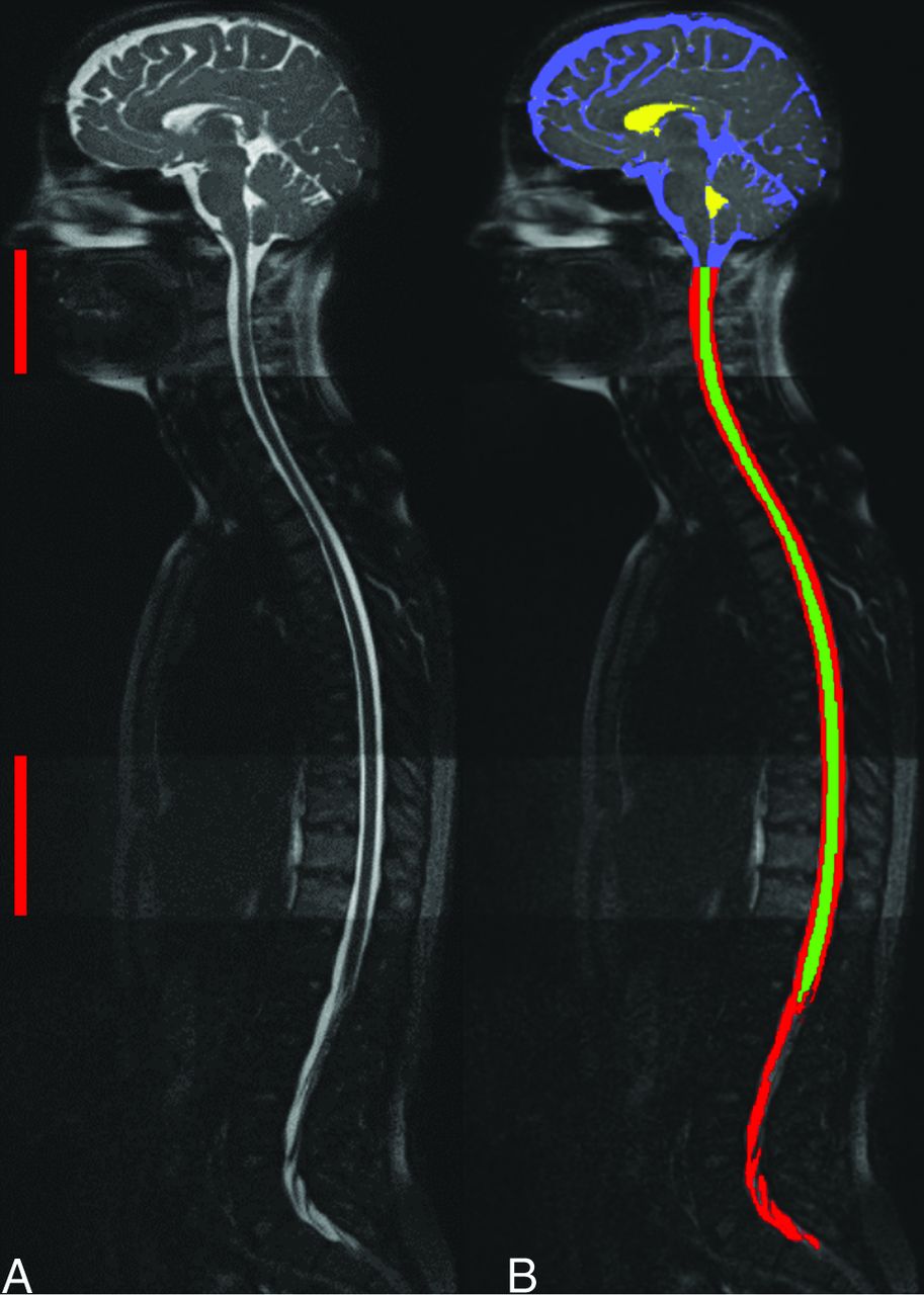
- Fig 2.
A mid-sagittal image demonstrating complete coverage of the CNS generated by merging 3 separate acquisitions (A) with overlapping coverage indicated by red bars on the left. B, Segmentation of cranial (blue), ventricular (yellow), and spinal (red) CSF and cord (green).
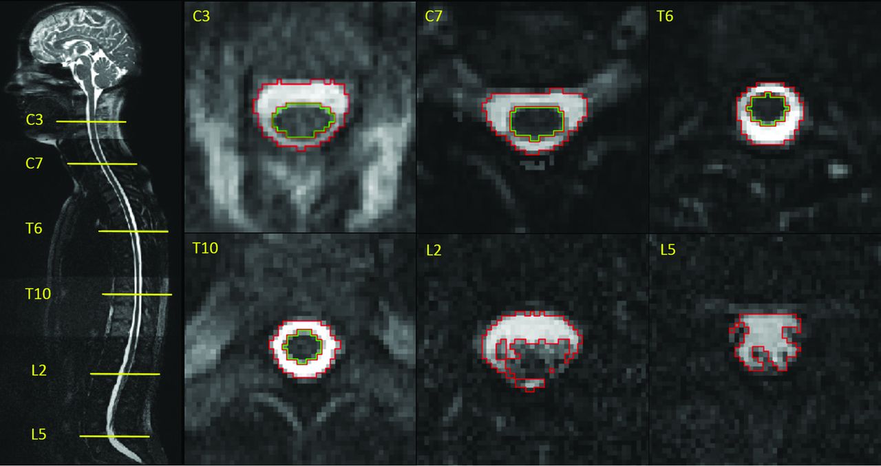
- Fig 3.
Sample CSF (red outline) and cord (green) segmentations along the spinal column at the level of C3, C7, T6, T10, L2, and L5 vertebrae.
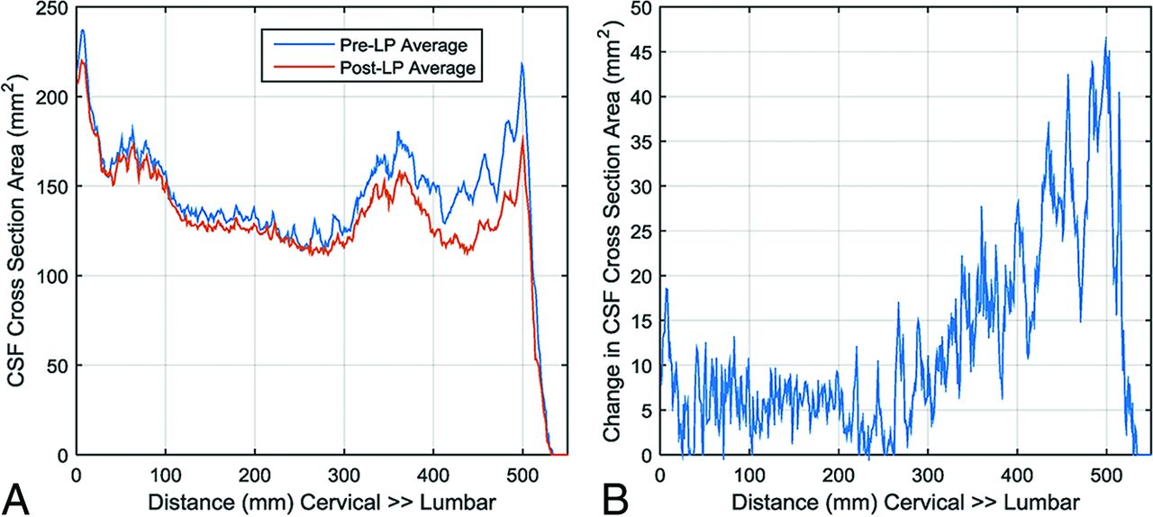
- Fig 4.
Average CSF cross-sectional area before and after lumbar puncture (A) and average change in CSF cross-sectional area following CSF withdrawal with respect to the distance from the foramen magnum to the caudal end of the thecal sac (B).
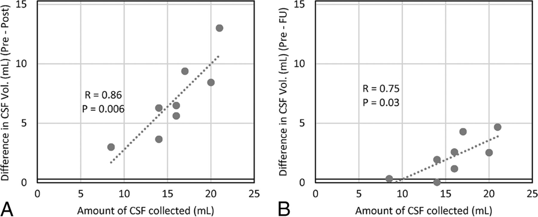
- Fig 5.
The relationship between the CSF volume withdrawn during lumbar puncture and CSF volume change in the spinal canal between the pre- and post-lumbar puncture scan (A) and the pre- and the 2-week follow-up scan (B) as measured by the proposed method.


Tables
Age (yr) BMI Opening Pressure (cm H2O) EVC CSF Vol (mL) Ventricular CSF Vol (mL) SC CSF Vol (mL) Total CSF Vol (mL) Cord Vol (mL) Subject No. 1 28 34 39 164 18.9 86.7 269 20.8 2 37 33 45 169 15.6 83.1 268 23.5 3 19 26 17 157 16.4 76.8 250 21.9 4 33 27 29 171 11.6 77.3 260 20.7 5 28 43 34 213 12.8 67.6 293 19.9 6 32 40 34 166 28.9 89.8 285 23.9 7 30 41 25 176 15.8 68.9 261 18.8 8 22 30 40 98 9.2 69.9 177 18.6 Mean 29 ± 6 34 ± 6.7 33 ± 9.1 164 ± 31.8 16.2 ± 6.0 77.5 ± 8.4 258 ± 35.6 21.0 ± 2.0 Variability (SD/Mean) 19% 37% 11% 14% 10% Note:—BMI indicates body mass index; EVC, extraventricular cranial; SC, spinal canal; Vol, volume.
- Table 2:
Pre- to post-lumbar puncture changes in ICP and CSF volume differences in the various compartments, amounts of CSF withdrawn, and the calculated effective CSF production rates
Closing-Opening Pressure (cm H2O) EVC CSF Vol Change (mL) Ventricular CSF Vol Change (mL) SC CSF Vol Change (mL) Total CSF Vol Change (mL) Amount of CSF Collected (mL) Effective CSF Production Rate (mL/min) Subject No. 1 NA −1.6 0.0 −5.3 −6.9 −16.0 0.25 2 −26 −2.0 −0.1 −6.2 −8.2 −16.0 0.24 3 −3 −3.6 −0.1 −6.0 −9.6 −14.0 NAa 4 −15 0.7 0.2 −3.3 −2.5 −14.0 0.65 5 −16 2.0 0.1 −12.7 −10.5 −21.0 0.43 6 −20 −1.6 −0.6 −8.1 −10.3 −20.0 0.63 7 −12 −1.0 −0.2 −2.7 −3.9 −8.5 0.24 8 −28 0.8 0.1 −9.1 −8.2 −17.0 0.40 Mean −17 ± 8.6 −0.8 ± 1.8 −0.1 ± 0.2 −6.7 ± 3.2 −7.5 ± 2.9 −15.8 ± 3.9 0.41 ± 0.18 P value .002b .24 .45 .0007b .0002b


