
Article Figures & Data
Figures
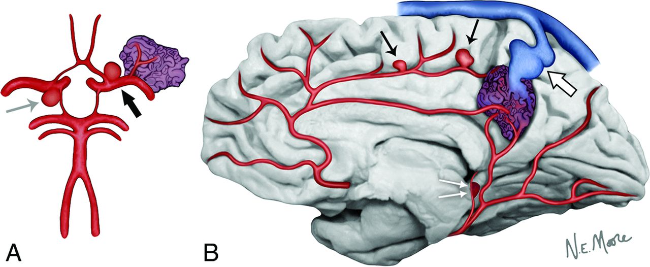
- Fig 1.
In this circle of Willis figure model (A), an AVM nidus in relation to a branch of the left middle cerebral artery is noted. At the left internal carotid artery bifurcation, a hemodynamically relevant aneurysm is located proximal to the feeding pedicle of the AVM nidus (proximal flow-related aneurysm) (black arrow). An unrelated aneurysm, with no hemodynamic connection to the AVM nidus, is present at the right posterior communicating artery origin (gray arrow). In this midsagittal view of the brain (B), distal flow-related aneurysms are seen to originate from the feeding arterial pedicles of the AVM nidus (black arrows). Arterial pseudoaneurysms are thought to be the result of the rupture of thin-walled small perforating arteries that supply the AVM and result from the unclotted portion of the hematoma still in communication with the vessel lumen and are very close to the ependymal surface (double white arrows). Finally, venous varices represent irregular, usually circumferential, enlargements of the venous outflow tract of the AVM nidus (large white arrow).
- Fig 2.
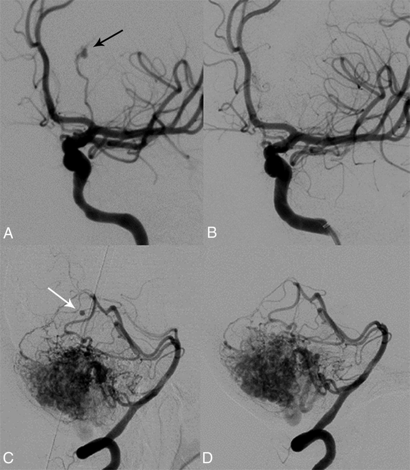
A 46-year-old woman with loss of consciousness and right hemiparesis due to an intracerebral hemorrhage in the left thalamus. Conventional angiography (oblique view) reveals a pseudoaneurysm (black arrow) of a left posterior communicating artery branch that was treated with N-butyl cyanoacrylate embolization (A). Complete pseudoaneurysm obliteration after endovascular treatment was achieved (B). Spontaneous regression of a pseudoaneurysm (white arrow) associated with a branch of the right superior cerebellar artery is noted in a 74-year-old woman with subarachnoid hemorrhage centered in the right ambient cistern (C and D).
- Fig 3.
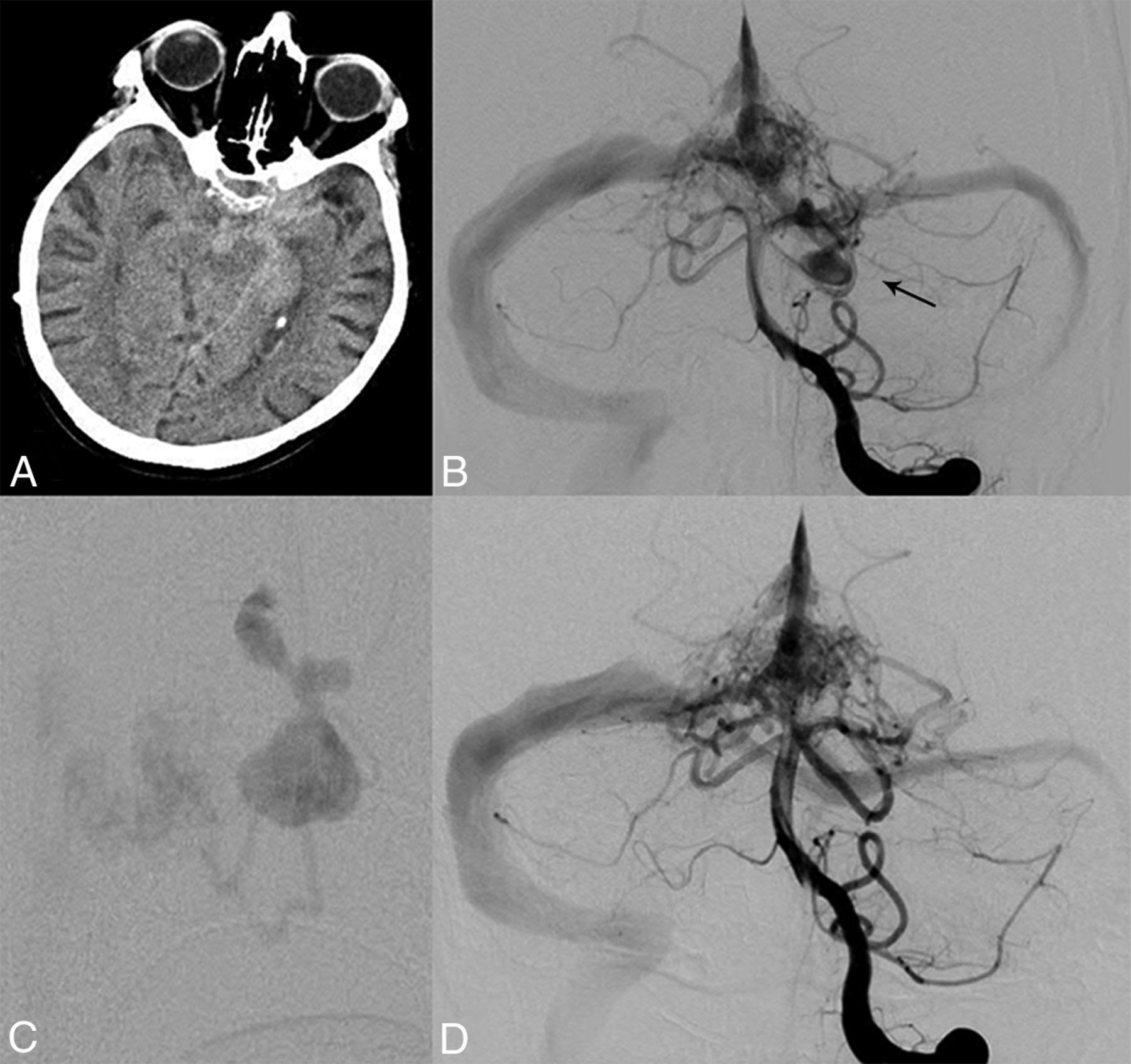
A 70-year-old man with subarachnoid hemorrhage centered in the prepontine cistern (A). Left vertebral artery angiography (anteroposterior view) reveals an AVM of the region of the torcula and a large irregular aneurysm of the left superior cerebellar artery (B and C). The presence of isolated subarachnoid hemorrhage suggests the aneurysm as the source of hemorrhage. The aneurysm was treated selectively with N-butyl cyanoacrylate embolization as noted on postprocedural angiography (anteroposterior view) (D), while the treatment of the AVM nidus was deferred.
- Fig 4.
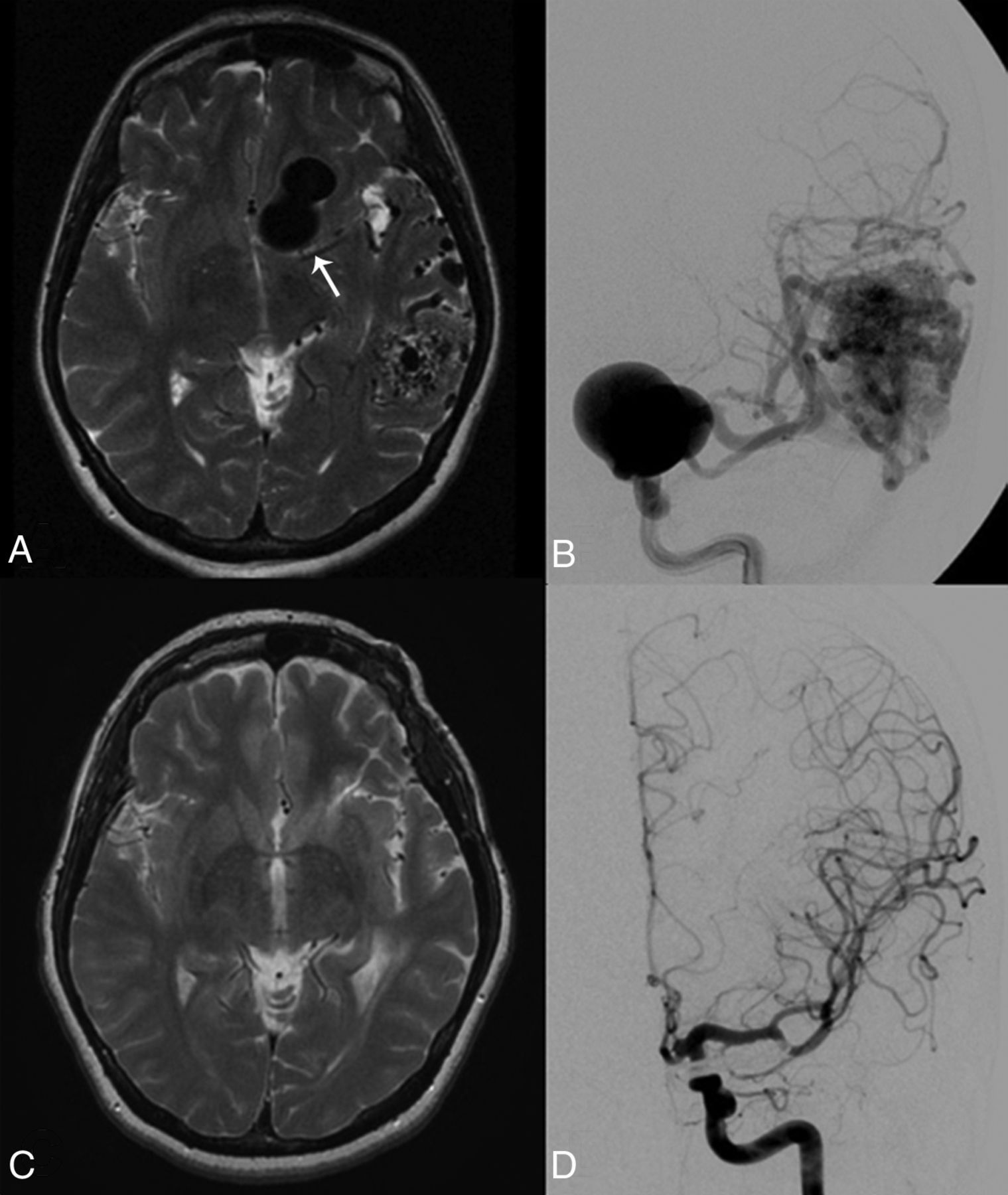
A 42-year-old woman who lost consciousness while dancing. MR imaging (T2 axial) reveals a large flow void suggestive of a giant left aneurysm (arrow) and an associated left temporal lobe AVM (A). Conventional angiography (anteroposterior projection) confirmed a giant left ICA aneurysm and the left temporal AVM (B). The aneurysm was treated with surgical clipping, and the patient underwent stereotactic radiosurgery for the AVM. Follow-up MR imaging (T2 axial) (C) and conventional angiography (D) 6 years later show complete exclusion of the aneurysm and obliteration of the AVM.


Jump to section
Related Articles
Cited By...
- Prevalence and Characteristics of Intracranial Aneurysms in Hereditary Hemorrhagic Telangiectasia
- Prevalence of Intracranial Aneurysms in Hereditary Hemorrhagic Telangiectasia: Report from a Single Reference Center
- Morphologic Change of Flow-Related Aneurysms in Brain Arteriovenous Malformations after Stereotactic Radiosurgery
- Morphologic Change of Flow-Related Aneurysms in Brain Arteriovenous Malformations after Stereotactic Radiosurgery