
Article Figures & Data
Figures
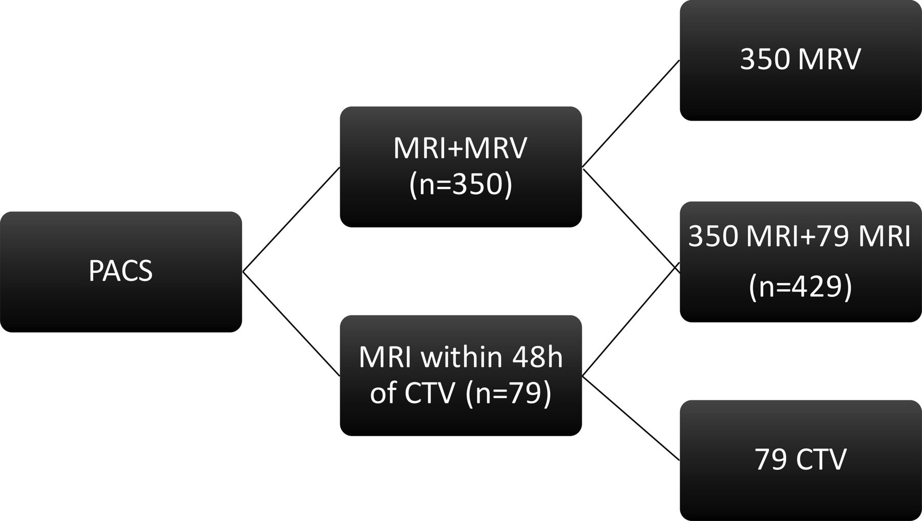
- Fig 1.
Patient selection and image separation.
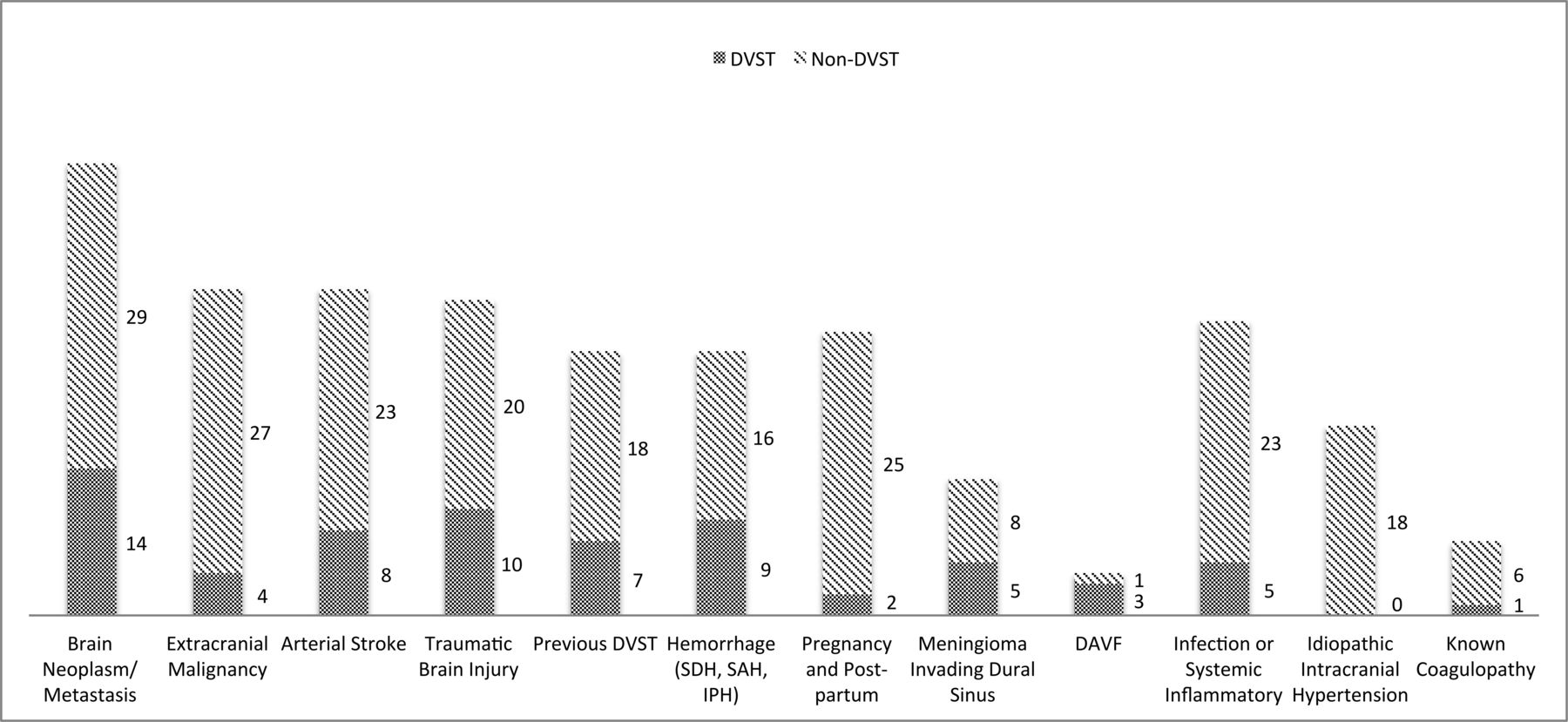
- Fig 2.
Etiology or underlying condition based on electronic medical records. SDH indicates subdural hemorrhage; IPH, intraparenchymal hemorrhage; DAVF, dural arteriovenous fistula.
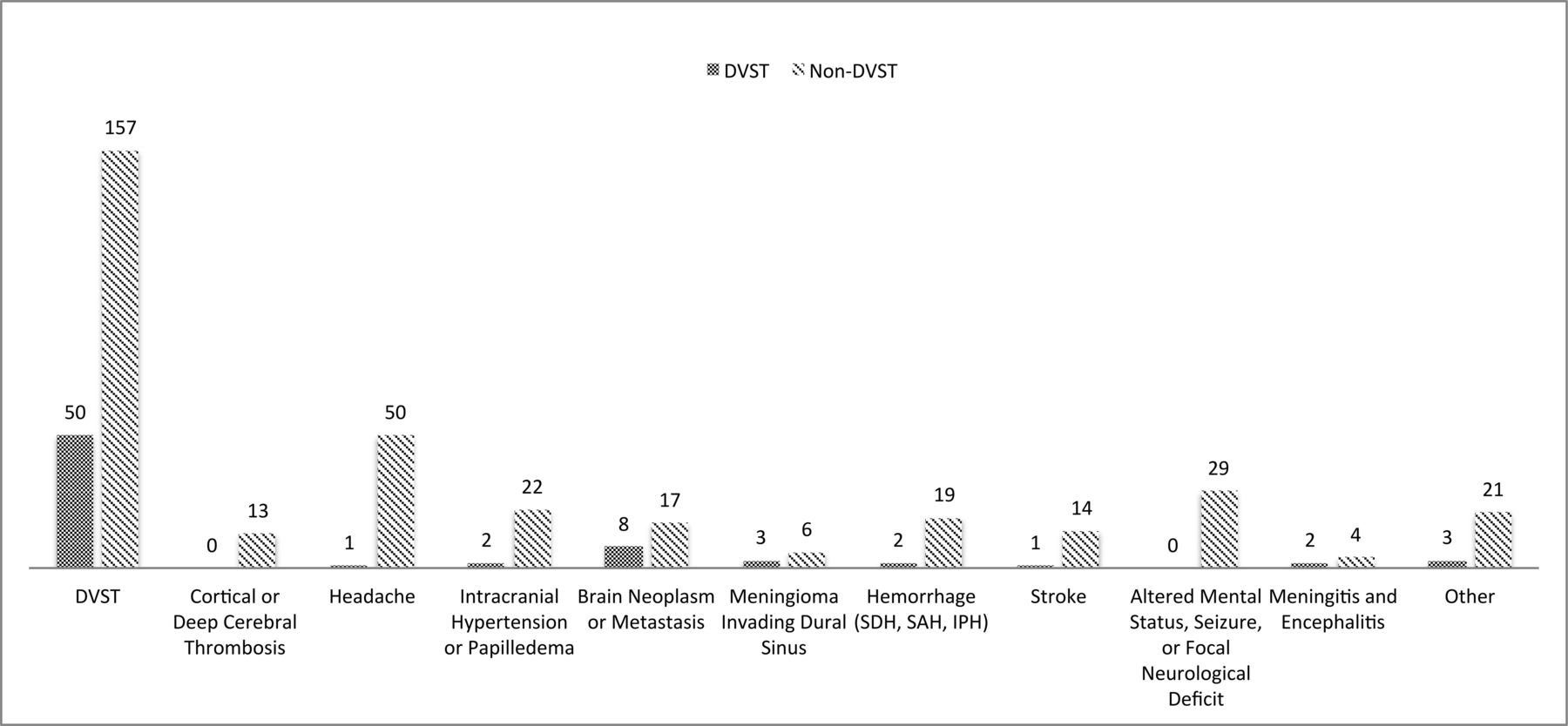
- Fig 3.
Indications for imaging based on the imaging requisition form. SDH indicates subdural hemorrhage; IPH, intraparenchymal hemorrhage.
- Fig 4.
MR imaging signs of dural venous sinus thrombosis from multiple cases. A, Hyperintense signal in the right transverse sinus on sagittal T1. B, Loss of flow void in the right transverse sinus on axial T2. C, Hyperintense signal in the right transverse sinus on FLAIR. D, Hyperintense signal in the right transverse sinus on DWI. E, Blooming artifacts in the right transverse sinus on GRE. F, Filling defect in the right transverse/sigmoid sinus on CE-SE-T1WI. G, Filling defect in the right transverse/sigmoid sinus on CE-3D-T1WI. H, Filling defect in the right transverse/sigmoid sinus on CTV. I, Filling defect in the right transverse/sigmoid sinus on MIP-CEMRV.


Tables
DVST (n = 72) Non-DVST (n = 357) Total (n = 429) Age (yr) (mean) 51.4 ± 16.8 45.8 ± 17.0 46.7 ± 17.1 Female (No.) (%) 45 (62.5) 239 (66.9) 284 (66.2) 3T MRI (No.) (%) 16 (22.2) 130 (36.4) 146 (34.0) - Table 4:
Interrater reliability for each MRI sequence individually and combined using the κ coefficient
κ Agreementa Sagittal T1 (n = 418) 0.28 Fair Axial T2 (n = 71) 0.34 Fair Axial GRE (n = 420) 0.40 Fair Axial FLAIR (n = 417) 0.34 Fair Axial DWI (n = 421) 0.33 Fair Axial CE-SE-T1WI (n = 241) 0.42 Moderate Axial CE-3D-T1WI (n = 199) 0.41 Moderate MRI total (n = 429) 0.50 Moderate ↵a Agreement was considered slight if κ values were 0–0.20; fair if, 0.21–0.40; moderate if, 0.41–0.60; substantial if, 0.61–0.80; and almost perfect if, 0.81–1.


