
Article Figures & Data
Figures
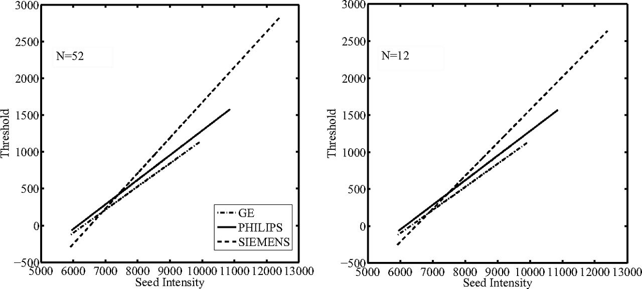
- Fig 1.
Threshold functions obtained after the training step for each different scanner manufacturer at the decreasing of the training set sample sizes (ie, number of patients included) as indicated. It is possible to observe that with decreasing sample size, the linear regression functions did not modify their trends.
- Fig 2.
Dice similarity coefficient values (top left), mean true-positive fraction/false-positive fraction values (top right), and mean false-negative fraction values (bottom left) are shown for each patient. In the bottom right, a scatterplot to compare manual lesion load with automatic lesion load is shown. The dashed line is the line of identity.
- Fig 3.
Example lesion segmentations for 2 patients (rows) from 2 different scanners by the proposed method (red) compared with the expert operator segmentation (blue). The corresponding T2-weighted images are shown in the right column.


Jump to section
Related Articles
Cited By...
- No citing articles found.