
Article Figures & Data
Figures
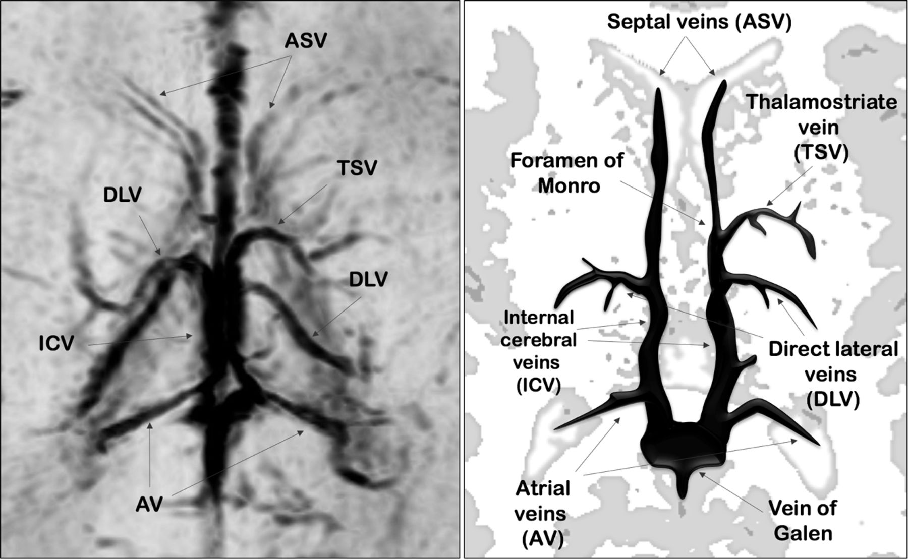
- Fig 1.
Axial-reformatted SWI venography and corresponding schematic representation of SV.
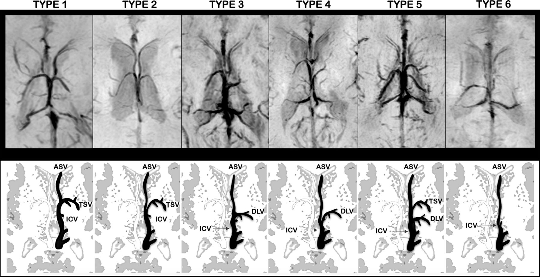
- Fig 2.
Axial-reformatted SWI venography and corresponding schematic representation of SV patterns. The left side of SWI venography and the black schematic vein represent the anatomic pattern. Type 1: The ASV joined the ICV at the level of the foramen of Monro and the TSV-ICV junction. The DLV was absent. Type 2: The ASV joined the ICV posterior to both the TSV-ICV junction and the foramen of Monro. The DLV was absent. Type 3: The ASV joined the ICV close to the site of DLV-ICV junction, posterior to the foramen of Monro. The TSV was absent. Type 4: The ASV joined the ICV posterior to both the foramen of Monro and DLV-ICV junction. The TSV was absent. Type 5: Both the TSV and DLV were present. Type 6: Both the TSV and DLV were absent. Atrial veins were not included in schematic representations.


Tables
Sequence Section Thickness (mm) Matrix Intersection (mm) TR (msec) TE (msec) FA (deg) SAR (W/kg) Bandwidth Axial T1-SE 3 232 × 110 1 780 16 NA <2.6 8.2 Axial T2-TSE 3 308 × 171 1 6923 140 NA 3.1 5.6 Coronal T2-TSE 3 232 × 171 1 6782 140 NA <3 6.7 3D T1 TFE 1 200 × 150 NA 9.8 4.6 10 <0.2 2.3 Axial DWI 4 108 × 104 0.4 2530 74 NA 0.3 12.9 Note:—FA indicates flip angle; NA, not available; SAR, specific absorption rate; SE, spin echo; TFE, turbo-field echo.
- Table 2:
Overall frequencies and percentages of SV patterns for each brain hemisphere in the 3 groups of neonatesa
Pattern VP MLP TN Total Hemispheres Left Right Left Right Left Right Type 1 28 (33.3%) 42 (50%) 15 (48.4%) 20 (64.5%) 36 (72%) 32 (64%) 173 Type 2 4 (4.8%) 6 (7%) 4 (12.9%) 0 (0%) 2 (4%) 3 (6%) 19 Type 3 36 (42.9%) 25 (29.8) 9 (29%) 7 (22.6%) 10 (20%) 13 (26%) 100 Type 4 7 (8.2%) 4 (4.8%) 3 (9.7%) 1 (3.2%) 2 (4%) 2 (4%) 19 Type 5 5 (6%) 2 (2.4%) 0 (0%) 2 (6.5%) 0 (0%) 0 (0%) 9 Type 6 4 (4.8%) 5 (6%) 0 (0%) 1 (3.2%) 0 (0%) 0 (0%) 10 Note:—MLP indicates moderate-to-late preterm neonates; TN, term neonates; VP, very preterm neonates.
↵a 84 VP neonates; 31 MLP neonates; 50 TN neonates.
- Table 3:
Frequencies and percentages of interhemispheric symmetry and asymmetry patterns in the 3 groups of neonates
VP MLP TN Total Neonates Pattern Interhemispheric symmetry 34 (40.5%) 15 (48.4%) 33 (66%) 82 Interhemispheric asymmetry 50 (59.5%) 16 (51.6%) 17 (34%) 83 Total neonates 84 31 50 165 Note:—MLP indicates moderate-to-late preterm neonates; TN, term neonates; VP, very preterm neonates.


