
Article Figures & Data
Figures
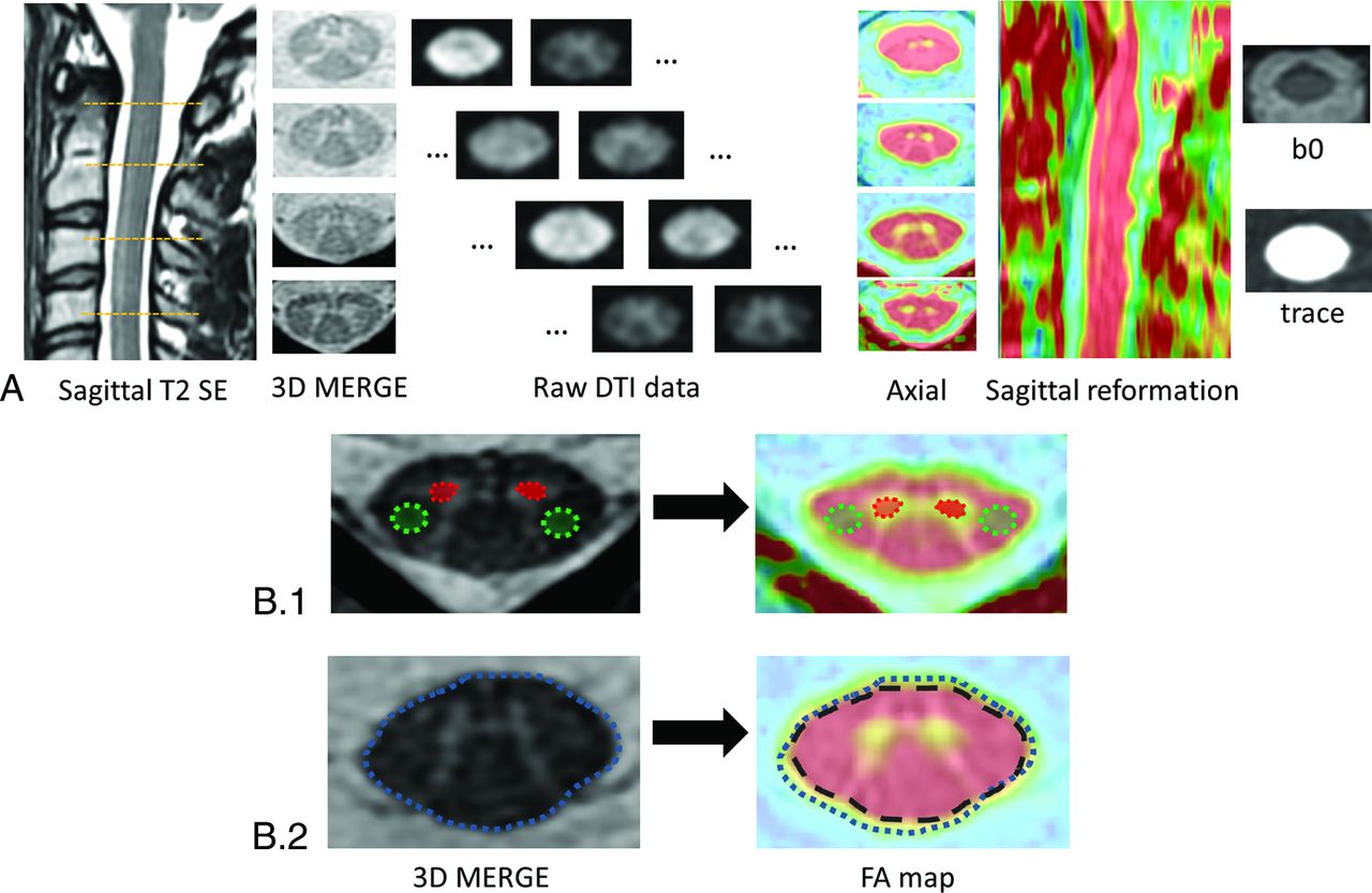
- Fig 1.
Postprocessing pipeline and analyses. A, Four sections were analyzed in detail: C1, C2, C3, and C4. Raw data diffusion-weighted images were treated for eddy current correction before generation of DTI parameters. We focused on FA because this parameter is the most commonly used. Fusions of FA and 3D-MERGE were created at these 4 levels as well as reconstructed on the sagittal orientation to facilitate the identification of distortions and pixel misregistration. B, ROI positioning: ROIs (B1) were manually delineated on 3D-MERGE, on the right and left anterior horns of the cord (red area) for gray matter, and on the right and left corticospinal tract (green area) for white matter, and then propagated on the coregistered FA map. B2, If needed, ROIs were manually adjusted to account for FA map distortion. Furthermore, because of the partial volume effect at the interface between CSF and the FA map, ROIs of the full section (blue dotted line, whose surface corresponded to S[Full Section − Merge]) were adjusted to remove the pixels subject to artifacts at the periphery of the ROI (black dashed line, whose area corresponded to S[Full Section − FA]).
- Fig 2.
Examples of MR images available for qualitative analysis. All the images came from the same subject. Cervical levels are located on 3D T2-MERGE and sagittal T2-spin echo. Fusion of FA − 3D MERGE clearly shows that f-FOV DTI and r-FOV 3N/20D are more distorted and more blurred with less anatomic precision than the other r-FOV images.
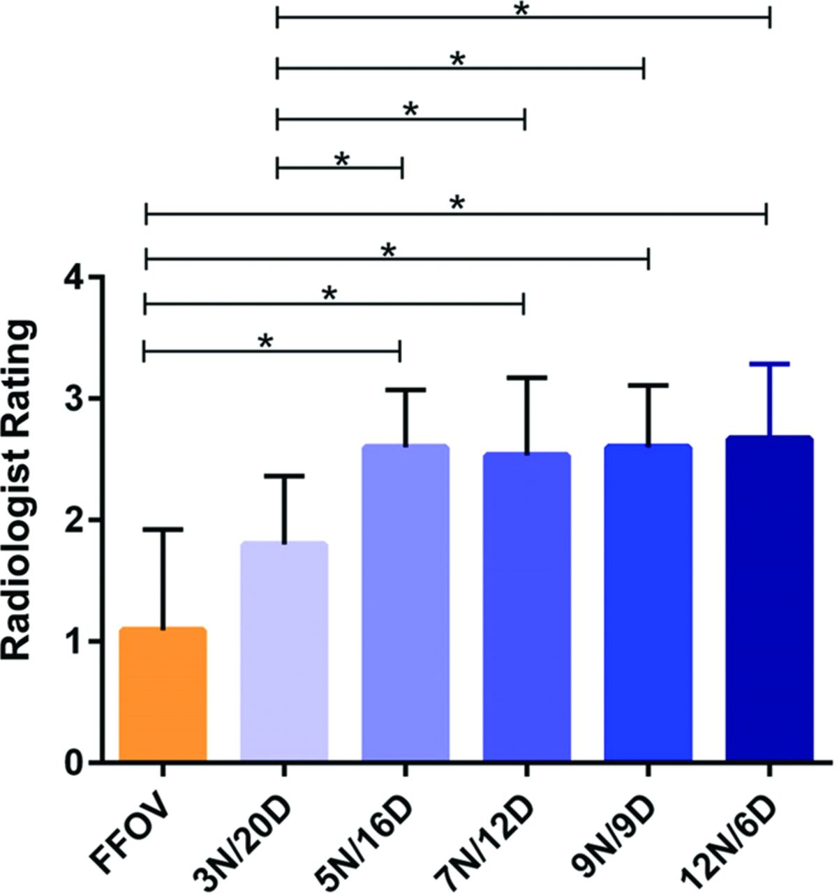
- Fig 3.
Qualitative analysis. Radiologists determined a rate for each sequence, for each subject, from 1 (nondiagnostic) to 4 (good). Mean rates ± SDs for the sequence are represented. Superimposed black lines indicate which sequences are statistically different with P < .05 (asterisk).
- Fig 4.
Percentages of sections with artifacts unusable for DTI analysis due to susceptibility artifacts or poor SNR. Superimposed black lines indicate which sequences are statistically different with P < .05 (asterisk) and P < .005 (double asterisks).
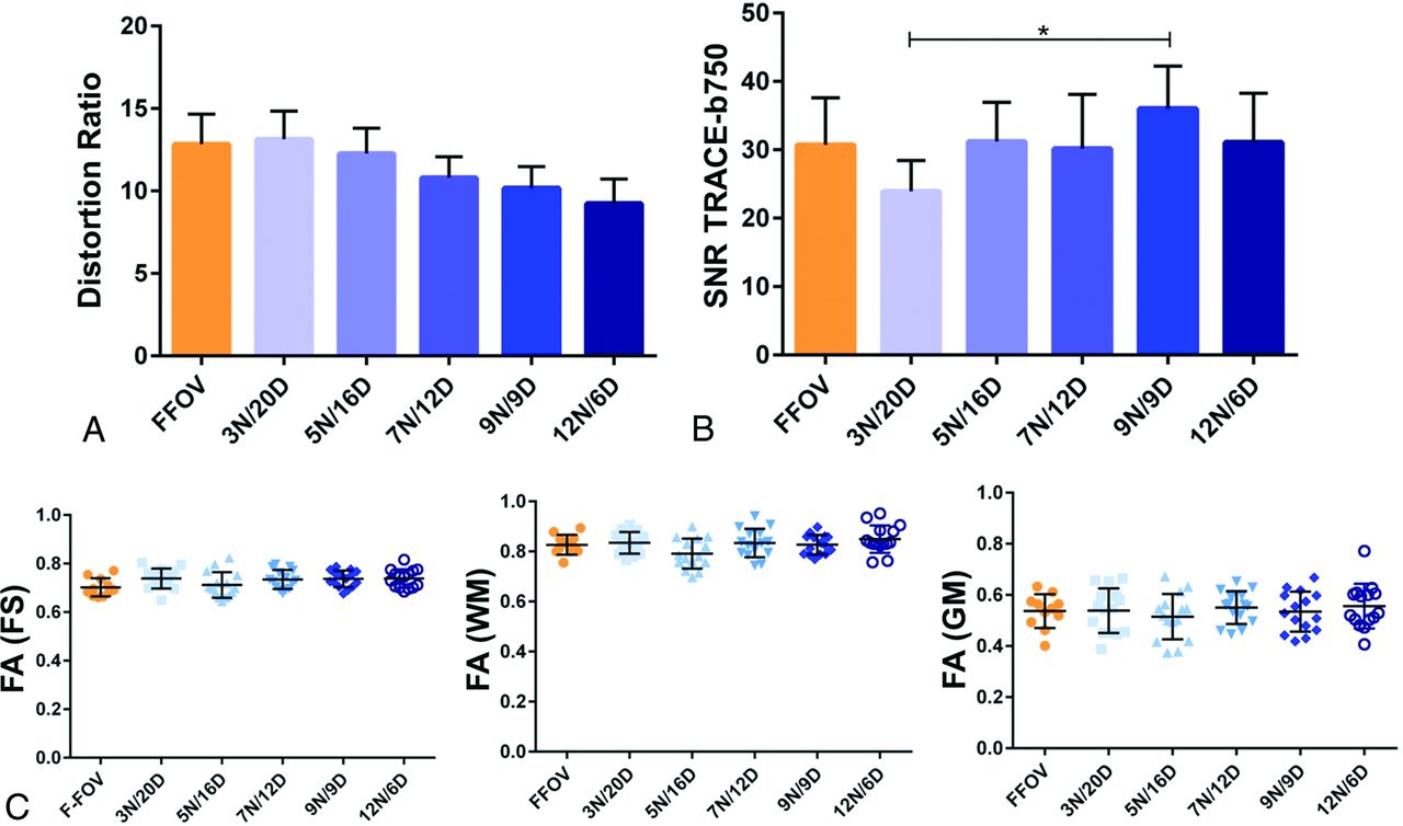
- Fig 5.
Quantitative comparisons on ROI-based analyses. A, The distortion ratio. B, The SNR on the trace image at b=750 s/mm2. C, Representation of the dispersion of FA values, depending on the DTI sequence and, successively, a full section of the spinal cord (FS), WM, and GM. Mean rates ± SDs for the sequence are represented. Superimposed black lines indicate which sequences are statistically different with P < .05 (asterisk).
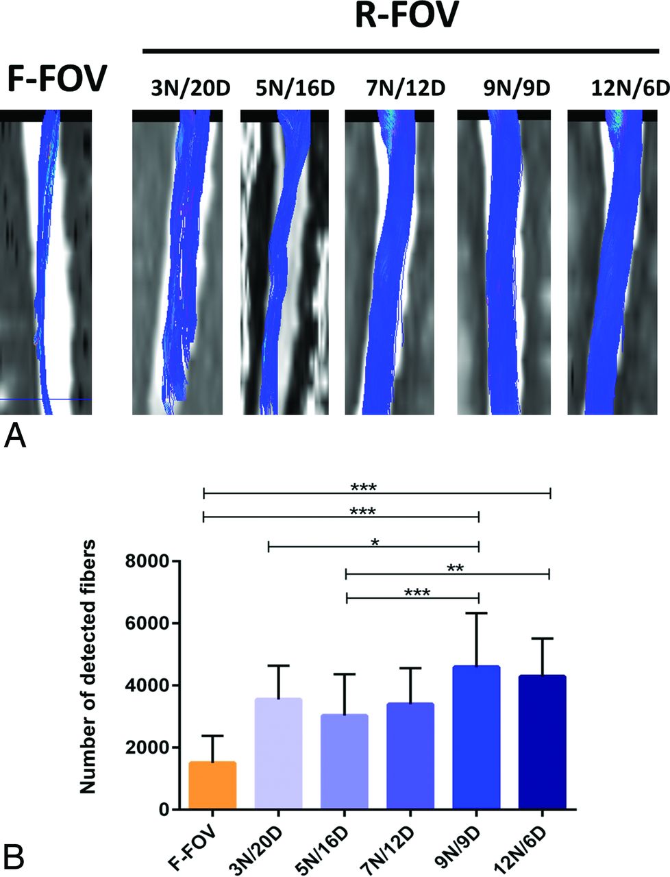
- Fig 6.
Tractography-based analyses. A, The reconstructed tractograms for the whole DTI dataset. For each DTI sequence, 2 similar seed ROIs were placed on the anatomic sequence, at the C1 and C3 levels, and then propagated on diffusion data. Care was taken to exclude abnormal fiber detection (ie, in the CSF). Qualitatively, r-FOV sequences clearly exhibit better tractogram definitions. B, The number of detected fibers between the 2 seeds. P < .05 (asterisk), P < .005 (double asterisks), P < .0005 (triple asterisks).


Jump to section
Related Articles
Cited By...
- No citing articles found.