
Article Figures & Data
Figures
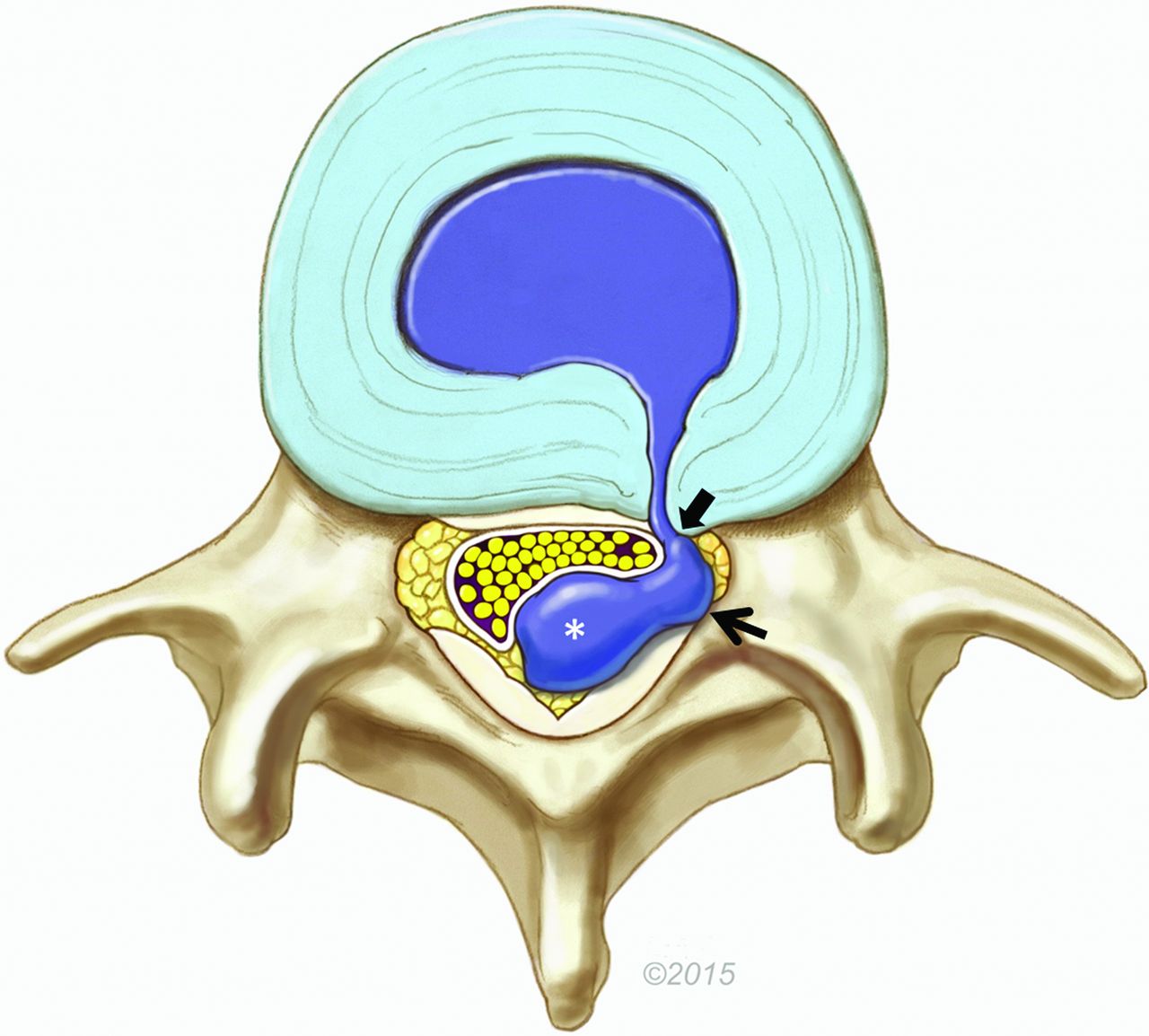
- Fig 1.
Illustration of a dorsal lumbar disc herniation. The bulk of the herniated disc material is located in the dorsal epidural space (asterisk), causing mass effect on the thecal sac and cauda equina. However, a lateral epidural component is also present (arrow). In this example, disc material extends into the ventrolateral epidural space directly to a rent in the annulus, contiguous with the parent disc (block arrow). On MR imaging, the soft tissue ventrally/laterally can represent disc material and/or granulation tissue. Reproduced with permission from the Mayo Foundation for Medical Education and Research.
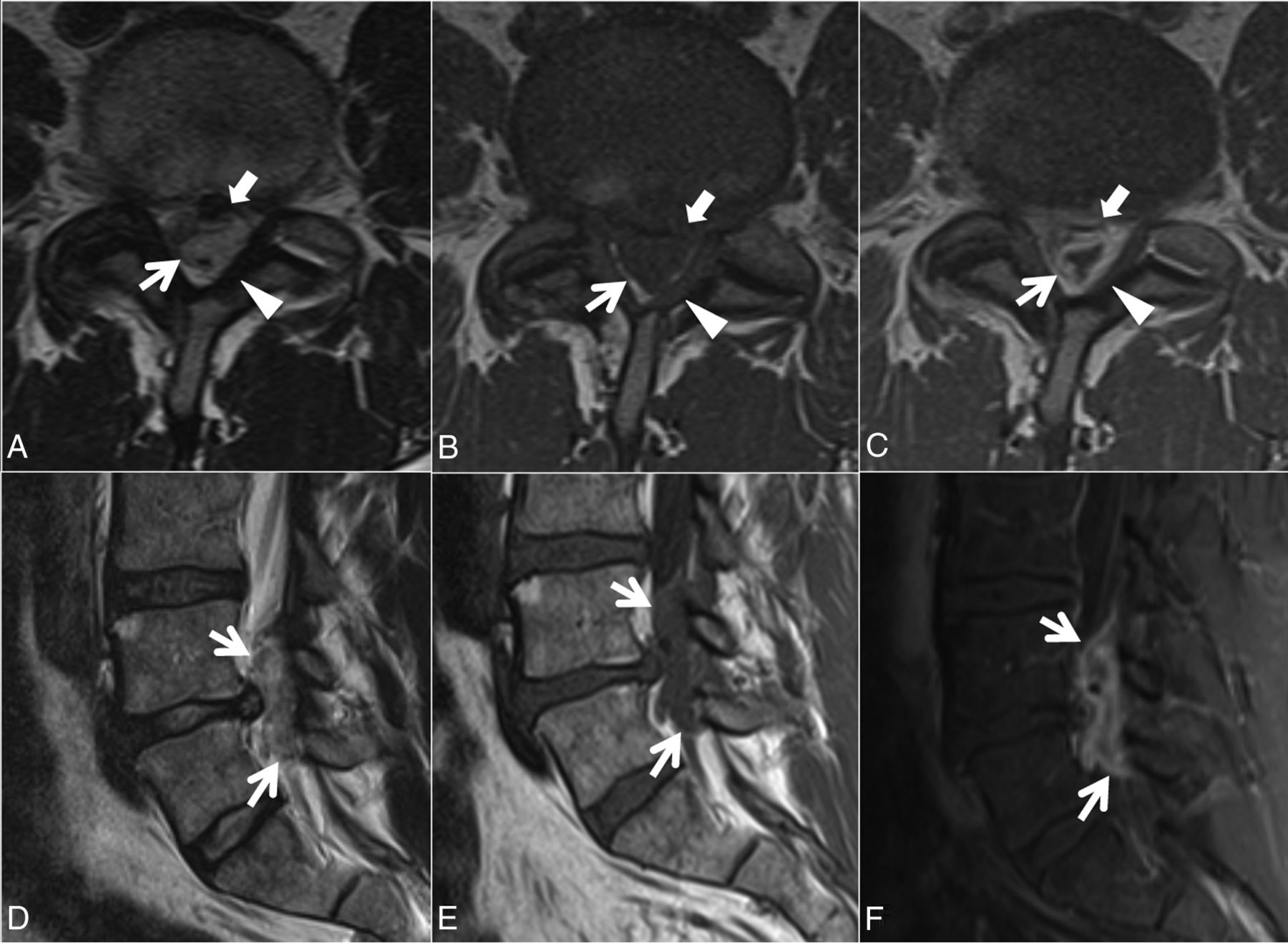
- Fig 2.
Case 1. A 48-year-old man with 3 weeks of back pain radiating to the right lower extremity and paresthesias on the dorsum of the right foot. Axial T2- (A), T1- (B), and postcontrast T1-weighted (C) and sagittal T2- (D), T1- (E), and postcontrast fat-suppressed T1-weighted (F) images. A heterogeneous predominantly T1-isointense, T2-hyperintense 3.5-cm maximal dimension mass (white arrows, A–F) in the dorsal and left lateral epidural fat partially abutting the left ligamentum flavum (arrowheads, A–C) contributes to severe L4–5 spinal stenosis, with rightward displacement and effacement of the thecal sac. There is no definite connection to the left L4–5 facet joint. The left lateral epidural fat is effaced, and the dorsal mass is contiguous with the dorsal margin of the L4–5 disc (white block arrow in A–C). The mass peripherally enhances (C and F). The radiologist's interpretation favored epidural abscess. At the operation, the dorsal disc herniation was an inflammatory-appearing mass with considerable adhesion to the undersurface of the lamina. Also contributing to the L4–5 stenosis are a disc protrusion and ligamentum flavum redundancy.
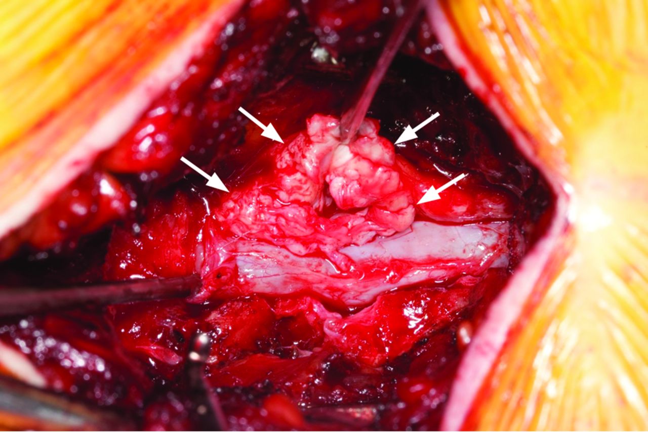
- Fig 3.
Case 1. Intraoperative photo after an L4–5 laminectomy demonstrates a left-sided mass (arrows) pathologically proved to be disc material.
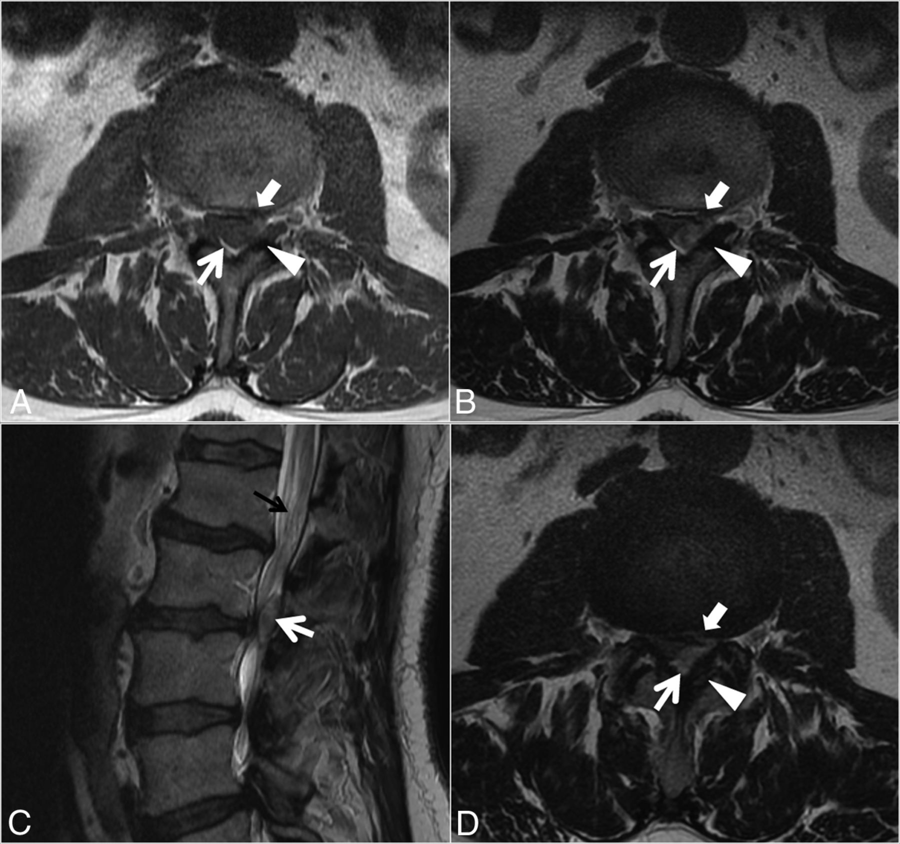
- Fig 4.
Case 2. A 77-year-old man with 4 months of low back pain and intermittent radiation into the right-more-than-left lower extremities, as well as weakness and an acute inability to ambulate. Sagittal T1- (A) and T2-weighted (B) and L3 inferior endplate–level axial T1- (C) and T2-weighted (D) images. A heterogeneous predominantly T1-isointense, mildly T2-hyperintense, 1.9-cm maximal-dimension mass (white arrows, A–D) in the dorsal and right lateral epidural fat abutting the right ligamentum flavum (arrowheads, C–D) contributes to severe L3–4 spinal stenosis, with leftward displacement and effacement of the thecal sac. There is no definite connection to the right L3–4 facet joint. The right lateral epidural fat is effaced, and the dorsal mass is contiguous with the dorsal margin of the L3–4 disc (white block arrow in C–D). Differential considerations in the radiologist's interpretation included focal epidural hematoma (particularly given the acute clinical presentation), dorsal migration of a disc fragment, an unusual-appearing synovial cyst or other degenerative cyst, and sequela of the recent epidural injection. At the operation, the dorsal disc herniation was indeed traced back to a large annular defect at the right lateral margin of the L3–4 disc. Also contributing to the L3–4 stenosis are slight retrolisthesis of L3 on L4, a disc bulge, and ligamentum flavum redundancy. Tortuosity of the cauda equina (black arrows, B) is compatible with the high-grade stenosis. An L1–2 left subarticular disc extrusion causing advanced lateral recess narrowing is also present at the superior aspect of the sagittal images (A and B).
- Fig 5.
Case 3. A 69-year-old man with 2 weeks of low back pain radiating into the thighs and progressive right lower extremity weakness. Axial T1- (A) and T2-weighted (B and D; D is 1 section below A and B) and sagittal T2-weighted (C) images. A heterogeneous predominantly T1-isointense, T2-hyperintense, 2.0-cm maximal-dimension mass (white arrows, A–D) in the dorsal and left lateral epidural fat abutting the left ligamentum flavum (arrowheads, A, B, and D) contributes to severe L2–3 spinal stenosis, with rightward displacement and effacement of the thecal sac. There is no definite connection to the left L2–3 facet joint. The left lateral epidural fat is effaced, and the dorsal mass is contiguous with the dorsal margin of the L2–3 disc (white block arrows in A, B, and D, best seen in D). The radiologist's diagnosis was a synovial cyst. At surgery, the dorsal disc herniation was traced back to the ventral aspect of the canal, and the disc was probed without other fragments identified. Also contributing to the L2–3 stenosis are a developmentally narrow canal, a disc bulge, and ligamentum flavum redundancy. Mild tortuosity of the cauda equina (black arrow, C) is compatible with the high-grade stenosis.
- Fig 6.
Case 4. A 61-year-old man with 5 days of low back pain and 2 days of progressive right lower extremity weakness. Axial T1- (A) and T2-weighted (B and D; D is 1 section above A and B) and sagittal T2-weighted (C) images. A heterogeneous predominantly T1-isointense, mildly T2-hypointense, 1.3-cm maximal-dimension mass (white arrows, A–D) in the dorsal and left lateral epidural fat abutting the left ligamentum flavum (arrowheads, A, B, and D) contributes to severe L3–4 spinal stenosis, with rightward displacement and effacement of the thecal sac. There is no definite connection to the left L3–4 facet joint. The left lateral epidural fat is effaced, and the dorsal mass is contiguous with the dorsal margin of the L3–4 disc (white block arrows in A, B, and D, best seen in D). The radiologist's favored diagnosis was a small epidural hematoma, with differential considerations of a sequestered disc fragment or degenerative facet-related lesion. At the operation, the dorsal disc herniation was followed back to the ventral aspect of the canal and no other fragments were identified. Also contributing to the L3–4 stenosis are a disc bulge and ligamentum flavum redundancy. Mild tortuosity of the cauda equina (black arrow, C) is compatible with the high-grade stenosis.


Tables
Clinical and MRI features of 5 patients with dorsal epidural disc herniations
Case No./Age (yr)/Sex Symptoms Side/Level Lateral Soft Tissue Ventral Soft Tissue, Abutting Disc Gadolinium Enhancement Radiologist's Favored Diagnosis 1/48/M 3-Week back pain, radiating to right lower extremity, paresthesias on dorsum of right foot, urinary hesitancy Left/L4–5 Yes Yes Peripheral Epidural abscess 2/77/M 4-Month low back pain, intermittent radiation into right > left lower extremities, weakness; acutely unable to ambulate Right/L3–4 Yes Yes NA Epidural hematoma 3/69/M 2-Week low back pain radiating into thighs, progressive right lower extremity weakness Left/L2–3 Yes Yes NA Synovial cyst 4/61/M 5-Day low back pain, 2-day progressive right lower extremity weakness Left/L3–4 Yes Yes NA Epidural hematoma 5/35/M 1-Week back and bilateral lower extremity pain, weakness Left/L4–5 Yes Yes Peripheral NA 6a/60/M 3-Week progressive severe right lower extremity radicular pain, resulting in hospital admission for pain control Right/L4–5 Yes Yes Peripheral Epidural abscess Note:—NA indicates not applicable.
↵a Images from case 6 appear in Diehn et al.8


Jump to section
Related Articles
Cited By...
- No citing articles found.