Advertisement

Main menu

  • Home
  • Content
    • Current Issue
    • Accepted Manuscripts
    • Article Preview
    • Past Issue Archive
    • AJNR Case Collection
    • Case of the Week Archive
    • Classic Case Archive
    • Case of the Month Archive
  • Special Collections
    • Spinal CSF Leak Articles (Jan 2020-June 2024)
    • 2024 AJNR Journal Awards
    • Most Impactful AJNR Articles
  • Multimedia
    • AJNR Podcast
    • AJNR Scantastics
    • Video Articles
  • For Authors
    • Submit a Manuscript
    • Author Policies
    • Fast publishing of Accepted Manuscripts
    • Graphical Abstract Preparation
    • Manuscript Submission Guidelines
    • Imaging Protocol Submission
    • Submit a Case for the Case Collection
  • About Us
    • About AJNR
    • Editorial Board
  • More
    • Become a Reviewer/Academy of Reviewers
    • Subscribers
    • Permissions
    • Alerts
    • Feedback
    • Advertisers
    • ASNR Home
  • Other Publications
    • ajnr

User menu

  • Alerts
  • Log in

Search

  • Advanced search
American Journal of Neuroradiology

American Journal of Neuroradiology

ASHNR American Society of Functional Neuroradiology ASHNR American Society of Pediatric Neuroradiology ASSR
  • Alerts
  • Log in

Advanced Search

  • Home
  • Content
    • Current Issue
    • Accepted Manuscripts
    • Article Preview
    • Past Issue Archive
    • AJNR Case Collection
    • Case of the Week Archive
    • Classic Case Archive
    • Case of the Month Archive
  • Special Collections
    • Spinal CSF Leak Articles (Jan 2020-June 2024)
    • 2024 AJNR Journal Awards
    • Most Impactful AJNR Articles
  • Multimedia
    • AJNR Podcast
    • AJNR Scantastics
    • Video Articles
  • For Authors
    • Submit a Manuscript
    • Author Policies
    • Fast publishing of Accepted Manuscripts
    • Graphical Abstract Preparation
    • Manuscript Submission Guidelines
    • Imaging Protocol Submission
    • Submit a Case for the Case Collection
  • About Us
    • About AJNR
    • Editorial Board
  • More
    • Become a Reviewer/Academy of Reviewers
    • Subscribers
    • Permissions
    • Alerts
    • Feedback
    • Advertisers
    • ASNR Home
  • Follow AJNR on Twitter
  • Visit AJNR on Facebook
  • Follow AJNR on Instagram
  • Join AJNR on LinkedIn
  • RSS Feeds

Welcome to the new AJNR, Updated Hall of Fame, and more. Read the full announcements.


AJNR is seeking candidates for the position of Associate Section Editor, AJNR Case Collection. Read the full announcement.


Letter

The Utility of Molecular Imaging for Investigating Patients with Visual Hallucinations

S. Clifford, Y.M. Purcell and R.P. Killeen
American Journal of Neuroradiology November 2016, 37 (11) E77-E78; DOI: https://doi.org/10.3174/ajnr.A4937
S. Clifford
aDepartment of Radiology St. Vincent's University Hospital Dublin, Ireland
  • Find this author on Google Scholar
  • Find this author on PubMed
  • Search for this author on this site
  • ORCID record for S. Clifford
Y.M. Purcell
aDepartment of Radiology St. Vincent's University Hospital Dublin, Ireland
  • Find this author on Google Scholar
  • Find this author on PubMed
  • Search for this author on this site
  • ORCID record for Y.M. Purcell
R.P. Killeen
bDepartment of Radiology St. Vincent's University Hospital Dublin, Ireland
cDepartment of Radiology Royal Victoria Eye and Ear Hospital Dublin, Ireland
  • Find this author on Google Scholar
  • Find this author on PubMed
  • Search for this author on this site
  • ORCID record for R.P. Killeen
  • Article
  • Figures & Data
  • Info & Metrics
  • Responses
  • References
  • PDF
Loading

We read with interest the recent article entitled “Distinguishing Neuroimaging Features in Patients Presenting with Visual Hallucinations” by Winton-Brown et al.1 They described the features of dementia with Lewy bodies (DLB) on multiplanar T1-weighted MR imaging.1 In our practice, we find the diagnosis of DLB challenging with anatomic imaging alone. Molecular imaging can be helpful for differentiating DLB from other causes of visual hallucinations and dementia.2⇓–4 We present examples of the imaging findings of DLB by using nigrostriatal dopamine terminal imaging with dopamine-transporter single-photon emission CT (DaTscan) with the radioligand [123I]FP-CIT (GE Healthcare, Buckinghamshire, United Kingdom) and FDG-PET. These imaging techniques may be considered in addition to anatomic imaging in cases in which a diagnosis of DLB is suspected but remains unclear.

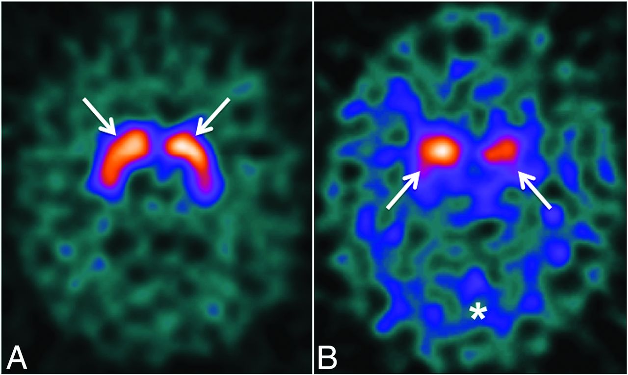
DaTscan is used for the early detection of Parkinson disease, to differentiate essential tremor and psychogenic-/neuroleptic-induced parkinsonism from presynaptic parkinsonian syndromes and to distinguish DLB from other forms of dementia. Nigrostriatal dopaminergic denervation is a crucial feature in Parkinson disease, parkinsonian disorders and DLB. [123I]FP-CIT has a high affinity for the dopamine transporter (DaT) and is used to visualize dopaminergic nerve terminals. DaTscan involves the acquisition of SPECT images 3–6 hours after a single injection of [123I]FP-CIT at a recommended dose of 111–185 MBq by using a dual-headed (or greater) gamma camera. In a normal DaTscan, the striatum is visible as symmetric, comma-shaped regions. Both the caudate and putamen show high uptake compared with the background (Fig 1A). In DLB, there is typically bilateral loss of DaT binding, predominantly in the putamen and caudate, more symmetric than in Parkinson disease (Fig 1B). A Phase III study by McKeith et al2 showed that DaTscan has a mean sensitivity of 77.7% for detecting clinically probable DLB and a specificity of 90.4% for excluding other causes of dementia, principally Alzheimer disease (AD).

Fig 1.
  • Download figure
  • Open in new tab
  • Download powerpoint
Fig 1.

A, Normal DaTscan findings with normal background activity. The arrows show normal radiotracer uptake within the caudate nuclei and putamen bilaterally. B, Abnormal DaTscan findings in a 68-year-old woman with visual hallucinations and cognitive impairment. There is reduced uptake within the caudate nuclei and putamina (arrows) bilaterally with increased background activity (asterisk). In the clinical context, these findings are consistent with DLB.

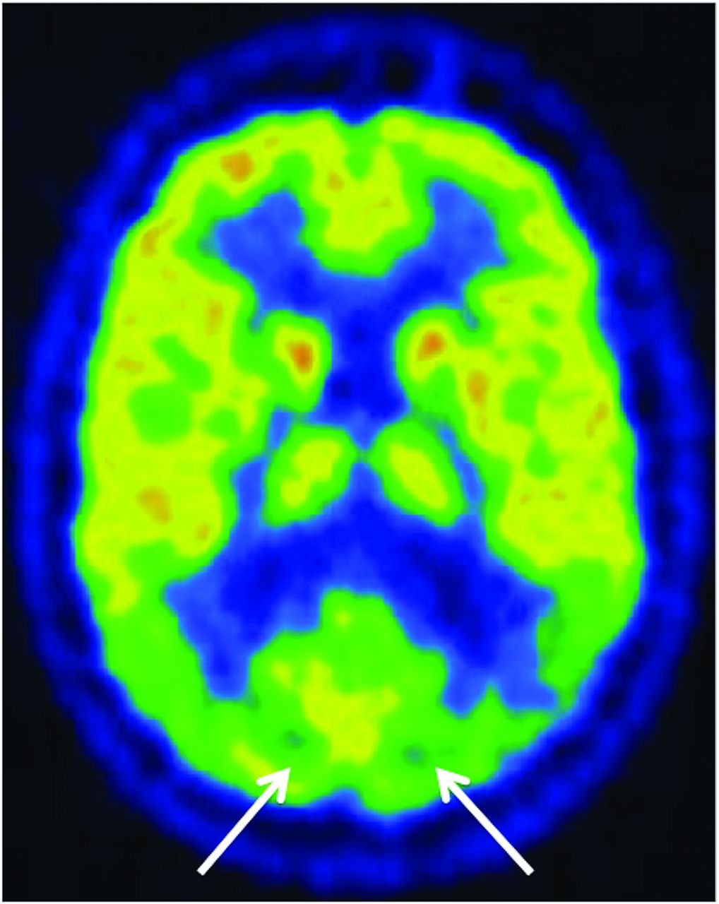
FDG-PET is a useful imaging technique in differentiating primary neurodegenerative disorders. The FDG metabolic signature of DLB is bilateral parietal and posterior temporal hypometabolism, posterior cingulate gyral hypometabolism, and involvement of the occipital lobes, which are spared in AD (Fig 2).3 Medial occipital lobe hypometabolism is a key feature of DLB that discriminates it from AD,4 with a reported sensitivity and specificity of 92%. If the occipital cortex is not involved, DLB and AD cannot be distinguished on the basis of their FDG metabolic pattern.3 When clinical and FDG-PET findings are indeterminate, DaTscan is complementary.

Fig 2.
  • Download figure
  • Open in new tab
  • Download powerpoint
Fig 2.

FDG-PET in the same patient as Fig 1B shows marked bilateral hypometabolism in the medial occipital lobes, which is a typical finding in DLB.3 These features distinguish DLB from AD; AD typically shows hypometabolism in the temporal-parietal regions and posterior cingulate gyrus but with normal medial occipital metabolism.

In conclusion, the integration of molecular imaging, including DaTscan and FDG-PET, can be useful in establishing a diagnosis of dementia with Lewy bodies in patients presenting with visual hallucinations.

Footnotes

  • Disclosures: Ronan P. Killeen—UNRELATED: Consultancy: Novartis, Comments: consultancy related to review of software for atrophy measures in multiple sclerosis; single fee under $1000; Royalties: Springer, Comments: FDG PET/CT in Clinical Oncology (book) by Jasna Mihailovic, Ronan P. Killeen, and Stanley J. Goldsmith, 2012; Stock/Stock Options: Dublin CyberKnife Financing Limited, Comments: owns shares in this company; it runs a CyberKnife for treatment of primarily brain tumors in Hermitage Medical Clinic, Dublin, Ireland.

References

  1. 1.↵
    1. Winton-Brown TT,
    2. Ting A,
    3. Mocellin R, et al
    . Distinguishing neuroimaging features in patients presenting with visual hallucinations. AJNR Am J Neuroradiol 2016;37:774–81 doi:10.3174/ajnr.A4636 pmid:26744445
    Abstract/FREE Full Text
  2. 2.↵
    1. McKeith I,
    2. O'Brien J,
    3. Walker Z, et al
    ; DLB Study Group. Sensitivity and specificity of dopamine transporter imaging with 123I-FP-CIT SPECT in dementia with Lewy bodies: a phase III, multicentre study. Lancet Neurol 2007;6:305–13 doi:10.1016/S1474-4422(07)70057-1 pmid:17362834
    CrossRefPubMed
  3. 3.↵
    1. Brown KJ,
    2. Bohnen NI,
    3. Wong KK, et al
    . Brain PET in suspected dementia: patterns of altered FDG metabolism. Radiographics 2014;34:684–701 doi:10.1148/rg.343135065 pmid:24819789
    CrossRefPubMed
  4. 4.↵
    1. Ishii K
    . PET approaches for diagnosis of dementia. AJNR Am J Neuroradiol 2014;35:2030–38 doi:10.3174/ajnr.A3695 pmid:23945233
    Abstract/FREE Full Text
  • © 2016 by American Journal of Neuroradiology
PreviousNext
Back to top

In this issue

American Journal of Neuroradiology: 37 (11)
American Journal of Neuroradiology
Vol. 37, Issue 11
1 Nov 2016
  • Table of Contents
  • Index by author
  • Complete Issue (PDF)
Advertisement
Print
Download PDF
Email Article

Thank you for your interest in spreading the word on American Journal of Neuroradiology.

NOTE: We only request your email address so that the person you are recommending the page to knows that you wanted them to see it, and that it is not junk mail. We do not capture any email address.

Enter multiple addresses on separate lines or separate them with commas.
The Utility of Molecular Imaging for Investigating Patients with Visual Hallucinations
(Your Name) has sent you a message from American Journal of Neuroradiology
(Your Name) thought you would like to see the American Journal of Neuroradiology web site.
CAPTCHA
This question is for testing whether or not you are a human visitor and to prevent automated spam submissions.
Cite this article
S. Clifford, Y.M. Purcell, R.P. Killeen
The Utility of Molecular Imaging for Investigating Patients with Visual Hallucinations
American Journal of Neuroradiology Nov 2016, 37 (11) E77-E78; DOI: 10.3174/ajnr.A4937

Citation Manager Formats

  • BibTeX
  • Bookends
  • EasyBib
  • EndNote (tagged)
  • EndNote 8 (xml)
  • Medlars
  • Mendeley
  • Papers
  • RefWorks Tagged
  • Ref Manager
  • RIS
  • Zotero
0 Responses
Respond to this article
Share
Bookmark this article
The Utility of Molecular Imaging for Investigating Patients with Visual Hallucinations
S. Clifford, Y.M. Purcell, R.P. Killeen
American Journal of Neuroradiology Nov 2016, 37 (11) E77-E78; DOI: 10.3174/ajnr.A4937
del.icio.us logo Twitter logo Facebook logo Mendeley logo
  • Tweet Widget
  • Facebook Like
  • Google Plus One
Purchase

Jump to section

  • Article
    • Footnotes
    • References
  • Figures & Data
  • Info & Metrics
  • Responses
  • References
  • PDF

Related Articles

  • Reply:
  • PubMed
  • Google Scholar

Cited By...

  • No citing articles found.
  • Crossref
  • Google Scholar

This article has not yet been cited by articles in journals that are participating in Crossref Cited-by Linking.

Similar Articles

Advertisement

Indexed Content

  • Current Issue
  • Accepted Manuscripts
  • Article Preview
  • Past Issues
  • Editorials
  • Editors Choice
  • Fellow Journal Club
  • Letters to the Editor

Cases

  • Case Collection
  • Archive - Case of the Week
  • Archive - Case of the Month
  • Archive - Classic Case

Special Collections

  • Special Collections

Resources

  • News and Updates
  • Turn around Times
  • Submit a Manuscript
  • Author Policies
  • Manuscript Submission Guidelines
  • Evidence-Based Medicine Level Guide
  • Publishing Checklists
  • Graphical Abstract Preparation
  • Imaging Protocol Submission
  • Submit a Case
  • Become a Reviewer/Academy of Reviewers
  • Get Peer Review Credit from Publons

Multimedia

  • AJNR Podcast
  • AJNR SCANtastic
  • Video Articles

About Us

  • About AJNR
  • Editorial Board
  • Not an AJNR Subscriber? Join Now
  • Alerts
  • Feedback
  • Advertise with us
  • Librarian Resources
  • Permissions
  • Terms and Conditions

American Society of Neuroradiology

  • Not an ASNR Member? Join Now

© 2025 by the American Society of Neuroradiology All rights, including for text and data mining, AI training, and similar technologies, are reserved.
Print ISSN: 0195-6108 Online ISSN: 1936-959X

Powered by HighWire