
Article Figures & Data
Figures
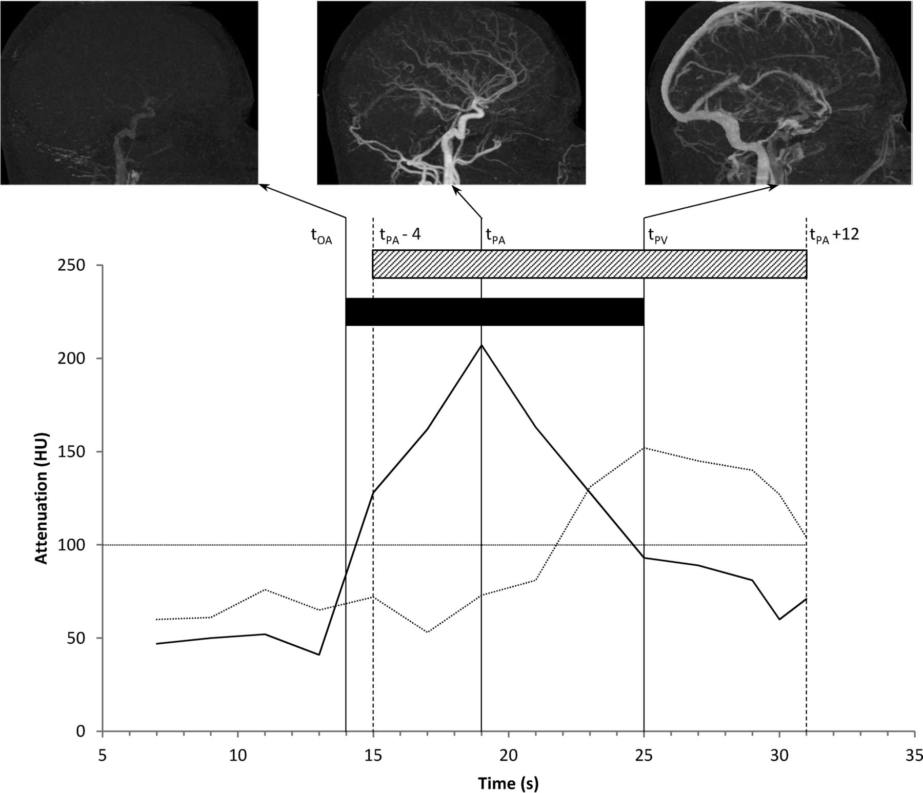
- Fig 1.
Schematic representation showing typical time-attenuation curves for arterial (solid line) and venous (dotted line) profiles. The tOA was set to the nearest integer second in which the arterial attenuation crossed 100 HU (14 seconds in this figure, but note that measurements were only made on odd-numbered seconds). The tPA and tPV represent times of peak arterial and venous attenuation, respectively. The gray bar indicates the exposure time for the estimated duration protocol (16 seconds), and the black bar indicates the exposure time for the measured-duration protocol. Subtracted MIP images illustrate typical appearances of the acquisition at key phases.
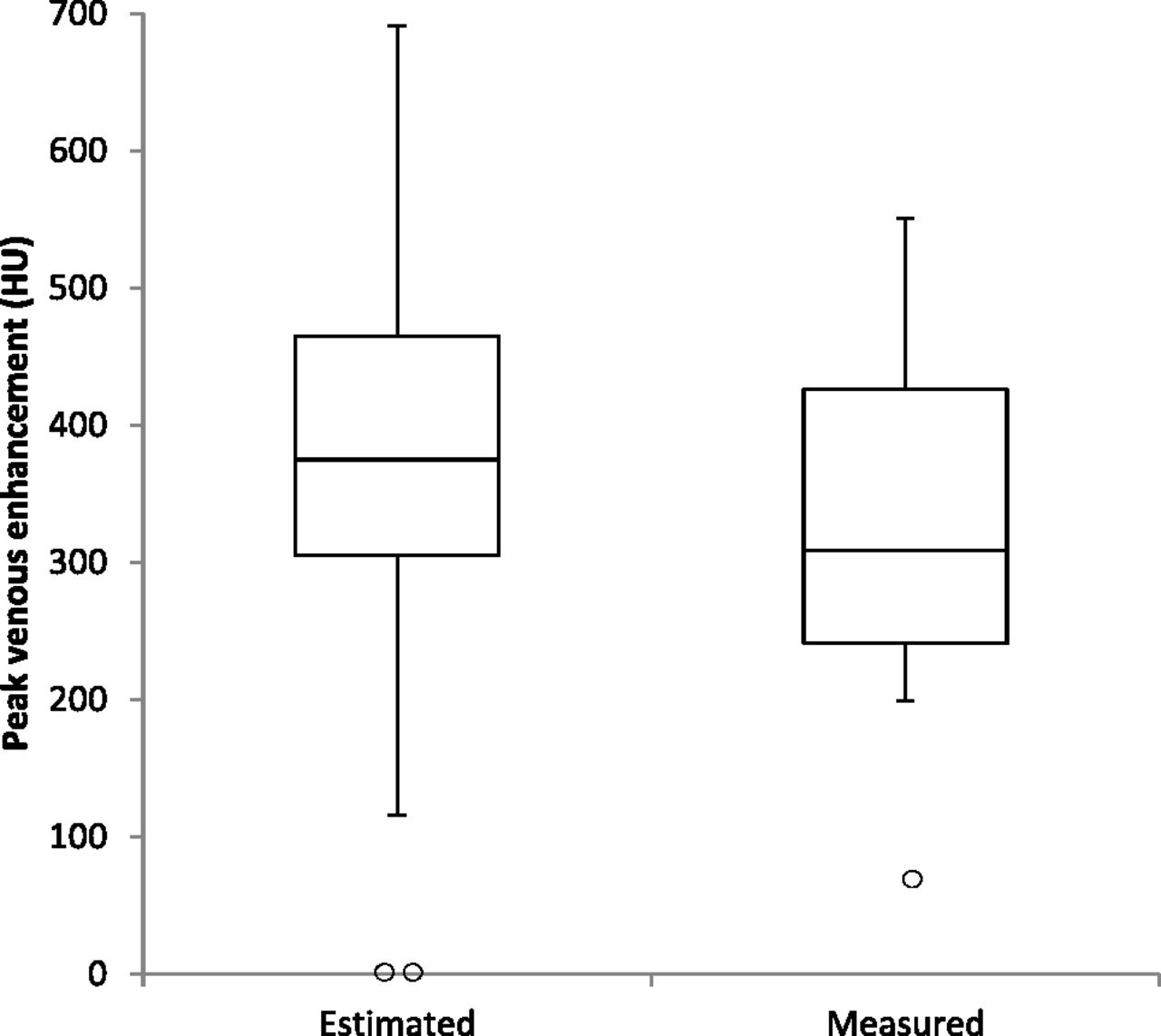
- Fig 2.
Boxplot illustrating distribution of peak venous enhancement between the estimated- and measured-duration scan protocols. Two outliers are shown in the estimated-duration group, in which no venous enhancement was present due to inadequate time coverage, and 1 outlier with very poor venous enhancement is shown in the measured-duration group.
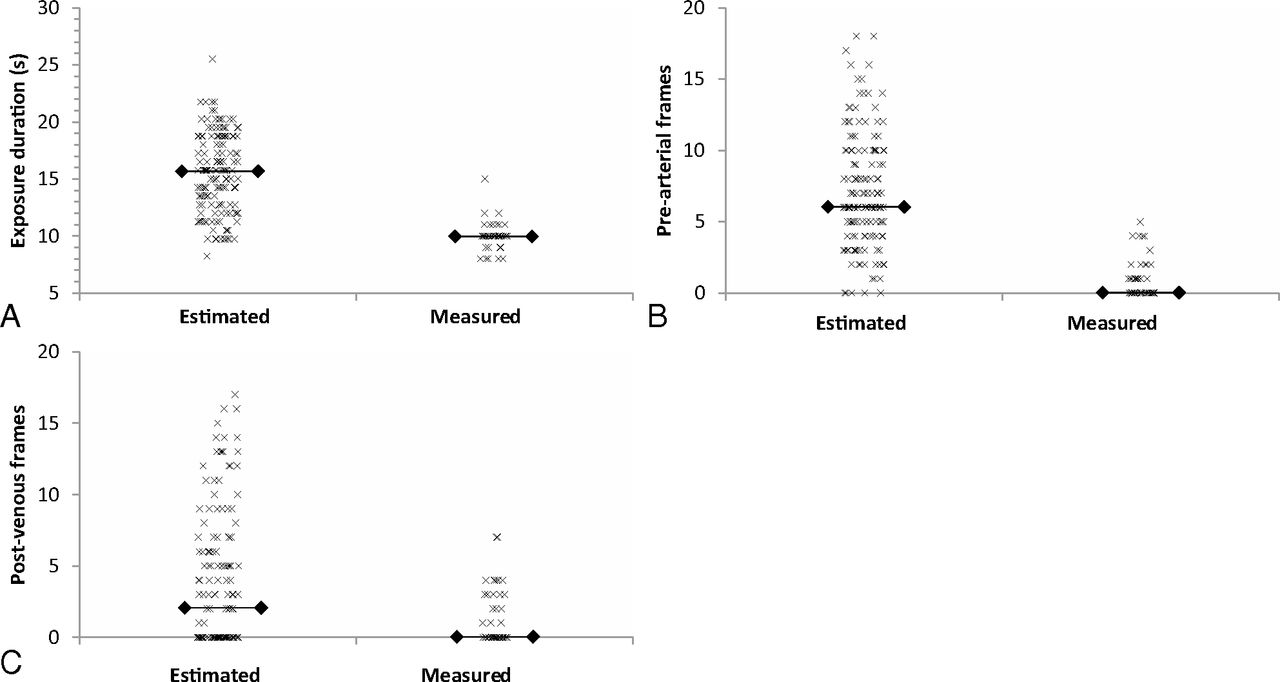
- Fig 3.
Scatterplots indicating exposure duration (A) and the number of nondiagnostic volumes due to the start of the acquisition before contrast arrival (B) and the end of the acquisition following contrast washout (C). Horizontal lines indicate the median.


Tables
Estimated Duration (n = 151) Measured Duration (n = 53) Demographics Age (yr) 53.4 (19–87) 51.8 (21–79) Sex (M/F) 67:84 25:28 Indications (No.) (%) Intraparenchymal hemorrhage 47 (31%) 20 (38%) Suspected vascular lesion 37 (25%) 11 (21%) Tinnitus 21 (14%) 5 (9%) Tumor vascular assessment 11 (7%) 5 (9%) Subdural hemorrhage 5 (3%) 0 (0%) Subarachnoid hemorrhage 4 (3%) 0 (0%) Untreated AVM/F assessment 7 (5%) 5 (9%) Treated AVM/F assessment 6 (4%) 3 (6%) Venous stenosis/thrombosis (including assessment of idiopathic intracranial hypertension) 8 (5%) 3 (6%) Other 5 (3%) 1 (2%) Note:—AVM/F indicates arteriovenous malformation or fistula.
Estimated Duration (n = 151) Measured Duration (n = 53) P Value Adjusted P Value Exposure and dose Exposure time (sec) 15.75 (8.25–25.5) 10.0 (8–12) <.001 <.001b No. of volumes 31 (26–36) 19 (19–19) Dose-length product (mGy × cm) 3021 (2536–3502) 1473 (1459–1526) Volume CT dose index (mGy) 185 (162–223) 88.9 (88.9–88.9) Effective dose (mSv) 6.9 (5.8–8.0) 3.4 (3.4–3.5) Study quality Maximum venous enhancement (HU) 372 (352–392) 324 (294–353) .019 .076 Adequate time coverage (No.) 134 (88.7%) 49 (92.5%) .444 1.00 Quality score 5 (5–5) 5 (5–5) .837 1.00 No. of prearterial volumes 6 (4–10) 0 (0–1) <.001c <.001b,c No. of postpeak venous volumes 2 (0–6) 0 (0–2) <.001c <.001b,c Diagnostic performance Positive findings (No.) 58 (38.4%) 21 (39.6%) No. having DSA 56 (37.1%) 22 (41.5%) Intermodality concordance between 4D-CTA and DSA (No.) 51 (91.0%) 22 (100.0%) Cohen κ statistic κ = 0.88 κ = 1.0 ↵a Noncount values are shown as median (interquartile range), except for exposure time shown as median (range) and enhancement shown as mean (95% CI). Values for dose-length product include the entire examination, including test-bolus acquisition. CT dose index values include only whole-head acquisitions.
↵b Significant at the 5% level after Bonferroni correction.
↵c Post hoc test.
- Table 3:
Comparison of the currently reported techniques with 4D-CTA techniques (excluding combined 4D-CTA/CTP techniques) reported by other groups
Report Exposure Duration (sec) Rotation Time (ms) Exposure Parameters Effective Dose (mSv) Brouwer et al (2010)2 22 NS 80 kV, 120 mA 5.1 Hoogenboom et al (2012)11 15 500 80 kV, 240 mA NS Fujiwara et al (2013)3 12a 500 80 kV, 200 mA 5.2 Willems et al (2011)1 and (2012)5 22 1000 80 kV, 100 mA 5.2 D'Orazio et al (2014)10 15 350 80 kV, 120 mA 5.62 This study Estimated duration 15.6 (16 with option to adjust) 750 80 kV, 150 mA 6.9 Measured exposure 10.1 (mean) 1000 80 kV, 100 mA 3.4 Note:—NS indicates not stated.
↵a Ten-second continuous exposure followed by 4 intermittent exposures.


