Advertisement

Main menu

  • Home
  • Content
    • Current Issue
    • Accepted Manuscripts
    • Article Preview
    • Past Issue Archive
    • AJNR Case Collection
    • Case of the Week Archive
    • Classic Case Archive
    • Case of the Month Archive
  • Special Collections
    • Spinal CSF Leak Articles (Jan 2020-June 2024)
    • 2024 AJNR Journal Awards
    • Most Impactful AJNR Articles
  • Multimedia
    • AJNR Podcast
    • AJNR Scantastics
    • Video Articles
  • For Authors
    • Submit a Manuscript
    • Author Policies
    • Fast publishing of Accepted Manuscripts
    • Graphical Abstract Preparation
    • Manuscript Submission Guidelines
    • Imaging Protocol Submission
    • Submit a Case for the Case Collection
  • About Us
    • About AJNR
    • Editorial Board
  • More
    • Become a Reviewer/Academy of Reviewers
    • Subscribers
    • Permissions
    • Alerts
    • Feedback
    • Advertisers
    • ASNR Home
  • Other Publications
    • ajnr

User menu

  • Alerts
  • Log in

Search

  • Advanced search
American Journal of Neuroradiology

American Journal of Neuroradiology

ASHNR American Society of Functional Neuroradiology ASHNR American Society of Pediatric Neuroradiology ASSR
  • Alerts
  • Log in

Advanced Search

  • Home
  • Content
    • Current Issue
    • Accepted Manuscripts
    • Article Preview
    • Past Issue Archive
    • AJNR Case Collection
    • Case of the Week Archive
    • Classic Case Archive
    • Case of the Month Archive
  • Special Collections
    • Spinal CSF Leak Articles (Jan 2020-June 2024)
    • 2024 AJNR Journal Awards
    • Most Impactful AJNR Articles
  • Multimedia
    • AJNR Podcast
    • AJNR Scantastics
    • Video Articles
  • For Authors
    • Submit a Manuscript
    • Author Policies
    • Fast publishing of Accepted Manuscripts
    • Graphical Abstract Preparation
    • Manuscript Submission Guidelines
    • Imaging Protocol Submission
    • Submit a Case for the Case Collection
  • About Us
    • About AJNR
    • Editorial Board
  • More
    • Become a Reviewer/Academy of Reviewers
    • Subscribers
    • Permissions
    • Alerts
    • Feedback
    • Advertisers
    • ASNR Home
  • Follow AJNR on Twitter
  • Visit AJNR on Facebook
  • Follow AJNR on Instagram
  • Join AJNR on LinkedIn
  • RSS Feeds

Welcome to the new AJNR, Updated Hall of Fame, and more. Read the full announcements.


AJNR is seeking candidates for the position of Associate Section Editor, AJNR Case Collection. Read the full announcement.


Research ArticleHead & Neck

CT and MR Imaging in the Diagnosis of Scleritis

M.C. Diogo, M.J. Jager and T.A. Ferreira
American Journal of Neuroradiology December 2016, 37 (12) 2334-2339; DOI: https://doi.org/10.3174/ajnr.A4890
M.C. Diogo
aFrom the Department of Neuroradiology (M.C.D.), Centro Hospitalar de Lisboa Central, Lisbon, Portugal
  • Find this author on Google Scholar
  • Find this author on PubMed
  • Search for this author on this site
  • ORCID record for M.C. Diogo
M.J. Jager
bDepartments of Ophthalmology (M.J.J.)
  • Find this author on Google Scholar
  • Find this author on PubMed
  • Search for this author on this site
  • ORCID record for M.J. Jager
T.A. Ferreira
cRadiology (T.A.F.), Leiden University Medical Center, Leiden, the Netherlands.
  • Find this author on Google Scholar
  • Find this author on PubMed
  • Search for this author on this site
  • ORCID record for T.A. Ferreira
  • Article
  • Figures & Data
  • Info & Metrics
  • Responses
  • References
  • PDF
Loading

Article Figures & Data

Figures

  • Tables
  • Fig 1.
    • Download figure
    • Open in new tab
    • Download powerpoint
    Fig 1.

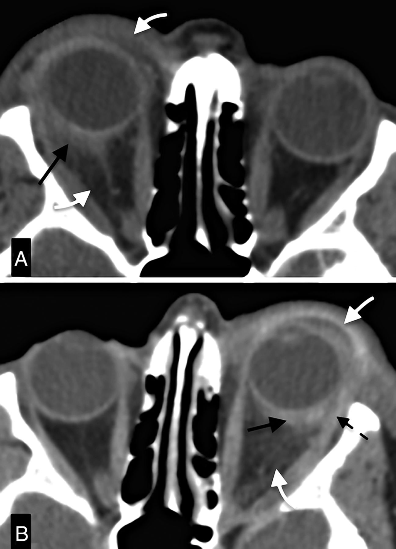
    Asynchronous IOID with scleritis. A, CECT depicts outward, eccentric thickening and enhancement of the right globe wall with focal periscleral cellulitis (black arrow), compatible with posterior scleritis. There is associated pre- and postseptal cellulitis (white arrow) and proptosis. B, CECT 18 months after examination (A) shows almost identical findings in the left orbit. Black and white arrows point to the scleritis and cellulitis, respectively. Notice the complete resolution of the alterations of the right orbit. Also, notice involvement of the tendon of the lateral rectus anteriorly (dashed arrow).

  • Fig 2.
    • Download figure
    • Open in new tab
    • Download powerpoint
    Fig 2.

    Bilateral inflammatory isolated scleritis: axial MR images. A, Post-Gd-DTPA T1-weighted image with fat saturation shows bilateral enhancement of the outer aspect of the sclera (white arrows) extending to the optic nerve sheath, depicting scleritis. There is also focal periscleral cellulitis. Notice the absence of ocular anomalies on the precontrast T1WI (B).

  • Fig 3.
    • Download figure
    • Open in new tab
    • Download powerpoint
    Fig 3.

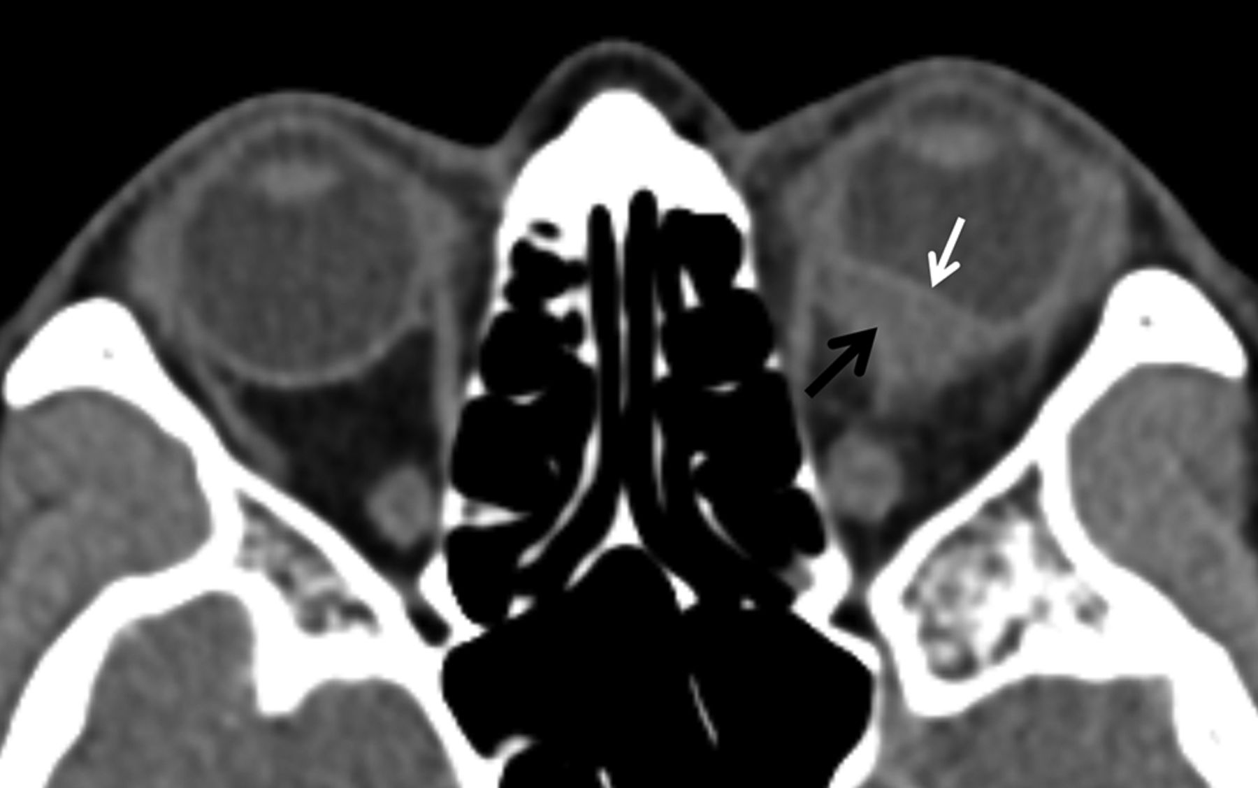
    Nodular inflammatory scleritis mimicking uveal melanoma. CECT depicts a posterior globe wall mass (black arrow) deviating the choroid-retinal layer internally (white arrow), and hence, most probably arising from the sclera. Also notice the presence of slight periscleral cellulitis.

  • Fig 4.
    • Download figure
    • Open in new tab
    • Download powerpoint
    Fig 4.

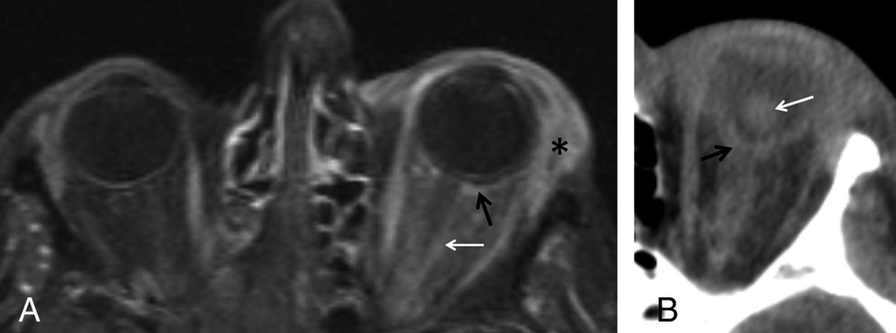
    Infectious orbital process with scleritis followed by panophthalmitis. Post-Gd-DTPA axial T1-weighted spectral presaturation with inversion recovery image of the orbit (A) depicts scleral enhancement (black arrow) and extensive pre- and postseptal cellulitis, with involvement of the optic nerve sheath (white arrow) and dacryoadenitis (asterisk). CECT performed 48 hours later (B) shows lens luxation (white arrow) and inward folding of the globe wall with volume loss (black arrow), depicting globe rupture.

  • Fig 5.
    • Download figure
    • Open in new tab
    • Download powerpoint
    Fig 5.

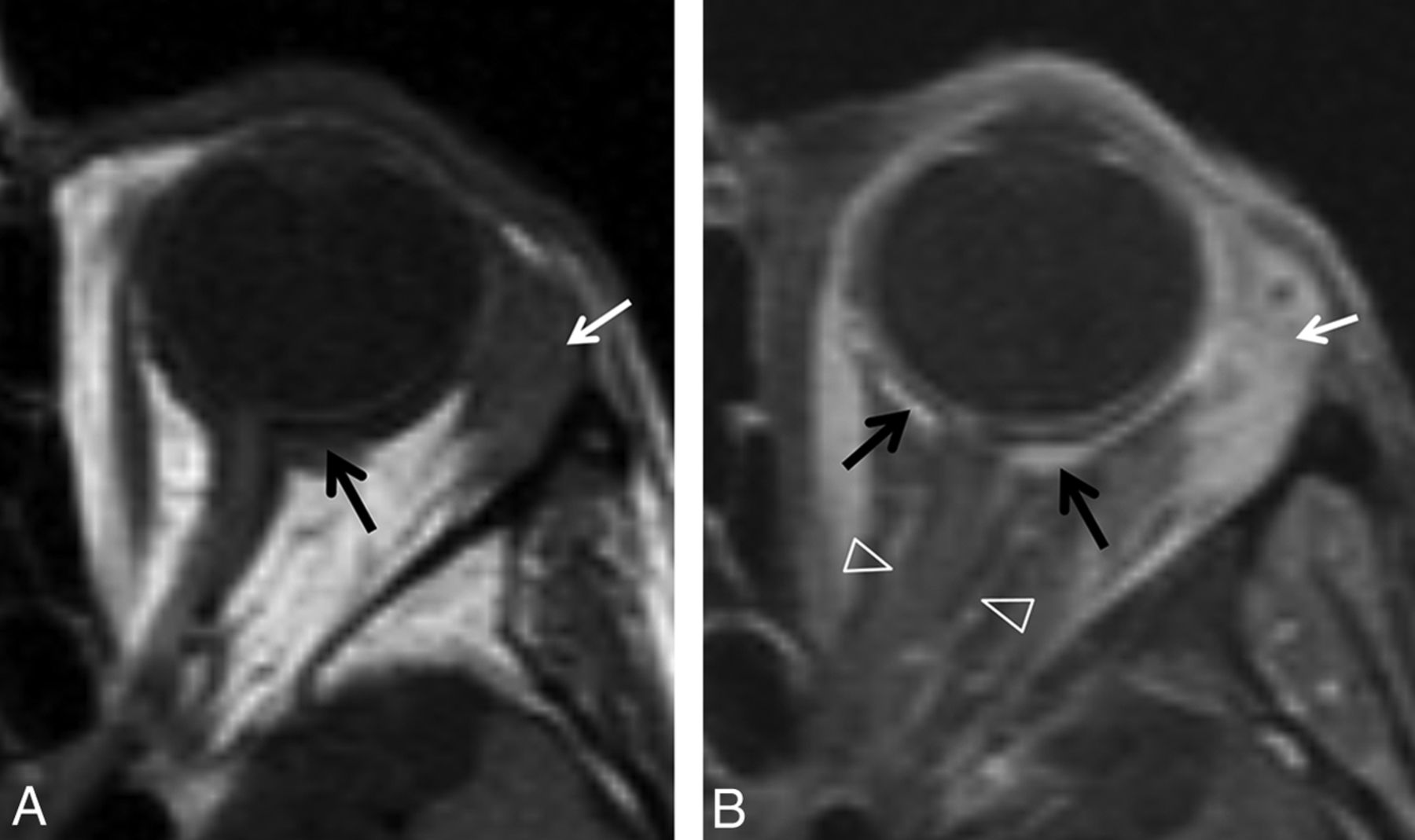
    Orbital inflammation with scleritis in patient with granulomatosis with polyangiitis (Wegener). T1WI (A) shows eccentric focal thickening of the sclera (black arrow), with coexisting enlargement of the lacrimal gland (white arrow), both enhancing on the post-Gd-DTPA T1-weighted spectral presaturation with inversion recovery image (B). Enhancement extends to the optic nerve sheath (open arrow heads). This illustrates posterior scleritis with optic perineuritis, cellulitis, and dacryoadenitis.

  • Fig 6.
    • Download figure
    • Open in new tab
    • Download powerpoint
    Fig 6.

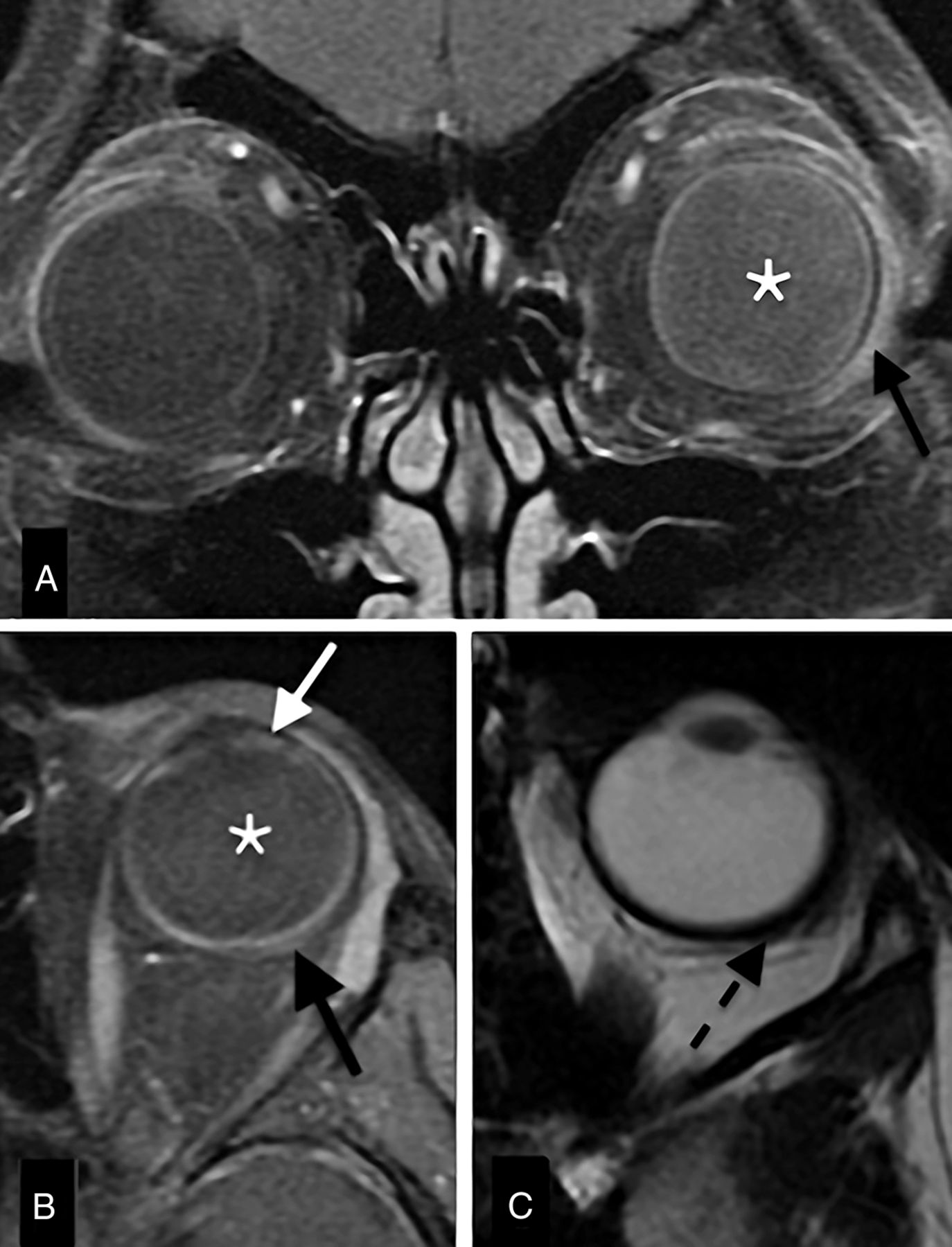
    Scleritis with vitritis and uveitis. Enhanced coronal (A) and axial (B) T1-weighted spectral presaturation with inversion recovery images and axial T2WI (C) with a sclerouveitis. There is increased signal intensity of the vitreous on the left (vitritis; A, B, asterisk), with slight focal enhancement of the iris/cilliary body (uveitis; white arrow) and concurrent slight focal scleral outward thickening (C, black dashed arrow) and enhancement (B, black solid arrow).

  • Fig 7.
    • Download figure
    • Open in new tab
    • Download powerpoint
    Fig 7.

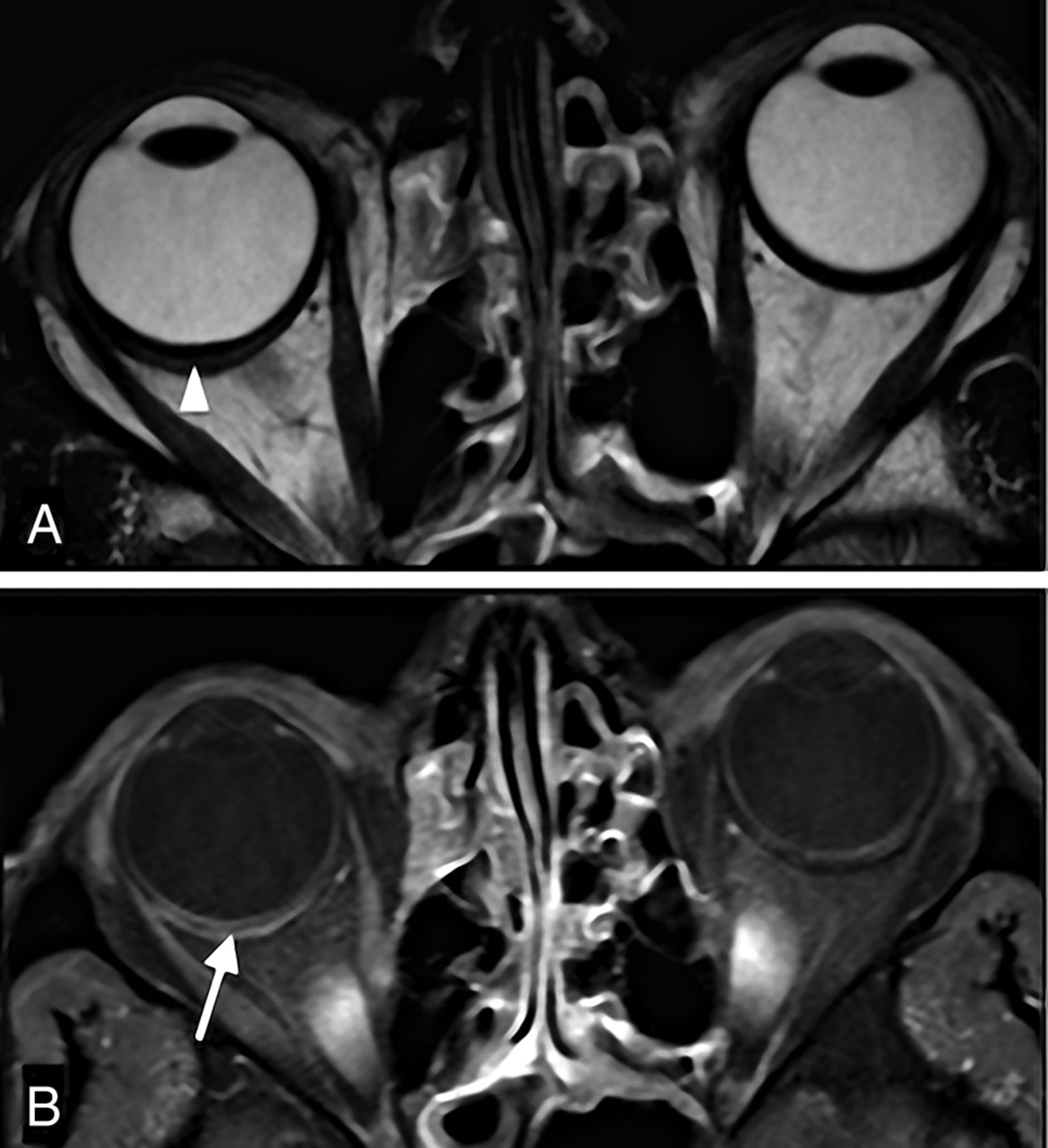
    Isolated inflammatory scleritis: orbital MR imaging. T2-weighted (A) and Gd-DTPA enhanced T1-weighted spectral presaturation with inversion recovery (B) images depict scleral thickening and enhancement (B, white arrow). There is a linear hyperintense (fluid) collection between the sclera and the choroid/retina, representing a suprachoroidal effusion (A, white arrowhead).

Tables

  • Figures
    • View popup
    Table 1:

    Clinical data of patients with scleritis analyzed by CT and/or MR imaging

    PtDuration of SymptomsaUS Performed, DiagnosisClinical DiagnosisbImaging ModalitySystemic DiseaseFinal Diagnosis
    15 moNoInfectious cellulitisCTDown syndromeIOID with scleritis
    12 moNoInfectious cellulitisCTDown syndromeIOID with scleritis
    23 moYes, disc edemaOptic neuritisMRINot foundBilateral idiopathic scleritis
    32 moNoIntraorbital massCTNot foundIdiopathic scleritis
    42 moYes, uveal massChoroidal melanomaCTNot foundNodular idiopathic scleritis
    5AcuteNoInfectious cellulitisCT, MRIDRM; Colon carcinomaInfectious orbital disease with panophthalmitis
    66 moNoInfectious cellulitisCT, MRIDown syndromeIOID with sclerouveitis
    71 moYes, scleritisScleritis or tumorMRIGranulomatosis with polyangiitisAutoimmune orbital inflammation with scleritis
    82 moNoUveitisCT, MRIJIAAutoimmune sclerouveitis
    93 wkYes, inconclusiveOptic pathway conditionCT, MRINoneIdiopathic scleritis
    104 wkNoOptic pathway conditionMRINoneIdiopathic scleritis
    • Note:—DRM indicates dermatomyositis; JIA, juvenile idiopathic arthritis; Pt, patient; US, ultrasonography.

    • ↵a Duration of symptoms refers to the time elapsed between onset of symptoms of scleritis (pain, vision disturbances) and the time of imaging.

    • ↵b Diagnosis after ophthalmologic evaluation and ultrasound and before CT and/or MRI.

    • View popup
    Table 2:

    Imaging findings in 11 cases with scleritis on CT (n = 8) and MRI (n = 8)

    Imaging ModalityImaging FindingNo. (%)
    CTEccentric enhancement of the globe wall8 (100)
    Eccentric thickening of the sclerouveal rim8 (100)
    Periscleral cellulitis6 (75)
    Pre/postseptal cellulitis4 (50)
    Nodular scleral tickening1 (13)
    MRScleral enhancement8 (100)
    Scleral thickening6 (75)
    Focal periscleral cellulitis4 (50)
    Pre/postseptal cellulitis2 (25)
    Scleral thinning1 (13)
    Dacryoadenitis1 (13)
    Uveitis2 (25)
    Suprachoroidal effusion1 (13)
    Retinal detachment1 (13)
    Choroidal detachment1 (13)
PreviousNext
Back to top

In this issue

American Journal of Neuroradiology: 37 (12)
American Journal of Neuroradiology
Vol. 37, Issue 12
1 Dec 2016
  • Table of Contents
  • Index by author
  • Complete Issue (PDF)
Advertisement
Print
Download PDF
Email Article

Thank you for your interest in spreading the word on American Journal of Neuroradiology.

NOTE: We only request your email address so that the person you are recommending the page to knows that you wanted them to see it, and that it is not junk mail. We do not capture any email address.

Enter multiple addresses on separate lines or separate them with commas.
CT and MR Imaging in the Diagnosis of Scleritis
(Your Name) has sent you a message from American Journal of Neuroradiology
(Your Name) thought you would like to see the American Journal of Neuroradiology web site.
CAPTCHA
This question is for testing whether or not you are a human visitor and to prevent automated spam submissions.
Cite this article
M.C. Diogo, M.J. Jager, T.A. Ferreira
CT and MR Imaging in the Diagnosis of Scleritis
American Journal of Neuroradiology Dec 2016, 37 (12) 2334-2339; DOI: 10.3174/ajnr.A4890

Citation Manager Formats

  • BibTeX
  • Bookends
  • EasyBib
  • EndNote (tagged)
  • EndNote 8 (xml)
  • Medlars
  • Mendeley
  • Papers
  • RefWorks Tagged
  • Ref Manager
  • RIS
  • Zotero
0 Responses
Respond to this article
Share
Bookmark this article
CT and MR Imaging in the Diagnosis of Scleritis
M.C. Diogo, M.J. Jager, T.A. Ferreira
American Journal of Neuroradiology Dec 2016, 37 (12) 2334-2339; DOI: 10.3174/ajnr.A4890
del.icio.us logo Twitter logo Facebook logo Mendeley logo
  • Tweet Widget
  • Facebook Like
  • Google Plus One
Purchase

Jump to section

  • Article
    • Abstract
    • ABBREVIATIONS:
    • Case Series
    • Discussion
    • Conclusions
    • Footnotes
    • References
  • Figures & Data
  • Info & Metrics
  • Responses
  • References
  • PDF

Related Articles

  • No related articles found.
  • PubMed
  • Google Scholar

Cited By...

  • No citing articles found.
  • Crossref (32)
  • Google Scholar

This article has been cited by the following articles in journals that are participating in Crossref Cited-by Linking.

  • Systemic lupus erythematosus and ocular involvement: an overview
    Rosanna Dammacco
    Clinical and Experimental Medicine 2018 18 2
  • CT and MR imaging of orbital inflammation
    Teresa A. Ferreira, P. Saraiva, S. W. Genders, M. V. Buchem, G. P. M. Luyten, J-W Beenakker
    Neuroradiology 2018 60 12
  • Scleritis: Differentiating infectious from non-infectious entities
    SomasheilaI Murthy, Swapnali Sabhapandit, S Balamurugan, Pranesh Subramaniam, Maite Sainz-de-la-Maza, Manisha Agarwal, Carlos Parvesio
    Indian Journal of Ophthalmology 2020 68 9
  • Current Approach for the Diagnosis and Management of Noninfective Scleritis
    Parthopratim Dutta Majumder, Rupesh Agrawal, Peter McCluskey, Jyotirmay Biswas
    Asia-Pacific Journal of Ophthalmology 2021 10 2
  • Understanding Ocular Findings and Manifestations of Systemic Lupus Erythematosus: Update Review of the Literature
    Wojciech Luboń, Małgorzata Luboń, Przemysław Kotyla, Ewa Mrukwa-Kominek
    International Journal of Molecular Sciences 2022 23 20
  • Choroidal involvement in non-infectious posterior scleritis
    Sandra Vermeirsch, Ilaria Testi, Carlos Pavesio
    Journal of Ophthalmic Inflammation and Infection 2021 11 1
  • Imaging of orbital infectious and inflammatory disease in children
    Usha D. Nagaraj, Bernadette L. Koch
    Pediatric Radiology 2021 51 7
  • Magnetic Resonance Imaging in the Clinical Care for Uveal Melanoma Patients—A Systematic Review from an Ophthalmic Perspective
    Myriam G. Jaarsma-Coes, Lisa Klaassen, Marina Marinkovic, Gregorius P. M. Luyten, T. H. Khanh Vu, Teresa A. Ferreira, Jan-Willem M. Beenakker
    Cancers 2023 15 11
  • Silent volumetric multi-contrast 7 Tesla MRI of ocular tumors using Zero Echo Time imaging
    Jan-Willem M. Beenakker, Joep Wezel, Jan Groen, Andrew G. Webb, Peter Börnert, Juri G. Gelovani
    PLOS ONE 2019 14 9
  • Using a novel MR imaging sign to differentiate retinal pigment epithelium from uveal melanoma
    Yaping Su, Xiaolin Xu, Wenbin Wei, Junfang Xian
    Neuroradiology 2020 62 3

More in this TOC Section

  • NI-RADS for HEAD&NECK Cancer Recurrence
  • WHO Classification Update: Nasal&Skull Base Tumors
  • Peritumoral Signal in Vestibular Schwannomas
Show more HEAD & NECK

Similar Articles

Advertisement

Indexed Content

  • Current Issue
  • Accepted Manuscripts
  • Article Preview
  • Past Issues
  • Editorials
  • Editors Choice
  • Fellow Journal Club
  • Letters to the Editor

Cases

  • Case Collection
  • Archive - Case of the Week
  • Archive - Case of the Month
  • Archive - Classic Case

Special Collections

  • Special Collections

Resources

  • News and Updates
  • Turn around Times
  • Submit a Manuscript
  • Author Policies
  • Manuscript Submission Guidelines
  • Evidence-Based Medicine Level Guide
  • Publishing Checklists
  • Graphical Abstract Preparation
  • Imaging Protocol Submission
  • Submit a Case
  • Become a Reviewer/Academy of Reviewers
  • Get Peer Review Credit from Publons

Multimedia

  • AJNR Podcast
  • AJNR SCANtastic
  • Video Articles

About Us

  • About AJNR
  • Editorial Board
  • Not an AJNR Subscriber? Join Now
  • Alerts
  • Feedback
  • Advertise with us
  • Librarian Resources
  • Permissions
  • Terms and Conditions

American Society of Neuroradiology

  • Not an ASNR Member? Join Now

© 2025 by the American Society of Neuroradiology All rights, including for text and data mining, AI training, and similar technologies, are reserved.
Print ISSN: 0195-6108 Online ISSN: 1936-959X

Powered by HighWire