
Article Figures & Data
Figures
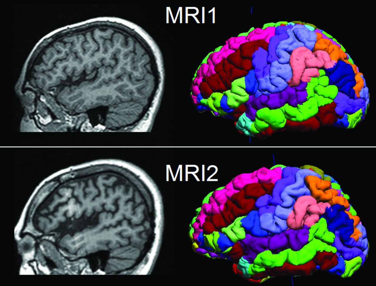
- Fig 1.
Illustration of 2 serial MRIs from the same patient (P6) and the SVReg output of BrainSuite. First row: MR imaging at 10 years of age. Second row: MR imaging at 17 years of age. Shown in the left column are the sagittal T1-weighted MPRAGE images. The right column is the cortical rendering of the SVReg labels, with different colors denoting different anatomic areas of the brain. Pronounced atrophy can be observed at the left peri-Sylvian area, and HRvol shows a decrease from 0.79 to 0.70.
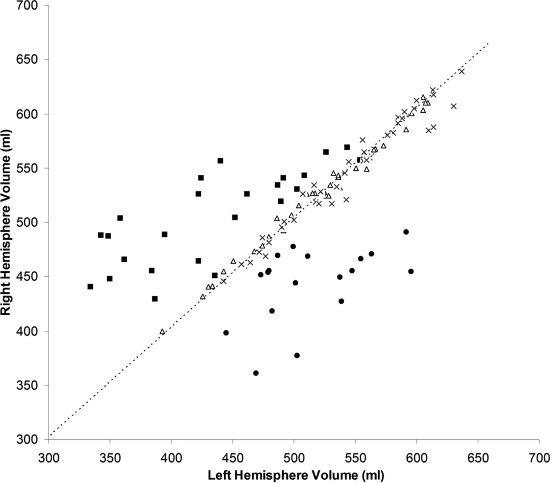
- Fig 2.
Absolute volume of the right hemisphere versus volume of the left hemisphere in patients with RE and 2 control groups. Triangles indicate healthy controls; crosses, controls with non-RE epilepsy; squares, patients with left RE; and circles, patients with right RE. The dashed line is the diagonal line representing hemispheres with equal volumes.
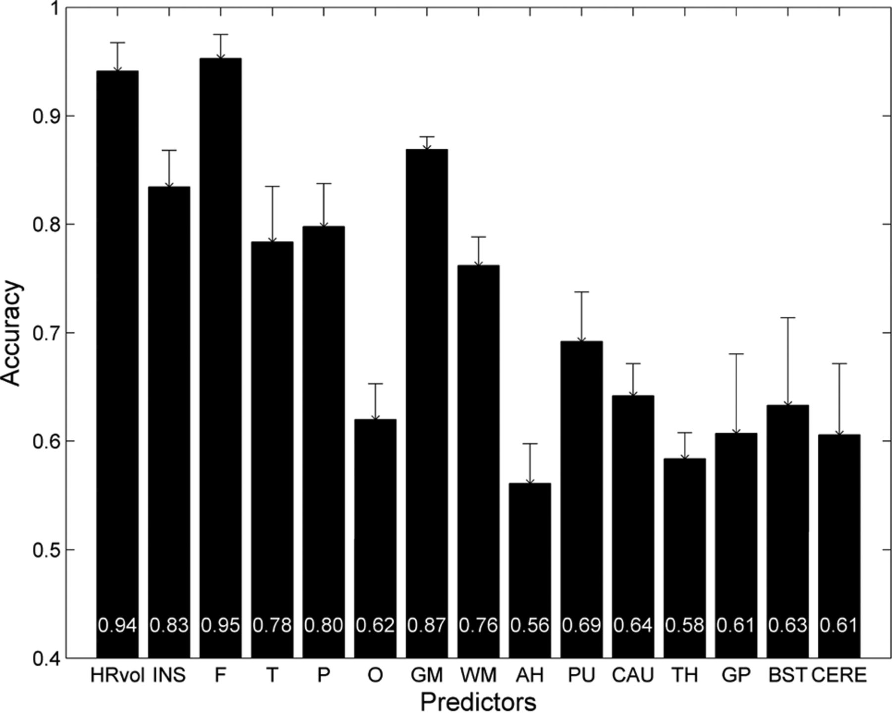
- Fig 3.
Mean accuracy across the 5 cross-validation runs of the logistic regression classifier for 15 volumetric ratio measures. Patients with RE were compared with controls with non-RE epilepsy with matching disease durations. Error bars denote the SD. The mean accuracy values for each measure were plotted at the bottom of the bars. INS indicates insula; F, frontal; T, temporal; P, parietal; O, occipital; AH, amygdala and hippocampus; PU, putamen; CAU, caudate nucleus; TH, thalamus; GP, globus pallidus; BST, brain stem; and CERE, cerebellum.
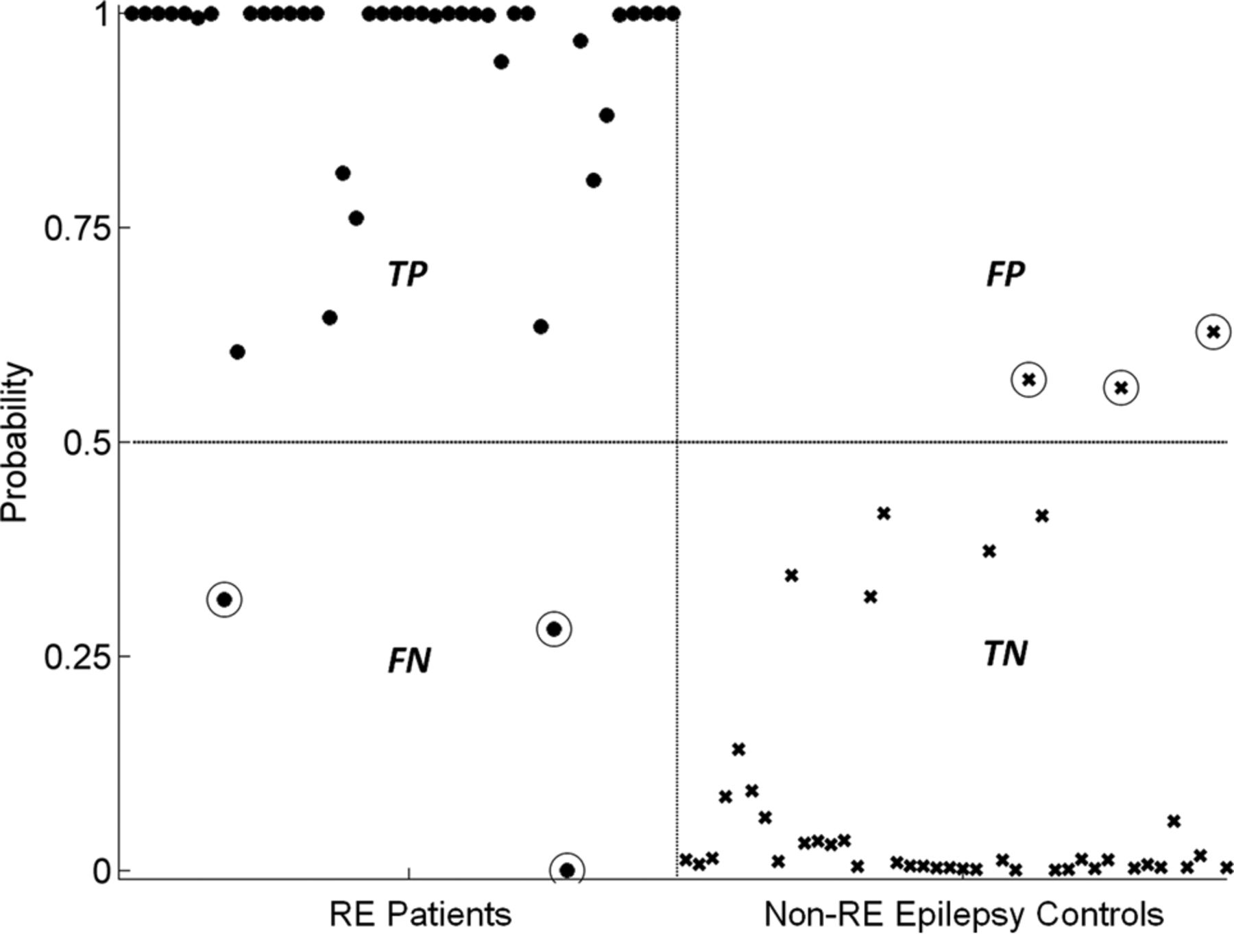
- Fig 4.
Performance of the classifier using HRvol. Patients with RE are denoted with dots, and controls with non-RE epilepsy were denoted with crosses. True-positives (TP) are defined as patients correctly identified as patients by the classifier. True-negatives (TN) are defined as controls correctly identified as controls. False-positives (FP) are defined as controls incorrectly identified as patients. False-negatives (FN) are defined as patients incorrectly identified as controls. Circled dots/crosses denote the subjects who were misclassified (3 FPs and 3 FNs).
- Fig 5.
Receiver operating characteristic analyses showing a highly discriminative classifier using HRvol.
- Fig 6.
Probability curves depicting the relationship between HRvol and the probability of RE. The solid curve was estimated on the basis of comparison of 42 scans from patients with RE and 42 scans from controls with non-RE epilepsy with the same disease duration. The thin dashed curve was additionally generated to correct for the difference in incidence of RE and non-RE epilepsy (1 in 1,000,000 versus 1 in 100).
- Fig 7.
A, HRvol plotted over epilepsy duration in the 9 patients with serial MR imaging. All except P5 show a decrease in HRvol for the observed time. The axis is broken from 14 to 20 years because there are no data points for these durations. B, Absolute hemispheric volume (right-sided versus left-sided plots) of the same 9 patients. The direction of each dotted arrow shows the progression of disease over time in each patient. A and B share the same symbol for each patient for direct comparison.


Tables
- Table 1:
Detailed demographics and clinical data of the 19 patients with RE and 2 control groups
Demographics Patients with RE (N = 19) Mean age and SD at epilepsy onset (median) (range) (yr) 7.3 ± 5.3; 8; 1.5–22 Mean disease duration and SD at first MRI (median) (range) (yr) 4.0 ± 4.8; 2.9; 0.1–20 Sex (No.) Female 11 Male 8 Handedness (No.) Right 13 Left 5 Ambidextrous 1 Surgery location (No.) Hemispherectomy 12 (10 SF) Frontal 1 (1 SF) Insular/opercular 1 Temporal 1 No surgery 4 No. of MRI scans Single scan 10 Multiple scans 9 (range, 2–7) Mean age and SD at MRI (N = 42 scans) (median) (range) (yr) RE scans 14.2 ± 8.0; 14.8; 3–43 Healthy controls 14.3 ± 8.0; 14.0; 3.6–43 Controls with non-RE epilepsy 16.9 ± 7.6; 15.5; 5–31 Mean disease duration and SD at MRI (N = 42 scans) (median) (range) (yr) RE scans 7.0 ± 5.6; 6.3; 0.8–21.4 Controls with non-RE epilepsy 7.6 ± 5.1; 6.5; 1–22 Note:—SF indicates seizure-free with >12 months postoperative follow-up.
- Table 2:
Regional atrophy difference in all the lobar, basal ganglia and mesial temporal structure regions
Region Ratio of Atrophy (±SD) Lobar Insula 0.77 ± 0.04 Frontal 0.84 ± 0.03 Temporal 0.88 ± 0.03 Parietal 0.87 ± 0.03 Occipital 0.90 ± 0.05 GM 0.85 ± 0.02 WM 0.88 ± 0.02 Basal ganglia and mesial temporal structures AH 0.90 ± 0.02 PU 0.88 ± 0.03 CAU 0.89 ± 0.06 TH 0.95 ± 0.04 GP 0.94 ± 0.02 Note:—AH indicates amygdala and hippocampus combined; PU, putamen; CAU, caudate nucleus; TH, thalamus; GP, globus pallidus.


Jump to section
Related Articles
Cited By...
- Acute seizure risk in patients with encephalitis: development and validation of clinical prediction models from two independent prospective multicentre cohorts
- Immunomodulation With Azathioprine Therapy in Rasmussen Syndrome: A Multimodal Evaluation
- A Large-scale Comparison of Cortical and Subcortical Structural Segmentation Methods in Alzheimers Disease: a Statistical Approach