
Article Figures & Data
Figures
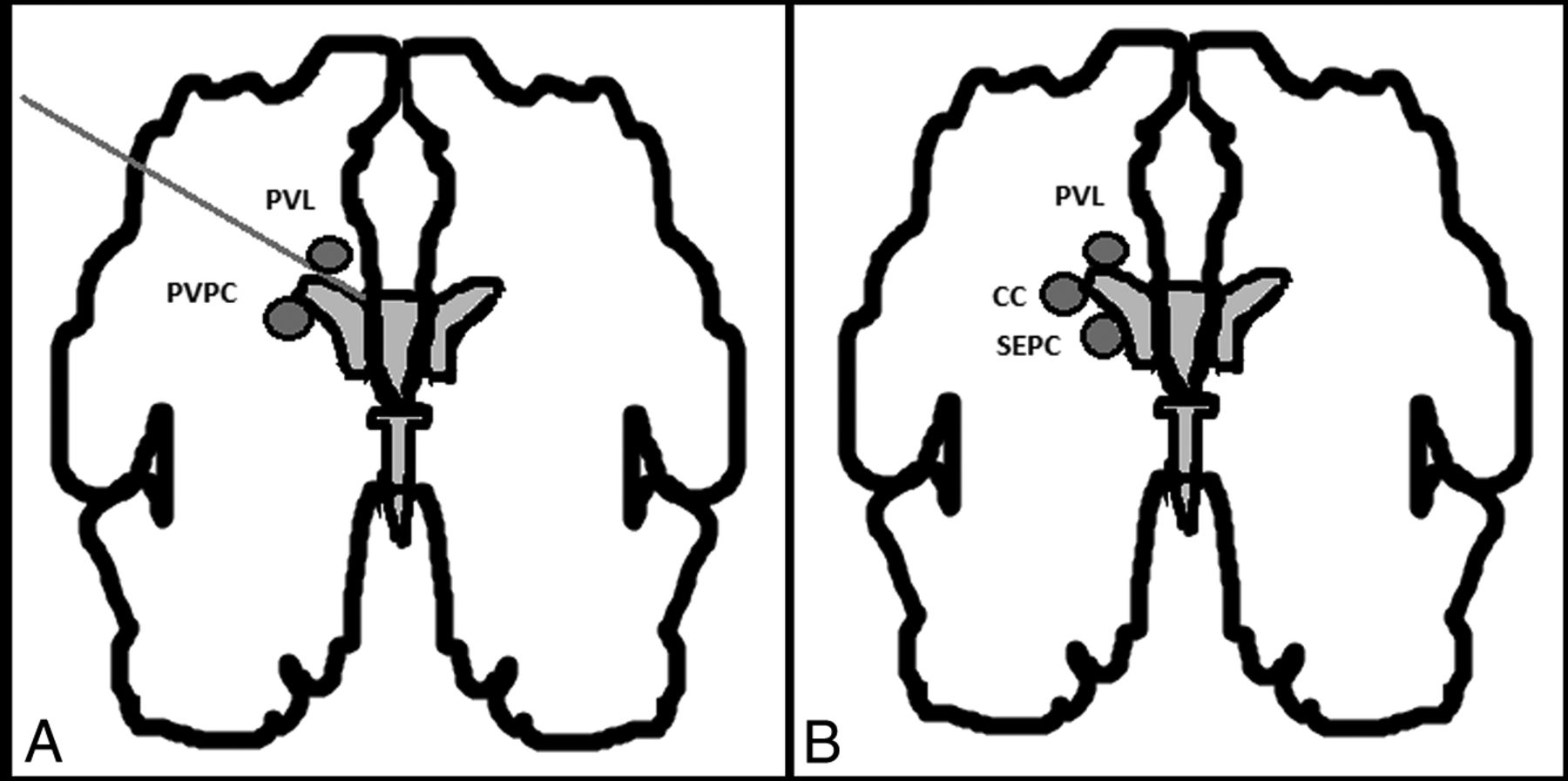
- Fig 1.
A, Schematic representation of the differential diagnosis between periventricular pseudocysts and periventricular leukomalacia. Originally published by Malinger et al.4 B, Differential diagnosis between the cystic lesions seen in periventricular leukomalacia (PVL), connatal cysts (CC), and subependymal cysts (SC). Malinger et al4 original publication modified by Epelman et al.6
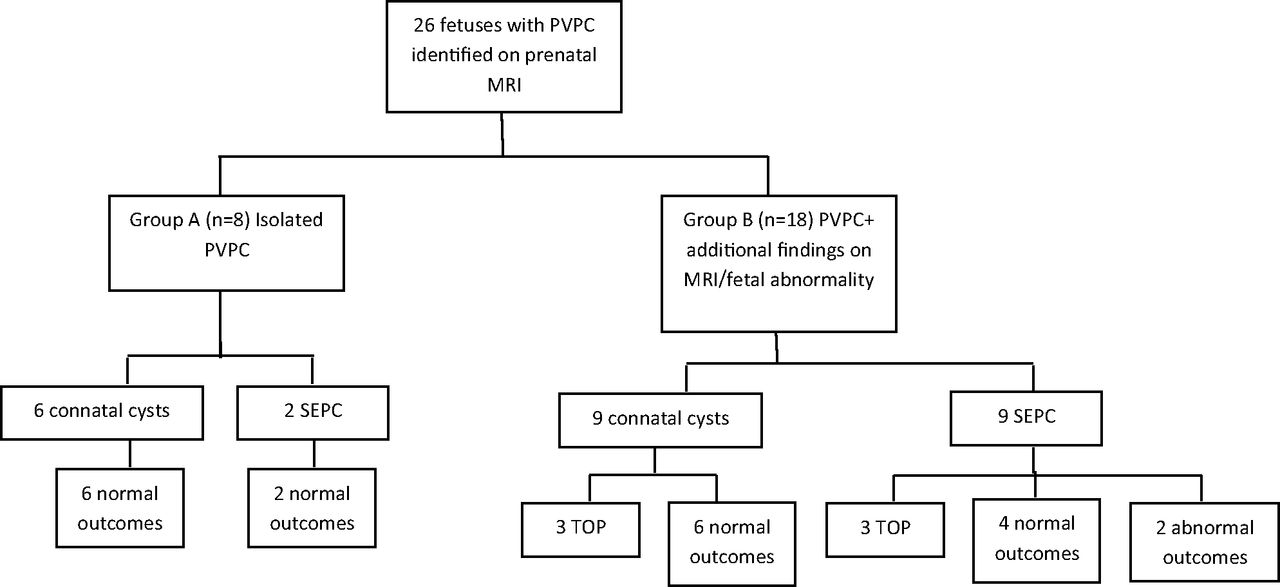
- Fig 2.
Flowchart illustrating the study design and outcome. Cases are divided to 2 groups: fetuses in group A had only PVPC on MR imaging, while fetuses on group B had additional findings on MR imaging or fetal abnormality. Fetal abnormality is defined as the presence of fetal infection, chromosomal abnormality, IUGR, abnormal echocardiogram findings, or other fetal malformation. The groups were further subdivided into connatal cysts or subependymal pseudocysts.
- Fig 3.
T2 MR imaging coronal view. A, Case 5, bilateral connatal cysts located at the external angle, anterior to the foramina of Monro. B, Case 17, bilateral subependymal pseudocysts located posterior to the foramina of Monro.


Tables
Characteristics Pregnancy and maternal Maternal age (yr) 32.5 (21–42) In vitro fertilization 3/26 (11.5%) Multiple pregnancies 2/26 (7.7%) Maternal infection 5/26 (19.2%) CMV 4/26 (15.3%) Toxoplasma 1/26 (3.8%) Maternal medical treatment 5/26 (19.2%) Maternal hypercoagulative disorder 4/26 (15.3%) TOP 6/26 (23.1%) Fetal and neonatal Male/female ratio (15:11) 1.4:1 Fetal abnormalities IUGR 4/26 (15.4%) CMV infection 2/26 (7.7%) Chromosomal aberration 2/26 (7.7%) Abnormal fetal echocardiogram findings 1/26 (3.8%) GA at MRI diagnosis (wk) 33 (29–38) Birth GA (wk) 38 (35–41) BW (g) 3210 (2445–4060) Apgar score At 1 min 9 (8–9) At 5 min 10 (8–10) Mode of delivery (n = 20) Vaginal delivery 11/20 (55%) Cesarean delivery 7/20 (35%) Assisted vaginal delivery 2/20 (10%) Note:—GA indicates gestational age, BW, birth weight.
↵a Data are expressed as median (range) or number (percentage).
- Table 2:
MRI morphologic features and neurodevelopmental outcome of connatal cysts and subependymal pseudocysts
MRI Morphologic Feature Connatal Cysts SEPC P Value Bilateral (No.) 15/15 (100%) 9/11 (82%) .17 Multilocular (No.) 14/15 (93.3%) 10/11 (91%) 1.00 Mean height 5.17 ± 1.03 5.61 ± 0.63 .22 Mean AP diameter 8.53 ± 2.76 8.97 ± 2.10 .65 Near the occipital horns (No.) 0 1/11 (9%) .42 Posterior to the caudothalamic notch (No.) 0 4/11 (36%) .02 Atypical morphology (No.) 1/15 (6.6%) 0 .42 Abnormal neurodevelopmental outcome (No.) 0 2/11 (18%) .15 TOP (No.) 3/15 (20%) 3/11 (27%) – Note:—AP indicates anteroposterior.


