Advertisement

Main menu

  • Home
  • Content
    • Current Issue
    • Accepted Manuscripts
    • Article Preview
    • Past Issue Archive
    • Video Articles
    • AJNR Case Collection
    • Case of the Week Archive
    • Case of the Month Archive
    • Classic Case Archive
  • Special Collections
    • AJNR Awards
    • Low-Field MRI
    • Alzheimer Disease
    • ASNR Foundation Special Collection
    • Photon-Counting CT
    • View All
  • Multimedia
    • AJNR Podcasts
    • AJNR SCANtastic
    • Trainee Corner
    • MRI Safety Corner
    • Imaging Protocols
  • For Authors
    • Submit a Manuscript
    • Submit a Video Article
    • Submit an eLetter to the Editor/Response
    • Manuscript Submission Guidelines
    • Statistical Tips
    • Fast Publishing of Accepted Manuscripts
    • Graphical Abstract Preparation
    • Imaging Protocol Submission
    • Author Policies
  • About Us
    • About AJNR
    • Editorial Board
    • Editorial Board Alumni
  • More
    • Become a Reviewer/Academy of Reviewers
    • Subscribers
    • Permissions
    • Alerts
    • Feedback
    • Advertisers
    • ASNR Home

User menu

  • Alerts
  • Log in

Search

  • Advanced search
American Journal of Neuroradiology

American Journal of Neuroradiology

ASHNR American Society of Functional Neuroradiology ASHNR American Society of Pediatric Neuroradiology ASSR
  • Alerts
  • Log in

Advanced Search

  • Home
  • Content
    • Current Issue
    • Accepted Manuscripts
    • Article Preview
    • Past Issue Archive
    • Video Articles
    • AJNR Case Collection
    • Case of the Week Archive
    • Case of the Month Archive
    • Classic Case Archive
  • Special Collections
    • AJNR Awards
    • Low-Field MRI
    • Alzheimer Disease
    • ASNR Foundation Special Collection
    • Photon-Counting CT
    • View All
  • Multimedia
    • AJNR Podcasts
    • AJNR SCANtastic
    • Trainee Corner
    • MRI Safety Corner
    • Imaging Protocols
  • For Authors
    • Submit a Manuscript
    • Submit a Video Article
    • Submit an eLetter to the Editor/Response
    • Manuscript Submission Guidelines
    • Statistical Tips
    • Fast Publishing of Accepted Manuscripts
    • Graphical Abstract Preparation
    • Imaging Protocol Submission
    • Author Policies
  • About Us
    • About AJNR
    • Editorial Board
    • Editorial Board Alumni
  • More
    • Become a Reviewer/Academy of Reviewers
    • Subscribers
    • Permissions
    • Alerts
    • Feedback
    • Advertisers
    • ASNR Home
  • Follow AJNR on Twitter
  • Visit AJNR on Facebook
  • Follow AJNR on Instagram
  • Join AJNR on LinkedIn
  • RSS Feeds

AJNR Awards, New Junior Editors, and more. Read the latest AJNR updates

Research ArticleNeurointervention

MR Angiographic–Guided Percutaneous Sclerotherapy for Venous Vascular Malformations: A Radiation Dose-Reduction Strategy

S.G. Imbesi, D.A. Green, A. Cho and R.S. Pakbaz
American Journal of Neuroradiology February 2016, 37 (2) 205-209; DOI: https://doi.org/10.3174/ajnr.A4518
S.G. Imbesi
aFrom the Department of Radiology (S.G.I., D.A.G., A.C.), University of California, San Diego Health System, San Diego, California
  • Find this author on Google Scholar
  • Find this author on PubMed
  • Search for this author on this site
D.A. Green
aFrom the Department of Radiology (S.G.I., D.A.G., A.C.), University of California, San Diego Health System, San Diego, California
  • Find this author on Google Scholar
  • Find this author on PubMed
  • Search for this author on this site
A. Cho
aFrom the Department of Radiology (S.G.I., D.A.G., A.C.), University of California, San Diego Health System, San Diego, California
  • Find this author on Google Scholar
  • Find this author on PubMed
  • Search for this author on this site
R.S. Pakbaz
bDepartment of Radiology (R.S.P.), VA San Diego Healthcare System, San Diego, California.
  • Find this author on Google Scholar
  • Find this author on PubMed
  • Search for this author on this site
  • Article
  • Figures & Data
  • Info & Metrics
  • Responses
  • References
  • PDF
Loading

Article Figures & Data

Figures

  • Fig 1.
    • Download figure
    • Open in new tab
    • Download powerpoint
    Fig 1.

    Early (A), mid (B), and late (C) phase direct needle puncture phlebography of a left facial venous malformation by using a traditional digital subtraction angiography technique shows appropriate needle localization for subsequent embolization. There is satisfactory sequential opacification of the venous channels of the lesion (arrow) and only faint flow into small draining veins (arrowheads), with no evidence of contrast extravasation or demonstration of large draining veins.

  • Fig 2.
    • Download figure
    • Open in new tab
    • Download powerpoint
    Fig 2.

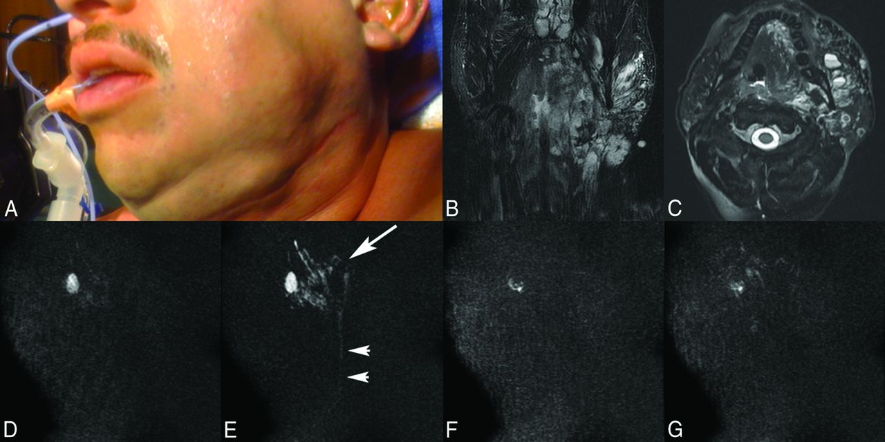
    A 36-year-old man with a left facial venous malformation who undergone 14 treatments during the past 3 years, the last 2 with MR imaging guidance. Overview of the direct-injection MR angiography technique for embolization of the lesion. A, Patient photograph before MR sclerotherapy shows left facial swelling from a persistent left facial venous malformation. Coronal (B) and axial (C) T2-weighted fat-saturated MR images demonstrate the typical imaging characteristics of a venous vascular malformation, including a mixed signal intensity but mostly bright trans-spatial lesion extending both superficial and deep to the left mandible composed of multiloculated blood-filled channels with fluid-fluid levels producing local mass effect. Pretreatment MR angiograms, early (D) and late (E) phase, show appropriate needle positioning within the loculated blood-filled channels of the venous malformation (arrow) with slow venous runoff into a small draining vein (arrowheads). Posttreatment MR angiograms, early (F) and late (G) phase, demonstrate pruning of the vascular channels of the venous malformation with markedly diminished flow into the draining vein, compatible with satisfactory embolization of the lesion.

  • Fig 3.
    • Download figure
    • Open in new tab
    • Download powerpoint
    Fig 3.

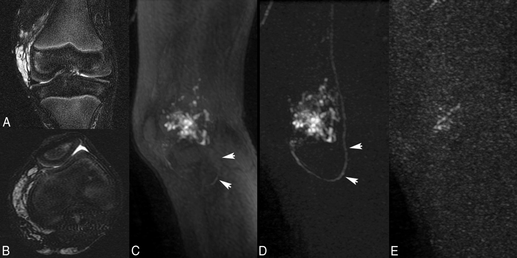
    A 6-year-old boy with a peripheral venous malformation of the left knee. Note depiction of the mask subtraction technique that results in more complete visualization of the vascular structures on MR angiography. Coronal (A) and axial (B) T2-weighted fat-saturated MR images demonstrate a multiloculated increased T2-signal venous vascular malformation in the left knee, particularly surrounding the medial and posterior portions of the distal femur. Pretreatment MR angiogram, without (C) and with (D) digital mask subtraction, shows opacification of the venous malformation but limited visualization of the draining vein (arrowheads) without mask subtraction; however, there is not only improved visualization of the venous malformation but also excellent visualization of the draining vein (arrowheads) achieved following mask subtraction of the images. E, Posttreatment MR angiogram again shows pruning of the vascular channels of the venous malformation with no significant flow into the draining vein, compatible with satisfactory embolization of the lesion.

  • Fig 4.
    • Download figure
    • Open in new tab
    • Download powerpoint
    Fig 4.

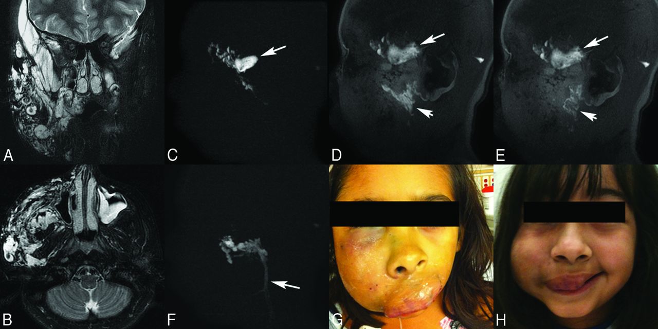
    A 10-year-old girl with a very large venous malformation of the right face, who has had >20 previous treatment episodes with traditional digital subtraction angiography guidance, now being treated with MR imaging guidance. These images demonstrate the necessity of angiography to assure proper needle positioning. Coronal (A) and axial (B) T2-weighted fat-saturated MR images show the very large venous vascular malformation extending essentially throughout all superficial and deep spaces of the right face, producing significant local mass effect. C, Pretreatment MR angiogram, initial needle placement, demonstrates opacification of the venous malformation but also a focus of stagnation of contrast material that remained on the late-phase images, indicating extravasation (arrow). Thus, this is not a safe location to treat, and sclerotherapy was not performed. Note that normal venous backflow from the needle hub was initially detected, but adequate needle placement can only be assured following angiography. Early (D) and late (E) phase pretreatment MR angiograms, second needle placement, again demonstrate opacification of the venous malformation but now also show runoff of the contrast material on the late-phase image, indicating safe needle placement for embolization (arrowheads). Note the continued presence of the previously extravasated contrast material from the first injection on this nonmask subtracted image (arrows). F, Early-phase pretreatment MR angiogram, third needle placement, depicts needle localization within the venous malformation; however, there is very rapid filling of an enlarged draining vein (arrow). Thus, this is not a safe location to treat, and sclerotherapy was not performed. Again, adequate needle placement can only be assured following angiographic visualization of the lesion. Patient photographs before (G) and following (H) MR sclerotherapy show significant improvement in right facial swelling; however, a persistent right facial venous malformation is still present, requiring additional multiple future treatment sessions.

PreviousNext
Back to top

In this issue

American Journal of Neuroradiology: 37 (2)
American Journal of Neuroradiology
Vol. 37, Issue 2
1 Feb 2016
  • Table of Contents
  • Index by author
  • Complete Issue (PDF)
Advertisement
Print
Download PDF
Email Article

Thank you for your interest in spreading the word on American Journal of Neuroradiology.

NOTE: We only request your email address so that the person you are recommending the page to knows that you wanted them to see it, and that it is not junk mail. We do not capture any email address.

Enter multiple addresses on separate lines or separate them with commas.
MR Angiographic–Guided Percutaneous Sclerotherapy for Venous Vascular Malformations: A Radiation Dose-Reduction Strategy
(Your Name) has sent you a message from American Journal of Neuroradiology
(Your Name) thought you would like to see the American Journal of Neuroradiology web site.
CAPTCHA
This question is for testing whether or not you are a human visitor and to prevent automated spam submissions.
Cite this article
S.G. Imbesi, D.A. Green, A. Cho, R.S. Pakbaz
MR Angiographic–Guided Percutaneous Sclerotherapy for Venous Vascular Malformations: A Radiation Dose-Reduction Strategy
American Journal of Neuroradiology Feb 2016, 37 (2) 205-209; DOI: 10.3174/ajnr.A4518

Citation Manager Formats

  • BibTeX
  • Bookends
  • EasyBib
  • EndNote (tagged)
  • EndNote 8 (xml)
  • Medlars
  • Mendeley
  • Papers
  • RefWorks Tagged
  • Ref Manager
  • RIS
  • Zotero
0 Responses
Respond to this article
Share
Bookmark this article
MR Angiographic–Guided Percutaneous Sclerotherapy for Venous Vascular Malformations: A Radiation Dose-Reduction Strategy
S.G. Imbesi, D.A. Green, A. Cho, R.S. Pakbaz
American Journal of Neuroradiology Feb 2016, 37 (2) 205-209; DOI: 10.3174/ajnr.A4518
del.icio.us logo Twitter logo Facebook logo Mendeley logo
  • Tweet Widget
  • Facebook Like
  • Google Plus One
Purchase

Jump to section

  • Article
    • Abstract
    • Discussion
    • Conclusions
    • References
  • Figures & Data
  • Info & Metrics
  • Responses
  • References
  • PDF

Related Articles

  • No related articles found.
  • PubMed
  • Google Scholar

Cited By...

  • No citing articles found.
  • Crossref
  • Google Scholar

This article has not yet been cited by articles in journals that are participating in Crossref Cited-by Linking.

More in this TOC Section

Neurointervention

  • A Retrospective Study in Tentorial DAVFs
  • Proximal Protection Devices for Carotid Stenting
  • Hydrocoils: an Updated Meta-Analysis
Show more Neurointervention

Head and Neck Imaging

  • Hydrops Herniation into the Semicircular Canals
  • MRI of the Parasellar Ligaments
  • ASL Sensitivity for Head and Neck Paraganglioma
Show more Head and Neck Imaging

Similar Articles

Advertisement

Indexed Content

  • Current Issue
  • Accepted Manuscripts
  • Article Preview
  • Past Issues
  • Editorials
  • Editor's Choice
  • Fellows' Journal Club
  • Letters to the Editor
  • Video Articles

Cases

  • Case Collection
  • Archive - Case of the Week
  • Archive - Case of the Month
  • Archive - Classic Case

More from AJNR

  • Trainee Corner
  • Imaging Protocols
  • MRI Safety Corner
  • Book Reviews

Multimedia

  • AJNR Podcasts
  • AJNR Scantastics

Resources

  • Turnaround Time
  • Submit a Manuscript
  • Submit a Video Article
  • Submit an eLetter to the Editor/Response
  • Manuscript Submission Guidelines
  • Statistical Tips
  • Fast Publishing of Accepted Manuscripts
  • Graphical Abstract Preparation
  • Imaging Protocol Submission
  • Evidence-Based Medicine Level Guide
  • Publishing Checklists
  • Author Policies
  • Become a Reviewer/Academy of Reviewers
  • News and Updates

About Us

  • About AJNR
  • Editorial Board
  • Editorial Board Alumni
  • Alerts
  • Permissions
  • Not an AJNR Subscriber? Join Now
  • Advertise with Us
  • Librarian Resources
  • Feedback
  • Terms and Conditions
  • AJNR Editorial Board Alumni

American Society of Neuroradiology

  • Not an ASNR Member? Join Now

© 2025 by the American Society of Neuroradiology All rights, including for text and data mining, AI training, and similar technologies, are reserved.
Print ISSN: 0195-6108 Online ISSN: 1936-959X

Powered by HighWire