
Article Figures & Data
Figures
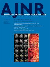
- Fig 1.
IAC diverticula on temporal bone CT. A, CT of the right temporal bone in a 41-year-old woman with vertigo. A CSF-density saccular outpouching of the anterior IAC medial to the cochlea is compatible with an IAC diverticulum (black arrow). B, CT of the left temporal bone in a 67-year-old woman undergoing evaluation of SNHL. A smaller diverticulum or notch is present along the anterior IAC in a location similar to that of the larger diverticula in A (black arrow).
- Fig 2.
IAC diverticulum with classic imaging findings of otosclerosis on temporal bone CT in a 34-year-old woman evaluated for mixed hearing loss. There is a small IAC diverticulum along the anterior IAC (black arrow) with focal lucency of the otic capsule adjacent to the anterior oval window near the fissula ante fenestram (white arrow).


Tables
Characteristic Total Population (n = 807) IAC Diverticulum Otosclerosis Present (n = 43) Not Present (n = 764) P Value Present (n = 39) Not Present (n = 768) P Value Median age (range) (yr) 52 (18–96) 61 (18–91) 52 (18–96) <.01a 52 (22–85) 52 (18–96) .69a Sex (No.) (%) .93b .13b Male 343 (42.5) 18 (41.9) 325 (42.5) 12 (30.8) 331 (43.1) Female 464 (57.5) 25 (58.1) 439 (57.5) 27 (69.2) 437 (56.9) Characteristic IAC Diverticulum Only (n = 36) Otosclerosis Only (n = 32) IAC Diverticulum + Otosclerosis (n = 7) None (n = 732) P Value Median age (range) (yr) 62 (18–91) 52 (18–91) 61 (30–70) 52 (18–96) <.01a Sex (No.) (%) .47b Male 15 (41.7) 9 (28.1) 3 (42.9) 316 (43.2) Female 21 (58.3) 23 (71.9) 4 (57.1) 416 (56.8) Temporal bone involvement (No.) (%) .02b Unilateral 9 (20.9) 15 (38.5) 5 (71.4) Bilateral 34 (79.1) 24 (61.5) 2 (28.6) Characteristic IAC Diverticulum Only (n = 43) Otosclerosis Only (n = 39) IAC Diverticulum + Otosclerosis (n = 7) P Value Hearing loss (No.) (%) <.01a None 4 (9%) 7 (18%) 1 (14%) SNHL 27 (63%) 10 (26%) 2 (29%) PTA (average) (range) (dB) 53.5 (10.6–120) 55.3 (16.9–115) 97.5 (94.4–100.6) CHL (No.) (%) None 13 (33%) 1 (14%) Mixed (No.) (%) 12 (28%) 9 (23%) 3 (43%) Note:—PTA indicates pure tone average.
↵a P value was calculated for comparison of groups with the Fisher exact test.


Jump to section
Related Articles
Cited By...
- Internal Auditory Canal Diverticula among Pediatric Patients: Prevalence and Assessment for Hearing Loss and Anatomic Associations
- Isolated Internal Auditory Canal Diverticula: A Normal Anatomic Variant Not Associated with Sensorineural Hearing Loss
- Cavitary Plaques in Otospongiosis: CT Findings and Clinical Implications