
Article Figures & Data
Figures
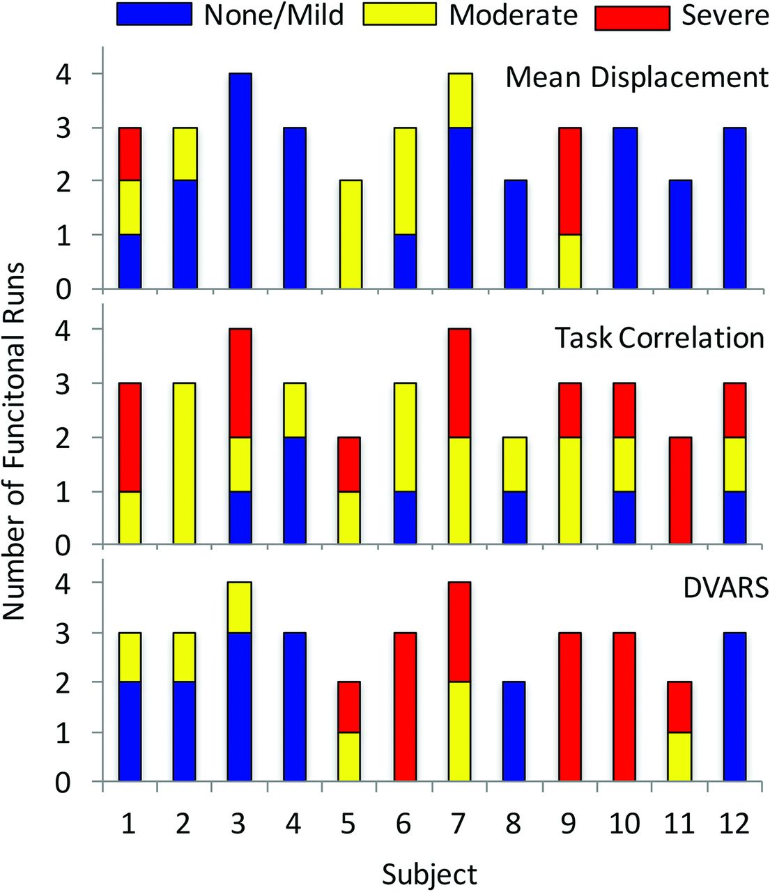
- Fig 1.
Distribution of motion parameters in each individual subject showing the number of functional runs characterized as having a none/mild, moderate, or severe rating in each of the 3 recorded parameters (DVARS, task-correlated motion, and MD).
- Fig 2.
Distribution of diagnostic and nondiagnostic scans for each correction method per subject.
- Fig 3.
Processed fMRI data from 3 different subjects. Each case shows ICA-denoised data in blue, motion-scrubbed data in red, and overlapping areas of ICA-denoised data and motion-scrubbed data in green. The first case (A) is a motor finger task in a patient with a left parietal glioma. The primary motor cortex for the right finger (arrow) is only seen after ICA denoising. Likewise, the supplementary motor area (arrowhead) shows a slight increase in statistical significance. The second subject (B) is undergoing a motor face task with severe task-correlated motion and severe DVARS showing no major change in the primary motor face cortex (arrow); however, the number of motion-related false-positives (noise) is markedly reduced. The third case (C) is a semantic decision task in which no meaningful activation is present on the motion-scrubbed data. Expected areas of activation in the anterior and posterior language areas are clearly present after ICA denoising.
- Fig 4.
A and B, Images for a subject undergoing a motor finger task showing areas in which the z score increased (red) after ICA denoising compared with motion scrubbing. The arrow illustrates increased statistical significance in the left finger primary motor cortex adjacent to the tumor. The location of the primary motor cortex for the left finger was confirmed surgically and is shown on the intraoperative map (C), where the crosshairs correspond to primary finger motor cortex. The white “blobs” in C show the thresholded maps for the realignment-only data where there is no activation in the area of the primary motor cortex.
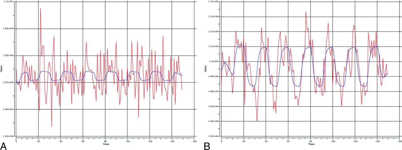
- Fig 5.
Signal intensity with time from 1 voxel is shown before (A) and after (B) ICA denoising with the expected hemodynamic response in blue. One can appreciate the amount of overlapping noise from a variety of artifacts resulting in a poor fit with the ideal curve. These noise effects are largely removed after ICA denoising, and the subsequent denoised time course shows a substantially improved fit with the task design.


Jump to section
Related Articles
Cited By...
- Recommended Resting-State fMRI Acquisition and Preprocessing Steps for Preoperative Mapping of Language and Motor and Visual Areas in Adult and Pediatric Patients with Brain Tumors and Epilepsy
- Identifying brain areas for tactile perception through natural touch with MR-compatible tactile stimulus delivery system