
Article Figures & Data
Figures
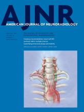
- Fig 1.
The radiologic presentation of a 64-year-old man with a FTAVF. A, Spinal angiogram shows the FTAVF at the level of S2 (arrow). Note the faint filum terminale artery (arrowhead) from the T10 intercostal artery, converging with the draining vein upwardly. B, Internal iliac artery angiogram demonstrates the extra supply of the FTAVF lesion in A. Note the same appearance of the drainage vein (arrow) as in A. C, The T1 contrast-enhanced image demonstrates the abnormally dilated and tortuous vessels situated on the surface of the spinal cord (arrow). D, T2-weighted image of the thoracic spine shows cord edema extending to the upper thoracic spinal cord.
- Fig 2.
The radiologic presentation of a 34-year-old man with concomitant rAVS and conus AVM. A, Left L3 lumbar artery angiogram shows the nidus-type conus AVM at the level of L1 (arrow). B, Right internal iliac artery angiogram demonstrates an rAVS (arrow), which shares the draining vein of the AVM. C, Spinal CT angiography shows the draining vein of the rAVS and its connection to the conus AVM. D, Cast of liquid embolic agent with complete occlusion of the rAVS from the right internal iliac artery.
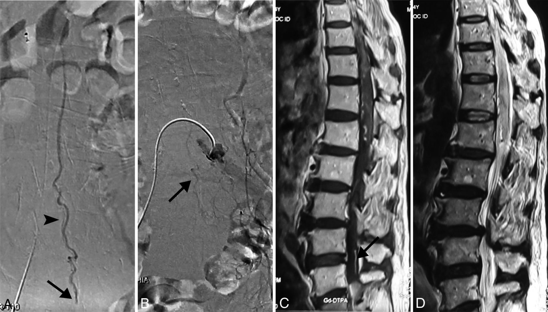
- Fig 3.
The radiologic presentation of a 64-year-old man with an SDAVF below the conus associated with a tethered cord. A, Left internal iliac artery angiogram shows the SDAVF at the level of S2 (arrow). B, Embolic material cast reveals that the embolic agent is approaching the proximal venous end (arrowhead). C, The patient also has a tethered cord (white arrow) on the T1-weighted MR image.


Tables
Classification No. of Patients (%) Feeding Artery FTAVF 11 (22.9%) Artery of the filum terminale FTAVF with extra supply 3 (6.2%) FTAVF with conus AVM 1 (2.1%) rAVS 7 (14.6%) Radicular artery of the segmental artery rAVS with conus AVM 3 (6.3%) SDAVF 30 (62.5%) Dural branch from the lumbar artery or lateral sacral artery - Table 2:
Comparison of demographics, clinical presentation, and MRI findings of 3 types of spinal arteriovenous shunts below the conusa
Characteristics FTAVF (n = 11) rAVS (n = 7) SDAVF (n = 30) All (n = 48) P Valueb Demographic Age (mean) (yr) 52.9 ± 12.6 37.4 ± 15.7 55.6 ± 14.9 52.4 ± 15.5 .017c Female sex 3 (27.3%) 3 (42.9%) 6 (20%) 12 (25%) .443 Clinical symptoms Duration of symptoms (mean) (mo) 12.1 ± 7.8 9.9 ± 9.8 13.1 ± 28.0 12.4 ± 22.6 .396 Back pain 3 (27.3%) 3 (42.9%) 2 (6.7%) 8 (17.0%) .028c Progressive paraparesis 11 (100.0%) 6 (85.7%) 27 (90.0%) 44 (91.7%) .598 Progressive hypesthesia 7 (63.6%) 6 (85.7%) 20 (66.7%) 33 (68.8%) .739 Numbness 5 (45.5%) 3 (42.9%) 16 (53.3%) 24 (50%) .844 Bowel/bladder dysfunction 8 (72.7%) 4 (57.1%) 26 (86.7%) 38 (79.2%) .167 MRI findings Spinal cord edema 11 (100%) 5 (71.4%) 28 (93.3%) 44 (91.7%) .129 Engorged vein 10 (90.9%) 6 (85.7%) 29 (96.7%) 45 (93.8%) .313 Spinal cord tethering 6 (54.5%) 0 (0.0%) 7 (23.3%) 13 (27.1%) .032c Sacral lipoma 5 (45.5%) 0 (0.0%) 5 (16.7%) 10 (20.8%) .061 - Table 3:
Comparison of treatment and clinical outcomes of spinal arteriovenous shunts below the conus
FTAVF rAVS SDAVF All P Valuea Treatment option (No.) Embolization 0 5 16 21 .002b Surgery 9 1 8 18 E+S 2 1 6 9 Mean FUc 24.0 ± 26.1 21.3 ± 5.3 27.6 ± 20.6 25.7 ± 20.2 .54 Complete obliteration (No.) (%) 9 (100%) 6 (100%) 19 (82.6%) 34 (89.4%) .476 Median ALS grade pretreatmentc 9 7.5 7 7.5 .273 Median ALS grade at last FUc 5 4 5 4.5 P valued .023b .063 <.001b <.001b


Jump to section
Related Articles
Cited By...
- Long-term outcome in a cohort of 36 patients with sacral dural arteriovenous fistulae after endovascular embolisation or microsurgery
- Intraoperative direct venous puncture and embolization for arteriovenous shunt below the conus medullaris: a prospective cohort study
- Cauda Equina and Filum Terminale Arteriovenous Fistulas: Anatomic and Radiographic Features
- Republished: Transradial approach in the treatment of a sacral dural arteriovenous fistula: a technical note
- Transradial approach in the treatment of a sacral dural arteriovenous fistula: a technical note
- Concomitant conus medullaris arteriovenous shunts and sacral dural arteriovenous fistulas: pathophysiological links related to the venous drainage of the lesions in a series of five cases
- Clinical outcomes and prognostic factors in patients with spinal dural arteriovenous fistulas : a prospective cohort study in two Chinese centres