Advertisement

Main menu

  • Home
  • Content
    • Current Issue
    • Accepted Manuscripts
    • Article Preview
    • Past Issue Archive
    • Video Articles
    • AJNR Case Collection
    • Case of the Week Archive
    • Case of the Month Archive
    • Classic Case Archive
  • Special Collections
    • AJNR Awards
    • Low-Field MRI
    • Alzheimer Disease
    • ASNR Foundation Special Collection
    • Photon-Counting CT
    • View All
  • Multimedia
    • AJNR Podcasts
    • AJNR SCANtastic
    • Trainee Corner
    • MRI Safety Corner
    • Imaging Protocols
  • For Authors
    • Submit a Manuscript
    • Submit a Video Article
    • Submit an eLetter to the Editor/Response
    • Manuscript Submission Guidelines
    • Statistical Tips
    • Fast Publishing of Accepted Manuscripts
    • Graphical Abstract Preparation
    • Imaging Protocol Submission
    • Author Policies
  • About Us
    • About AJNR
    • Editorial Board
    • Editorial Board Alumni
  • More
    • Become a Reviewer/Academy of Reviewers
    • Subscribers
    • Permissions
    • Alerts
    • Feedback
    • Advertisers
    • ASNR Home

User menu

  • Alerts
  • Log in

Search

  • Advanced search
American Journal of Neuroradiology

American Journal of Neuroradiology

ASHNR American Society of Functional Neuroradiology ASHNR American Society of Pediatric Neuroradiology ASSR
  • Alerts
  • Log in

Advanced Search

  • Home
  • Content
    • Current Issue
    • Accepted Manuscripts
    • Article Preview
    • Past Issue Archive
    • Video Articles
    • AJNR Case Collection
    • Case of the Week Archive
    • Case of the Month Archive
    • Classic Case Archive
  • Special Collections
    • AJNR Awards
    • Low-Field MRI
    • Alzheimer Disease
    • ASNR Foundation Special Collection
    • Photon-Counting CT
    • View All
  • Multimedia
    • AJNR Podcasts
    • AJNR SCANtastic
    • Trainee Corner
    • MRI Safety Corner
    • Imaging Protocols
  • For Authors
    • Submit a Manuscript
    • Submit a Video Article
    • Submit an eLetter to the Editor/Response
    • Manuscript Submission Guidelines
    • Statistical Tips
    • Fast Publishing of Accepted Manuscripts
    • Graphical Abstract Preparation
    • Imaging Protocol Submission
    • Author Policies
  • About Us
    • About AJNR
    • Editorial Board
    • Editorial Board Alumni
  • More
    • Become a Reviewer/Academy of Reviewers
    • Subscribers
    • Permissions
    • Alerts
    • Feedback
    • Advertisers
    • ASNR Home
  • Follow AJNR on Twitter
  • Visit AJNR on Facebook
  • Follow AJNR on Instagram
  • Join AJNR on LinkedIn
  • RSS Feeds

AJNR Awards, New Junior Editors, and more. Read the latest AJNR updates

Research ArticleADULT BRAIN

Myelin Detection Using Rapid Quantitative MR Imaging Correlated to Macroscopically Registered Luxol Fast Blue–Stained Brain Specimens

J.B.M. Warntjes, A. Persson, J. Berge and W. Zech
American Journal of Neuroradiology June 2017, 38 (6) 1096-1102; DOI: https://doi.org/10.3174/ajnr.A5168
J.B.M. Warntjes
aFrom the Center for Medical Image Science and Visualization (J.B.M.W., A.P., W.Z.)
bDivision of Cardiovascular Medicine, Department of Medical and Health Sciences (J.B.M.W.), Linköping University, Linköping, Sweden
cSyntheticMR AB (J.B.M.W.), Linköping, Sweden
  • Find this author on Google Scholar
  • Find this author on PubMed
  • Search for this author on this site
  • ORCID record for J.B.M. Warntjes
A. Persson
aFrom the Center for Medical Image Science and Visualization (J.B.M.W., A.P., W.Z.)
  • Find this author on Google Scholar
  • Find this author on PubMed
  • Search for this author on this site
  • ORCID record for A. Persson
J. Berge
dInstitute of Forensic Medicine (J.B., W.Z.), Linköping, Sweden
  • Find this author on Google Scholar
  • Find this author on PubMed
  • Search for this author on this site
  • ORCID record for J. Berge
W. Zech
aFrom the Center for Medical Image Science and Visualization (J.B.M.W., A.P., W.Z.)
dInstitute of Forensic Medicine (J.B., W.Z.), Linköping, Sweden
eInstitute of Forensic Medicine (W.Z.), University of Bern, Bern, Switzerland.
  • Find this author on Google Scholar
  • Find this author on PubMed
  • Search for this author on this site
  • ORCID record for W. Zech
  • Article
  • Figures & Data
  • Info & Metrics
  • Responses
  • References
  • PDF
Loading

Article Figures & Data

Figures

  • Tables
  • Fig 1.
    • Download figure
    • Open in new tab
    • Download powerpoint
    Fig 1.

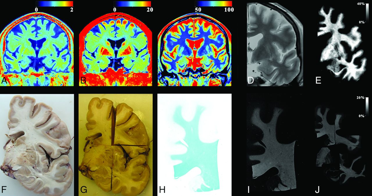
    The process used for myelin evaluation on a male subject, 69 years of age, acquired at a temperature of 10°C. The MR imaging quantification sequence provided the R1, R2, and PD maps of coronal slices of the cadaver (A–C). A synthetic proton density–weighted image was created for registration purposes using the R1, R2, and PD maps as input, resampled to 0.1 mm/pixel (D, zoomed in). The R1 map was corrected for temperature and then used, with the original R2 and PD maps, to generate the myelin partial volume map with the same algorithm as used for living subjects (E). For the histologic images, the brain was extracted and cut into coronal slices (F). Slices were fixated by using formaldehyde and cut into smaller pieces after fixation (G). The separate pieces of brain slices were stained with Luxol fast blue and photographed (H). The optical density of the photographs was converted to an intensity scale. These images were also resampled to 0.1 mm/pixel (I). All pieces were registered to the synthetic proton density–weighted image (J), so that each pixel from the slice photographs was at the same place as the corresponding MR imaging pixel. Finally, the resolution of both MR images and photographs was down-sampled to the original MR imaging resolution of 0.7 mm/pixel.

  • Fig 2.
    • Download figure
    • Open in new tab
    • Download powerpoint
    Fig 2.

    A, The observed mean T1 relaxation time in frontal white matter (squares), cortical gray matter (triangles), and ventricular CSF (dots) as a function of the core body temperature of all subjects. Added are the estimated slopes of T1 change per temperature. B, The observed mean T2 relaxation time in frontal WM (squares), cortical GM (triangles), and ventricular CSF (dots) as a function of core body temperature of all subjects.

  • Fig 3.
    • Download figure
    • Open in new tab
    • Download powerpoint
    Fig 3.

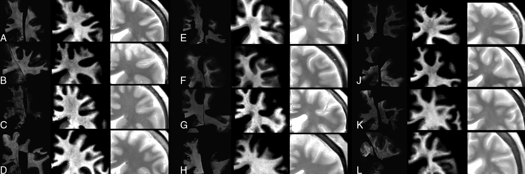
    Zoomed-in images of all subjects, ordered according to increasing age (A, 46; B, 47; C, 52; D, 55; E, 59; F, 61; G, 69; H, 70; I, 70; J, 71; K, 72; and L, 74 years of age). On the left, the registered LFB stains are shown as optical density, in the center is the MR imaging–detected myelin partial volume, and on the right is the synthetic PD-weighted images.

  • Fig 4.
    • Download figure
    • Open in new tab
    • Download powerpoint
    Fig 4.

    2D histograms of all subjects of the Luxol fast blue optical density values, plotted as a function of the observed R1 relaxation rate (A), R2 relaxation rate (B), and proton density (C). Also, the estimated myelin partial volume, based on temperature-corrected R1, R2, and PD is plotted (D). The intensity of the 2D histograms corresponds to the number of times a value occurs within each value interval. Intervals were divided into 40 × 40 bins.

Tables

  • Figures
    • View popup
    Table 1:

    The mean and SD for all subjects of the intercept and slopea

    InterceptSlopeSpearman ρ
    R11.19 ± 0.10 s−10.065 ± 0.009 s−1/%0.63 ± 0.12
    R212.31 ± 1.17 s−10.044 ± 0.131 s−1/%0.11 ± 0.28
    PD71.96 ± 4.22%−2.45 ± 0.53 %/%−0.73 ± 0.09
    Myelin1.50% ± 2.84%4.37% ± 1.73 %/%0.74% ± 0.11
    • ↵a Estimated by linear regression and the Spearman ρ of the LFB optical density as a function of (uncorrected) R1 and R2 relaxation, proton density, and (temperature-corrected) MRI-estimated myelin partial volume.

    • View popup
    Table 2:

    The mean and SD for all subjects of the position of highest value density for white matter and gray matter observed in the 2D histogramsa

    WMGMInterceptSlope
    R11.47 ± 0.08 s−11.27 ± 0.09 s−11.24 s−10.030 s−1/%
    R212.55 ± 1.25 s−112.11 ± 1.08 s−112.06 s−10.066 s−1/%
    PD52.01% ± 2.98%71.36% ± 3.95%73.46%−2.85 %/%
    Myelin30.98% ± 3.77%4.84% ± 2.30%2.01%3.84 %/%
    LFB7.54% ± 2.06%0.74% ± 0.65%––
    • ↵a Added are the derived slopes and intercepts when using the positions of R1, R2, PD, and myelin as a function of the position of LFB.

PreviousNext
Back to top

In this issue

American Journal of Neuroradiology: 38 (6)
American Journal of Neuroradiology
Vol. 38, Issue 6
1 Jun 2017
  • Table of Contents
  • Index by author
  • Complete Issue (PDF)
Advertisement
Print
Download PDF
Email Article

Thank you for your interest in spreading the word on American Journal of Neuroradiology.

NOTE: We only request your email address so that the person you are recommending the page to knows that you wanted them to see it, and that it is not junk mail. We do not capture any email address.

Enter multiple addresses on separate lines or separate them with commas.
Myelin Detection Using Rapid Quantitative MR Imaging Correlated to Macroscopically Registered Luxol Fast Blue–Stained Brain Specimens
(Your Name) has sent you a message from American Journal of Neuroradiology
(Your Name) thought you would like to see the American Journal of Neuroradiology web site.
CAPTCHA
This question is for testing whether or not you are a human visitor and to prevent automated spam submissions.
Cite this article
J.B.M. Warntjes, A. Persson, J. Berge, W. Zech
Myelin Detection Using Rapid Quantitative MR Imaging Correlated to Macroscopically Registered Luxol Fast Blue–Stained Brain Specimens
American Journal of Neuroradiology Jun 2017, 38 (6) 1096-1102; DOI: 10.3174/ajnr.A5168

Citation Manager Formats

  • BibTeX
  • Bookends
  • EasyBib
  • EndNote (tagged)
  • EndNote 8 (xml)
  • Medlars
  • Mendeley
  • Papers
  • RefWorks Tagged
  • Ref Manager
  • RIS
  • Zotero
0 Responses
Respond to this article
Share
Bookmark this article
Myelin Detection Using Rapid Quantitative MR Imaging Correlated to Macroscopically Registered Luxol Fast Blue–Stained Brain Specimens
J.B.M. Warntjes, A. Persson, J. Berge, W. Zech
American Journal of Neuroradiology Jun 2017, 38 (6) 1096-1102; DOI: 10.3174/ajnr.A5168
del.icio.us logo Twitter logo Facebook logo Mendeley logo
  • Tweet Widget
  • Facebook Like
  • Google Plus One
Purchase

Jump to section

  • Article
    • Abstract
    • ABBREVIATIONS:
    • Materials and Methods
    • Results
    • Discussion
    • Conclusions
    • Footnotes
    • References
  • Figures & Data
  • Info & Metrics
  • Responses
  • References
  • PDF

Related Articles

  • No related articles found.
  • PubMed
  • Google Scholar

Cited By...

  • Fast and reliable quantitative measures of white matter development with magnetic resonance fingerprinting
  • An automated pipeline for extracting histological stain area fraction for voxelwise quantitative MRI-histology comparisons
  • White Matter Abnormalities in Multiple Sclerosis Evaluated by Quantitative Synthetic MRI, Diffusion Tensor Imaging, and Neurite Orientation Dispersion and Density Imaging
  • Effect of Gadolinium on the Estimation of Myelin and Brain Tissue Volumes Based on Quantitative Synthetic MRI
  • Analysis of White Matter Damage in Patients with Multiple Sclerosis via a Novel In Vivo MR Method for Measuring Myelin, Axons, and G-Ratio
  • Crossref
  • Google Scholar

This article has not yet been cited by articles in journals that are participating in Crossref Cited-by Linking.

More in this TOC Section

  • Diagnostic Neuroradiology of Monoclonal Antibodies
  • Clinical Outcomes After Chiari I Decompression
  • Segmentation of Brain Metastases with BLAST
Show more Adult Brain

Similar Articles

Advertisement

Indexed Content

  • Current Issue
  • Accepted Manuscripts
  • Article Preview
  • Past Issues
  • Editorials
  • Editor's Choice
  • Fellows' Journal Club
  • Letters to the Editor
  • Video Articles

Cases

  • Case Collection
  • Archive - Case of the Week
  • Archive - Case of the Month
  • Archive - Classic Case

More from AJNR

  • Trainee Corner
  • Imaging Protocols
  • MRI Safety Corner
  • Book Reviews

Multimedia

  • AJNR Podcasts
  • AJNR Scantastics

Resources

  • Turnaround Time
  • Submit a Manuscript
  • Submit a Video Article
  • Submit an eLetter to the Editor/Response
  • Manuscript Submission Guidelines
  • Statistical Tips
  • Fast Publishing of Accepted Manuscripts
  • Graphical Abstract Preparation
  • Imaging Protocol Submission
  • Evidence-Based Medicine Level Guide
  • Publishing Checklists
  • Author Policies
  • Become a Reviewer/Academy of Reviewers
  • News and Updates

About Us

  • About AJNR
  • Editorial Board
  • Editorial Board Alumni
  • Alerts
  • Permissions
  • Not an AJNR Subscriber? Join Now
  • Advertise with Us
  • Librarian Resources
  • Feedback
  • Terms and Conditions
  • AJNR Editorial Board Alumni

American Society of Neuroradiology

  • Not an ASNR Member? Join Now

© 2025 by the American Society of Neuroradiology All rights, including for text and data mining, AI training, and similar technologies, are reserved.
Print ISSN: 0195-6108 Online ISSN: 1936-959X

Powered by HighWire