Advertisement

Main menu

  • Home
  • Content
    • Current Issue
    • Accepted Manuscripts
    • Article Preview
    • Past Issue Archive
    • AJNR Case Collection
    • Case of the Week Archive
    • Classic Case Archive
    • Case of the Month Archive
  • Special Collections
    • Spinal CSF Leak Articles (Jan 2020-June 2024)
    • 2024 AJNR Journal Awards
    • Most Impactful AJNR Articles
  • Multimedia
    • AJNR Podcast
    • AJNR Scantastics
    • Video Articles
  • For Authors
    • Submit a Manuscript
    • Author Policies
    • Fast publishing of Accepted Manuscripts
    • Graphical Abstract Preparation
    • Manuscript Submission Guidelines
    • Imaging Protocol Submission
    • Submit a Case for the Case Collection
  • About Us
    • About AJNR
    • Editorial Board
  • More
    • Become a Reviewer/Academy of Reviewers
    • Subscribers
    • Permissions
    • Alerts
    • Feedback
    • Advertisers
    • ASNR Home
  • Other Publications
    • ajnr

User menu

  • Alerts
  • Log in

Search

  • Advanced search
American Journal of Neuroradiology

American Journal of Neuroradiology

ASHNR American Society of Functional Neuroradiology ASHNR American Society of Pediatric Neuroradiology ASSR
  • Alerts
  • Log in

Advanced Search

  • Home
  • Content
    • Current Issue
    • Accepted Manuscripts
    • Article Preview
    • Past Issue Archive
    • AJNR Case Collection
    • Case of the Week Archive
    • Classic Case Archive
    • Case of the Month Archive
  • Special Collections
    • Spinal CSF Leak Articles (Jan 2020-June 2024)
    • 2024 AJNR Journal Awards
    • Most Impactful AJNR Articles
  • Multimedia
    • AJNR Podcast
    • AJNR Scantastics
    • Video Articles
  • For Authors
    • Submit a Manuscript
    • Author Policies
    • Fast publishing of Accepted Manuscripts
    • Graphical Abstract Preparation
    • Manuscript Submission Guidelines
    • Imaging Protocol Submission
    • Submit a Case for the Case Collection
  • About Us
    • About AJNR
    • Editorial Board
  • More
    • Become a Reviewer/Academy of Reviewers
    • Subscribers
    • Permissions
    • Alerts
    • Feedback
    • Advertisers
    • ASNR Home
  • Follow AJNR on Twitter
  • Visit AJNR on Facebook
  • Follow AJNR on Instagram
  • Join AJNR on LinkedIn
  • RSS Feeds

Welcome to the new AJNR, Updated Hall of Fame, and more. Read the full announcements.


AJNR is seeking candidates for the position of Associate Section Editor, AJNR Case Collection. Read the full announcement.


Research ArticleFUNCTIONAL
Open Access

Resting-State Seed-Based Analysis: An Alternative to Task-Based Language fMRI and Its Laterality Index

K.A. Smitha, K.M. Arun, P.G. Rajesh, B. Thomas and C. Kesavadas
American Journal of Neuroradiology June 2017, 38 (6) 1187-1192; DOI: https://doi.org/10.3174/ajnr.A5169
K.A. Smitha
aFrom the Departments of Imaging Sciences and Interventional Radiology (K.A.S., K.M.A., B.T., C.K.)
  • Find this author on Google Scholar
  • Find this author on PubMed
  • Search for this author on this site
  • ORCID record for K.A. Smitha
K.M. Arun
aFrom the Departments of Imaging Sciences and Interventional Radiology (K.A.S., K.M.A., B.T., C.K.)
  • Find this author on Google Scholar
  • Find this author on PubMed
  • Search for this author on this site
  • ORCID record for K.M. Arun
P.G. Rajesh
bNeurology (P.G.R.), Sree Chitra Tirunal Institute for Medical Sciences and Technology, Trivandrum, Kerala, India.
  • Find this author on Google Scholar
  • Find this author on PubMed
  • Search for this author on this site
  • ORCID record for P.G. Rajesh
B. Thomas
aFrom the Departments of Imaging Sciences and Interventional Radiology (K.A.S., K.M.A., B.T., C.K.)
  • Find this author on Google Scholar
  • Find this author on PubMed
  • Search for this author on this site
  • ORCID record for B. Thomas
C. Kesavadas
aFrom the Departments of Imaging Sciences and Interventional Radiology (K.A.S., K.M.A., B.T., C.K.)
  • Find this author on Google Scholar
  • Find this author on PubMed
  • Search for this author on this site
  • ORCID record for C. Kesavadas
  • Article
  • Figures & Data
  • Info & Metrics
  • Responses
  • References
  • PDF
Loading

Article Figures & Data

Figures

  • Tables
  • Fig 1.
    • Download figure
    • Open in new tab
    • Download powerpoint
    Fig 1.

    The bar chart presents the average value of the laterality indices obtained by using task-based fMRI for all the subjects in the Broca area, Brodmann area 6 (frontal area), and Wernicke area (temporal area).

  • Fig 2.
    • Download figure
    • Open in new tab
    • Download powerpoint
    Fig 2.

    A and B, Sagittal and axial images obtained after the 1-sample t test of the task-fMRI for the Broca area and Brodmann area 6. C and D, Wernicke area of 18 healthy controls.
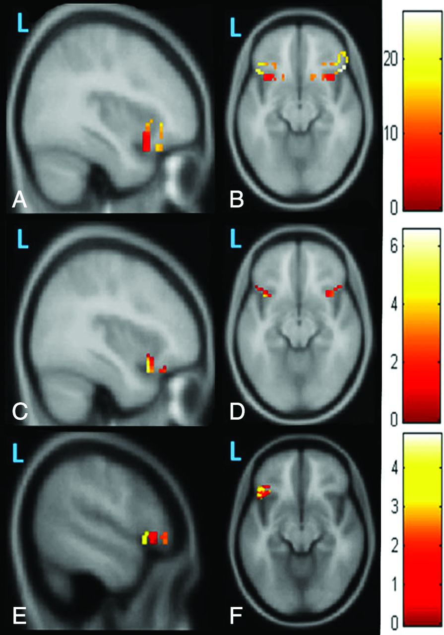
  • Fig 3.
    • Download figure
    • Open in new tab
    • Download powerpoint
    Fig 3.

    The first row shows the sagittal and axial images obtained after a 1-sample t test of the Z-maps of 18 functional connectivity images obtained with the Broca area as the seed area. The second row shows sagittal and axial images obtained after a 1-sample t test of the Z-maps of the amplitude of low-frequency fluctuations in 18 healthy controls. We observed limited correlated activity for functional connectivity (A and B) and ALFF (C and D); it seems to be fragmented and potentially less clinically reliable at the individual level. The third row shows the sagittal and axial images obtained after a 1-sample t test of the Z-maps of fractional amplitude of low-frequency fluctuations in 18 healthy controls. By contrast, the map of fALFF (E and F) appears more robust and potentially more reliable.

  • Fig 4.
    • Download figure
    • Open in new tab
    • Download powerpoint
    Fig 4.

    The regression line plotted with signals extracted from the T-map of the z scores of the fractional amplitude of low-frequency fluctuations from the Broca area of resting-state fMRI with the laterality index obtained from all ROIs of task-based fMRI.

Tables

  • Figures
    • View popup
    Table 1:

    The mean value of the laterality index obtained from Brodmann areas 44, 45, 47, 6, 21, and 22 using task-based fMRI for all subjects

    Language AreasMean LI of 18 Subjects
    Brodmann Area 440.375
    Brodmann Area 450.238
    Brodmann Area 470.430
    Brodmann Area 60.377
    Brodmann Area 210.166
    Brodmann Area 220.218
    • View popup
    Table 2:

    Correlation of the seed-based analysis metrics FC, ALFF, and fALFF of rsfMRI with the LI of task-based fMRI

    RegionCorrelation between the Z Score of FC and LICorrelation between the Z Score of ALFF and LICorrelation between the Z Score of fALFF and LI
    Broca area0.531 (P = .277)−0.153 (P = .771)0.849 (P = .032)
    Wernicke area and Brodmann area 6−0.752 (P = .085)0.182 (P = .729)0.372 (P = .467)
PreviousNext
Back to top

In this issue

American Journal of Neuroradiology: 38 (6)
American Journal of Neuroradiology
Vol. 38, Issue 6
1 Jun 2017
  • Table of Contents
  • Index by author
  • Complete Issue (PDF)
Advertisement
Print
Download PDF
Email Article

Thank you for your interest in spreading the word on American Journal of Neuroradiology.

NOTE: We only request your email address so that the person you are recommending the page to knows that you wanted them to see it, and that it is not junk mail. We do not capture any email address.

Enter multiple addresses on separate lines or separate them with commas.
Resting-State Seed-Based Analysis: An Alternative to Task-Based Language fMRI and Its Laterality Index
(Your Name) has sent you a message from American Journal of Neuroradiology
(Your Name) thought you would like to see the American Journal of Neuroradiology web site.
CAPTCHA
This question is for testing whether or not you are a human visitor and to prevent automated spam submissions.
Cite this article
K.A. Smitha, K.M. Arun, P.G. Rajesh, B. Thomas, C. Kesavadas
Resting-State Seed-Based Analysis: An Alternative to Task-Based Language fMRI and Its Laterality Index
American Journal of Neuroradiology Jun 2017, 38 (6) 1187-1192; DOI: 10.3174/ajnr.A5169

Citation Manager Formats

  • BibTeX
  • Bookends
  • EasyBib
  • EndNote (tagged)
  • EndNote 8 (xml)
  • Medlars
  • Mendeley
  • Papers
  • RefWorks Tagged
  • Ref Manager
  • RIS
  • Zotero
0 Responses
Respond to this article
Share
Bookmark this article
Resting-State Seed-Based Analysis: An Alternative to Task-Based Language fMRI and Its Laterality Index
K.A. Smitha, K.M. Arun, P.G. Rajesh, B. Thomas, C. Kesavadas
American Journal of Neuroradiology Jun 2017, 38 (6) 1187-1192; DOI: 10.3174/ajnr.A5169
del.icio.us logo Twitter logo Facebook logo Mendeley logo
  • Tweet Widget
  • Facebook Like
  • Google Plus One
Purchase

Jump to section

  • Article
    • Abstract
    • ABBREVIATIONS:
    • Materials and Methods
    • Results
    • Discussion
    • Conclusions
    • Acknowledgments
    • Footnotes
    • References
  • Figures & Data
  • Info & Metrics
  • Responses
  • References
  • PDF

Related Articles

  • No related articles found.
  • PubMed
  • Google Scholar

Cited By...

  • Resting-State Functional MRI for Determining Language Lateralization in Children with Drug-Resistant Epilepsy
  • Crossref (30)
  • Google Scholar

This article has been cited by the following articles in journals that are participating in Crossref Cited-by Linking.

  • Functional MRI for Surgery of Gliomas
    Antonella Castellano, Sara Cirillo, Lorenzo Bello, Marco Riva, Andrea Falini
    Current Treatment Options in Neurology 2017 19 10
  • Language network measures at rest indicate individual differences in naming decline after anterior temporal lobe resection
    Samantha Audrain, Alexander J. Barnett, Mary P. McAndrews
    Human Brain Mapping 2018 39 11
  • Mapping language with resting‐state functional magnetic resonance imaging: A study on the functional profile of the language network
    Paulo Branco, Daniela Seixas, São L. Castro
    Human Brain Mapping 2020 41 2
  • A Pilot Study of Changes in Medial Temporal Lobe Fractional Amplitude of Low Frequency Fluctuations after Sildenafil Administration in Patients with Alzheimer’s Disease
    Niyatee Samudra, Michael Motes, Hanzhang Lu, Min Sheng, Ramon Diaz-Arrastia, Michael Devous, John Hart, Kyle B. Womack
    Journal of Alzheimer’s Disease 2019 70 1
  • Functional Connectivity of Language Regions of Stroke Patients with Expressive Aphasia During Real-Time Functional Magnetic Resonance Imaging Based Neurofeedback
    Sujesh Sreedharan, KM Arun, PN Sylaja, Chandrasekharan Kesavadas, Ranganatha Sitaram
    Brain Connectivity 2019 9 8
  • Functional near-infrared spectroscopy is in moderate accordance with functional MRI in determining lateralisation of frontal language areas
    KM Arun, KA Smitha, PG Rajesh, Chandrasekharan Kesavadas
    The Neuroradiology Journal 2018 31 2
  • Resting fMRI as an alternative for task-based fMRI for language lateralization in temporal lobe epilepsy patients: a study using independent component analysis
    K. A. Smitha, K. M. Arun, P. G. Rajesh, Bejoy Thomas, Ashalatha Radhakrishnan, P. Sankara Sarma, C. Kesavadas
    Neuroradiology 2019 61 7
  • Resting-State Functional MRI for Determining Language Lateralization in Children with Drug-Resistant Epilepsy
    N.L. Phillips, A.S. Shatil, C. Go, A. Robertson, E. Widjaja
    American Journal of Neuroradiology 2021 42 7
  • A Functional Magnetic Resonance Imaging Approach for Language Laterality Assessment in Young Children
    Lisette Charbonnier, Mathijs A. H. Raemaekers, Philippe A. Cornelisse, Maxime Verwoert, Kees P. J. Braun, Nick F. Ramsey, Mariska J. Vansteensel
    Frontiers in Pediatrics 2020 8
  • Integrated multi‐echo denoising strategy improves identification of inherent language laterality
    Shiori Amemiya, Hiroshi Yamashita, Hidemasa Takao, Osamu Abe
    Magnetic Resonance in Medicine 2019 81 5

More in this TOC Section

  • Glutaric Aciduria Type 1: DK vs. Conventional MRI
  • Kurtosis and Epileptogenic Tubers: A Pilot Study
  • Choroid Plexus Calcification&Microglial Activation
Show more Functional

Similar Articles

Advertisement

Indexed Content

  • Current Issue
  • Accepted Manuscripts
  • Article Preview
  • Past Issues
  • Editorials
  • Editors Choice
  • Fellow Journal Club
  • Letters to the Editor

Cases

  • Case Collection
  • Archive - Case of the Week
  • Archive - Case of the Month
  • Archive - Classic Case

Special Collections

  • Special Collections

Resources

  • News and Updates
  • Turn around Times
  • Submit a Manuscript
  • Author Policies
  • Manuscript Submission Guidelines
  • Evidence-Based Medicine Level Guide
  • Publishing Checklists
  • Graphical Abstract Preparation
  • Imaging Protocol Submission
  • Submit a Case
  • Become a Reviewer/Academy of Reviewers
  • Get Peer Review Credit from Publons

Multimedia

  • AJNR Podcast
  • AJNR SCANtastic
  • Video Articles

About Us

  • About AJNR
  • Editorial Board
  • Not an AJNR Subscriber? Join Now
  • Alerts
  • Feedback
  • Advertise with us
  • Librarian Resources
  • Permissions
  • Terms and Conditions

American Society of Neuroradiology

  • Not an ASNR Member? Join Now

© 2025 by the American Society of Neuroradiology All rights, including for text and data mining, AI training, and similar technologies, are reserved.
Print ISSN: 0195-6108 Online ISSN: 1936-959X

Powered by HighWire