
Article Figures & Data
Figures
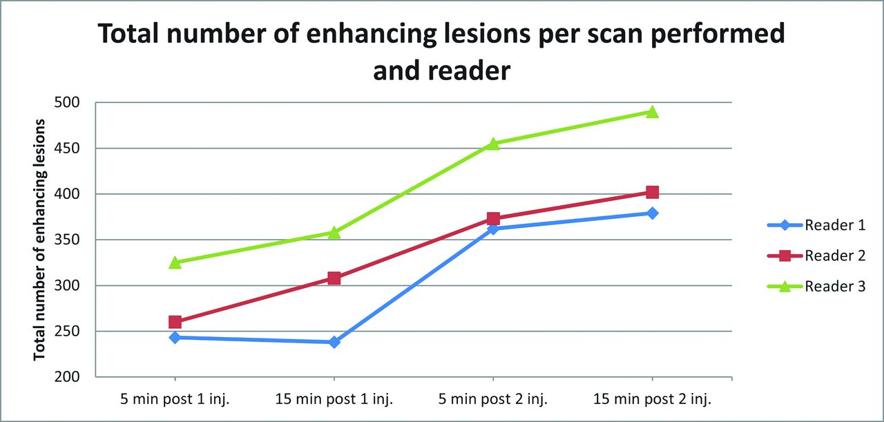
- Fig 1.
Total number of enhancing lesions per scan obtained and reader.
- Fig 2.
A 26-year-old woman with active relapsing-remitting multiple sclerosis. A small juxtacortical peri-Rolandic lesion located in the left brain hemisphere is identified on the 4 contrast-enhanced T1-weighted scans (arrows). Observe the increase in lesion size in the delayed single-dose scan (B) and cumulative-dose scans (C and D) compared with the early single-dose scan (A). Nonetheless, lesion detection is comparable in all 4 scan conditions.
- Fig 3.
A 26-year-old man presenting with clinically isolated syndrome. A small enhancing lesion located in the right temporal subcortical white matter is seen on the 2 cumulative-dose contrast-enhanced T1-weighted scans (arrows in C and D) but is initially missed on the 2 single-dose scans. Mild enhancement is seen in the early and delayed single-dose scans (arrows in A and B) only in retrospect.
- Fig 4.
A 25-year-old woman presenting with clinically isolated syndrome. A nodular enhancing lesion located in the right cerebellar hemisphere is seen on the 2 cumulative-dose contrast-enhanced T1-weighted scans (arrows in C and D) but was not identified on the early and delayed single-dose scans (A and B). The intensity of lesion enhancement is higher on the delayed cumulative-dose scan (D).
- Fig 5.
Percentage of patients with active lesions per scan performed and reader.


Tables
Patients with CIS (n = 26) Patients with MS (n = 89) Total (n = 115) Sex (No.) (%) Female 17 (65.4) 66 (74.2) 83 (72.2) Male 9 (34.6) 23 (25.8) 32 (27.8) Age (yr) Mean (SD) 32.9 (7.1) 36.0 (6.5) 35.3 (6.7) Median 31.0 36.0 35.0 Min, max (25.0, 47.0) (23.0, 50.0) (23.0, 50.0) EDSS score Mean (SD) 1.6 (1.1) 3.0 (1.7) 2.7 (1.7) Median 1.5 3 2.5 Min, max (0.0, 4.0) (0.0, 7.5) (0.0, 7.5) Note:—EDSS indicates Expanded Disability Status Scale; Min, minimum; max, maximum.
Scanb Lesion No. Reader 1 A 243 B 238 C 362 D 379 Reader 2 A 260 B 308 C 373 D 402 Reader 3 A 325 B 358 C 455 D 490 Total lesion No. (mean) A 828 (2.4) B 904 (2.6) C 1190 (3.5) D 1271 (3.7) Reader, Scanb No.c Total Median (mean) Minimum Maximumd 1 A 56 243 2.5 (4.3) 1.000 20.000 B 54 238 2.0 (4.4) 1.000 20.000 C 61 362 3.0 (5.9) 1.000 20.000 D 63 379 3.0 (6.0) 1.000 20.000 2 A 58 260 2.0 (4.5) 1.000 20.000 B 61 308 3.0 (5.0) 1.000 20.000 C 68 373 3.0 (5.5) 1.000 20.000 D 64 402 3.0 (6.3) 1.000 20.000 3 A 69 325 2.0 (4.7) 1.000 20.000 B 67 358 3.0 (5.3) 1.000 20.000 C 76 455 3.0 (6.0) 1.000 20.000 D 79 490 3.0 (6.2) 1.000 20.000 ↵a A higher number of enhancing lesions was detected for cumulative-versus-single dose scans (P < .001).
↵b A, early single dose; B, delayed single dose; C, early cumulative dose; D, delayed cumulative dose.
c No., number of patients with active lesions.
d The number of lesions was restricted to a maximum of 20 per patient.
- Table 4:
κ coefficients of the interreader agreement for the number of patients with active lesions
Postgadolinium T1-Weighted Scana κ (95% CI) Reader 1 vs Reader 2 Reader 1 vs Reader 3 Reader 2 vs Reader 3 A 0.79 (0.68–0.90) 0.71 (0.58–0.83) 0.67 (0.54–0.80) B 0.85 (0.75–0.94) 0.71 (0.59–0.83) 0.83 (0.72–0.93) C 0.81 (0.70–0.92) 0.70 (0.58–0.83) 0.78 (0.67–0.90) D 0.95 (0.89–1.00) 0.68 (0.55–0.81) 0.70 (0.57–0.82) ↵a A, early single dose; B, delayed single dose; C, early cumulative dose; D, delayed cumulative dose.


