Index by author
Liu, Y.
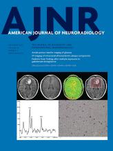
- EDITOR'S CHOICEAdult BrainOpen AccessRelationship between Glioblastoma Heterogeneity and Survival Time: An MR Imaging Texture AnalysisY. Liu, X. Xu, L. Yin, X. Zhang, L. Li and H. LuAmerican Journal of Neuroradiology September 2017, 38 (9) 1695-1701; DOI: https://doi.org/10.3174/ajnr.A5279
A group of 133 patients with primary glioblastoma who underwent postcontrast T1-weighted imaging (acquired before treatment) and whose data were filed with the survival times were selected from the Cancer Genome Atlas. On the basis of overall survival, the patients were divided into 2 groups: long-term (≥12 months, n = 67) and short-term (<12 months, n = 66) survival. To measure heterogeneity, the authors extracted 3 types of textures, co-occurrence matrix, run-length matrix, and histogram, reflecting local, regional, and global spatial variations, respectively. Then the support vector machine classification was used to determine how different texture types perform in differentiating the 2 groups. The results suggest that local and regional heterogeneity may play an important role in the survival stratification of patients with glioblastoma.
Longo, M.G.
- FELLOWS' JOURNAL CLUBAdult BrainYou have accessDiagnostic Performance of a 10-Minute Gadolinium-Enhanced Brain MRI Protocol Compared with the Standard Clinical Protocol for Detection of Intracranial Enhancing LesionsJ. Fagundes, M.G. Longo, S.Y. Huang, B.R. Rosen, T. Witzel, K. Heberlein, R.G. Gonzalez, P. Schaefer and O. RapalinoAmerican Journal of Neuroradiology September 2017, 38 (9) 1689-1694; DOI: https://doi.org/10.3174/ajnr.A5293
A total of 53 patients underwent MR imaging consisting of 5 basic fast precontrast sequences plus standard and accelerated versions of the same postcontrast T1WI sequences. Two neuroradiologists assessed the image quality and the final diagnosis for each set of postcontrast sequences and compared their performances. The 10-minute brain MR imaging protocol with contrast was comparable in diagnostic performance with the standard protocol in an inpatient motion-prone population, with the additional benefits of reducing acquisition times and image artifacts.
Lu, H.
- EDITOR'S CHOICEAdult BrainOpen AccessRelationship between Glioblastoma Heterogeneity and Survival Time: An MR Imaging Texture AnalysisY. Liu, X. Xu, L. Yin, X. Zhang, L. Li and H. LuAmerican Journal of Neuroradiology September 2017, 38 (9) 1695-1701; DOI: https://doi.org/10.3174/ajnr.A5279
A group of 133 patients with primary glioblastoma who underwent postcontrast T1-weighted imaging (acquired before treatment) and whose data were filed with the survival times were selected from the Cancer Genome Atlas. On the basis of overall survival, the patients were divided into 2 groups: long-term (≥12 months, n = 67) and short-term (<12 months, n = 66) survival. To measure heterogeneity, the authors extracted 3 types of textures, co-occurrence matrix, run-length matrix, and histogram, reflecting local, regional, and global spatial variations, respectively. Then the support vector machine classification was used to determine how different texture types perform in differentiating the 2 groups. The results suggest that local and regional heterogeneity may play an important role in the survival stratification of patients with glioblastoma.
Lu, J.
- EDITOR'S CHOICEAdult BrainOpen AccessIdentification and Quantitative Assessment of Different Components of Intracranial Atherosclerotic Plaque by Ex Vivo 3T High-Resolution Multicontrast MRIY. Jiang, W. Peng, B. Tian, C. Zhu, L. Chen, X. Wang, Q. Liu, Y. Wang, Z. Xiang, A.J. Degnan, Z. Teng, D. Saloner and J. LuAmerican Journal of Neuroradiology September 2017, 38 (9) 1716-1722; DOI: https://doi.org/10.3174/ajnr.A5266
Fifty-three intracranial arterial specimens with atherosclerotic plaques from 20 cadavers were imaged by 3T MR with T1, T2, and proton-density–weighted FSE and STIR sequences. The signal characteristics and areas of fibrous cap, lipid core, calcification, fibrous tissue, and healthy vessel wall were recorded on MR images and compared with histology. The signal intensity of the lipid core was significantly lower than that of the fibrous cap on T2-weighted, proton-density, and STIR sequences and was comparable on T1-weighted sequences. Optimal contrast between the lipid core and fibrous cap was found on T2-weighted images. Ex vivo 3T MR imaging can accurately identify and quantitatively assess intracranial atherosclerotic plaque components, providing a direct reference for in vivo intracranial plaque imaging.
Macmahon, P.J.
- Adult BrainOpen AccessCurrent and Emerging Therapies in Multiple Sclerosis: Implications for the Radiologist, Part 1—Mechanisms, Efficacy, and SafetyC. McNamara, G. Sugrue, B. Murray and P.J. MacMahonAmerican Journal of Neuroradiology September 2017, 38 (9) 1664-1671; DOI: https://doi.org/10.3174/ajnr.A5147
- Adult BrainOpen AccessCurrent and Emerging Therapies in Multiple Sclerosis: Implications for the Radiologist, Part 2—Surveillance for Treatment Complications and Disease ProgressionC. McNamara, G. Sugrue, B. Murray and P.J. MacMahonAmerican Journal of Neuroradiology September 2017, 38 (9) 1672-1680; DOI: https://doi.org/10.3174/ajnr.A5148
Magnussen, J.S.
- Spine Imaging and Spine Image-Guided InterventionsYou have accessProspective Comparison of Changes in Lumbar Spine MRI Findings over Time between Individuals with Acute Low Back Pain and Controls: An Exploratory StudyJ. Panagopoulos, J.S. Magnussen, J. Hush, C.G. Maher, M. Crites-Battie, J.G. Jarvik, T.S. Jensen and M.J. HancockAmerican Journal of Neuroradiology September 2017, 38 (9) 1826-1832; DOI: https://doi.org/10.3174/ajnr.A5357
Maher, C.G.
- Spine Imaging and Spine Image-Guided InterventionsYou have accessProspective Comparison of Changes in Lumbar Spine MRI Findings over Time between Individuals with Acute Low Back Pain and Controls: An Exploratory StudyJ. Panagopoulos, J.S. Magnussen, J. Hush, C.G. Maher, M. Crites-Battie, J.G. Jarvik, T.S. Jensen and M.J. HancockAmerican Journal of Neuroradiology September 2017, 38 (9) 1826-1832; DOI: https://doi.org/10.3174/ajnr.A5357
Majoie, C.B.L.M.
- Adult BrainOpen AccessValue of Thrombus CT Characteristics in Patients with Acute Ischemic StrokeJ. Borst, O.A. Berkhemer, E.M.M. Santos, A.J. Yoo, M. den Blanken, Y.B.W.E.M. Roos, E. van Bavel, W.H. van Zwam, R.J. van Oostenbrugge, H.F. Lingsma, A. van der Lugt, D.W.J. Dippel, H.A. Marquering and C.B.L.M. Majoie on behalf of the MR CLEAN investigatorsAmerican Journal of Neuroradiology September 2017, 38 (9) 1758-1764; DOI: https://doi.org/10.3174/ajnr.A5331
Maravilla, K.R.
- Adult BrainOpen AccessComparison of Gadoterate Meglumine and Gadobutrol in the MRI Diagnosis of Primary Brain Tumors: A Double-Blind Randomized Controlled Intraindividual Crossover Study (the REMIND Study)K.R. Maravilla, D. San-Juan, S.J. Kim, G. Elizondo-Riojas, J.R. Fink, W. Escobar, A. Bag, D.R. Roberts, J. Hao, C. Pitrou, A.J. Tsiouris, E. Herskovits and J.B. FiebachAmerican Journal of Neuroradiology September 2017, 38 (9) 1681-1688; DOI: https://doi.org/10.3174/ajnr.A5316
Marquering, H.A.
- Adult BrainOpen AccessValue of Thrombus CT Characteristics in Patients with Acute Ischemic StrokeJ. Borst, O.A. Berkhemer, E.M.M. Santos, A.J. Yoo, M. den Blanken, Y.B.W.E.M. Roos, E. van Bavel, W.H. van Zwam, R.J. van Oostenbrugge, H.F. Lingsma, A. van der Lugt, D.W.J. Dippel, H.A. Marquering and C.B.L.M. Majoie on behalf of the MR CLEAN investigatorsAmerican Journal of Neuroradiology September 2017, 38 (9) 1758-1764; DOI: https://doi.org/10.3174/ajnr.A5331
In this issue