Advertisement

Main menu

  • Home
  • Content
    • Current Issue
    • Accepted Manuscripts
    • Article Preview
    • Past Issue Archive
    • Video Articles
    • AJNR Case Collection
    • Case of the Week Archive
    • Case of the Month Archive
    • Classic Case Archive
  • Special Collections
    • AJNR Awards
    • Low-Field MRI
    • Alzheimer Disease
    • ASNR Foundation Special Collection
    • Photon-Counting CT
    • View All
  • Multimedia
    • AJNR Podcasts
    • AJNR SCANtastic
    • Trainee Corner
    • MRI Safety Corner
    • Imaging Protocols
  • For Authors
    • Submit a Manuscript
    • Submit a Video Article
    • Submit an eLetter to the Editor/Response
    • Manuscript Submission Guidelines
    • Statistical Tips
    • Fast Publishing of Accepted Manuscripts
    • Graphical Abstract Preparation
    • Imaging Protocol Submission
    • Author Policies
  • About Us
    • About AJNR
    • Editorial Board
    • Editorial Board Alumni
  • More
    • Become a Reviewer/Academy of Reviewers
    • Subscribers
    • Permissions
    • Alerts
    • Feedback
    • Advertisers
    • ASNR Home

User menu

  • Alerts
  • Log in

Search

  • Advanced search
American Journal of Neuroradiology

American Journal of Neuroradiology

ASHNR American Society of Functional Neuroradiology ASHNR American Society of Pediatric Neuroradiology ASSR
  • Alerts
  • Log in

Advanced Search

  • Home
  • Content
    • Current Issue
    • Accepted Manuscripts
    • Article Preview
    • Past Issue Archive
    • Video Articles
    • AJNR Case Collection
    • Case of the Week Archive
    • Case of the Month Archive
    • Classic Case Archive
  • Special Collections
    • AJNR Awards
    • Low-Field MRI
    • Alzheimer Disease
    • ASNR Foundation Special Collection
    • Photon-Counting CT
    • View All
  • Multimedia
    • AJNR Podcasts
    • AJNR SCANtastic
    • Trainee Corner
    • MRI Safety Corner
    • Imaging Protocols
  • For Authors
    • Submit a Manuscript
    • Submit a Video Article
    • Submit an eLetter to the Editor/Response
    • Manuscript Submission Guidelines
    • Statistical Tips
    • Fast Publishing of Accepted Manuscripts
    • Graphical Abstract Preparation
    • Imaging Protocol Submission
    • Author Policies
  • About Us
    • About AJNR
    • Editorial Board
    • Editorial Board Alumni
  • More
    • Become a Reviewer/Academy of Reviewers
    • Subscribers
    • Permissions
    • Alerts
    • Feedback
    • Advertisers
    • ASNR Home
  • Follow AJNR on Twitter
  • Visit AJNR on Facebook
  • Follow AJNR on Instagram
  • Join AJNR on LinkedIn
  • RSS Feeds

AJNR Awards, New Junior Editors, and more. Read the latest AJNR updates

Research ArticleAdult Brain
Open Access

Systematic Assessment of Multispectral Voxel-Based Morphometry in Previously MRI-Negative Focal Epilepsy

R. Kotikalapudi, P. Martin, J. Marquetand, T. Lindig, B. Bender and N.K. Focke
American Journal of Neuroradiology November 2018, 39 (11) 2014-2021; DOI: https://doi.org/10.3174/ajnr.A5809
R. Kotikalapudi
aFrom the Departments of Diagnostic and Interventional Neuroradiology (R.K., T.L., B.B.)
bNeurology and Epileptology (R.K., P.M., J.M., N.K.F.), Hertie Institute for Clinical Brain Research, University Hospital Tübingen, Tübingen, Germany
cDepartment of Clinical Neurophysiology (R.K., N.K.F.), University Hospital Göttingen, Göttingen, Germany.
  • Find this author on Google Scholar
  • Find this author on PubMed
  • Search for this author on this site
  • ORCID record for R. Kotikalapudi
P. Martin
bNeurology and Epileptology (R.K., P.M., J.M., N.K.F.), Hertie Institute for Clinical Brain Research, University Hospital Tübingen, Tübingen, Germany
  • Find this author on Google Scholar
  • Find this author on PubMed
  • Search for this author on this site
  • ORCID record for P. Martin
J. Marquetand
bNeurology and Epileptology (R.K., P.M., J.M., N.K.F.), Hertie Institute for Clinical Brain Research, University Hospital Tübingen, Tübingen, Germany
  • Find this author on Google Scholar
  • Find this author on PubMed
  • Search for this author on this site
  • ORCID record for J. Marquetand
T. Lindig
aFrom the Departments of Diagnostic and Interventional Neuroradiology (R.K., T.L., B.B.)
  • Find this author on Google Scholar
  • Find this author on PubMed
  • Search for this author on this site
  • ORCID record for T. Lindig
B. Bender
aFrom the Departments of Diagnostic and Interventional Neuroradiology (R.K., T.L., B.B.)
  • Find this author on Google Scholar
  • Find this author on PubMed
  • Search for this author on this site
  • ORCID record for B. Bender
N.K. Focke
bNeurology and Epileptology (R.K., P.M., J.M., N.K.F.), Hertie Institute for Clinical Brain Research, University Hospital Tübingen, Tübingen, Germany
cDepartment of Clinical Neurophysiology (R.K., N.K.F.), University Hospital Göttingen, Göttingen, Germany.
  • Find this author on Google Scholar
  • Find this author on PubMed
  • Search for this author on this site
  • ORCID record for N.K. Focke
  • Article
  • Figures & Data
  • Supplemental
  • Info & Metrics
  • Responses
  • References
  • PDF
Loading

Article Figures & Data

Figures

  • Tables
  • Fig 1.
    • Download figure
    • Open in new tab
    • Download powerpoint
    Fig 1.

    Diagnostic performance of different VBM models. A and B, The area under the curve for different smoothing kernels for gray matter concentration and volume. C, An example of an intersection plot for T1 VBM for the concordant rate and specificity against statistical cutoffs. D, An example of a receiver operating characteristic curve for T1 VBM at 12-mm smoothing for GMC. The Euclidean distance is calculated from the optimized T-threshold (3.7 in GMC analysis), where CR, SP = 38.5, 33.9 to CR, SP = 100, 100. False positive rate = 100-SP.

  • Fig 2.
    • Download figure
    • Open in new tab
    • Download powerpoint
    Fig 2.

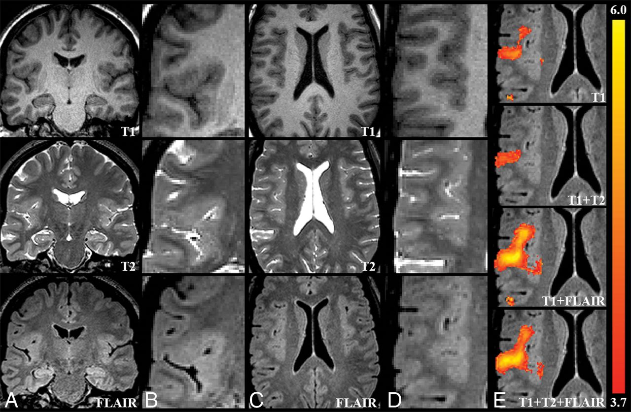
    Sample case (patient 4). A and C, An overview in the native space T1, T2, and FLAIR images. B and D, Zoomed-in images focusing on a possible epileptogenic lesion. A subtle disruption of cortical morphology is visible in this figure, more prominent on FLAIR images. This finding is concordant with the clinical hypothesis supported by noninvasive and intracranial EEG, indicating seizure onset in the right frontal lobe. The possible lesion was detected as increased GM by all VBM models, but the extent and effect size were clearly better for the VBM models, including FLAIR (E).

  • Fig 3.
    • Download figure
    • Open in new tab
    • Download powerpoint
    Fig 3.

    Sample case (patient 13). A, A VBM finding detected only by T1+FLAIR in the left temporoinsular region overlaid on the native space FLAIR image. B, Magnified images of T1, T2, and FLAIR suggesting a blurred gray-white matter junction (arrow). This finding is concordant with the clinical hypothesis of seizure onset.

Tables

  • Figures
    • View popup
    Table 1:

    GMC analysis for VBM models at 12 mm and a T-threshold of 3.7a

    ModelConcordant Rate (%)Specificity (%)Discordant Rate (%)Euclidean Distance (Range, 0–141.14)Concordant Rate/Discordant Rate
    T138.533.984.690.290.46
    T1+T246.240.353.880.370.86
    T1+FLAIR46.237.161.582.770.75
    T1+T2+FLAIR46.235.576.984.000.60
    • ↵a For VBM GMC automated analysis (prior to visual interpretation), values of concordant rate, specificity, discordant rate, Euclidean distance from CR = SP = 100, and concordant/discordant ratio are provided for all models: namely, T1, T1+T2, T1+FLAIR, and T1+T2+FLAIR. The smoothing level and T-threshold are 12 mm and 3.7, respectively.

    • View popup
    Table 2:

    GMV analysis for VBM models at 12 mm and a T-threshold of 3.0a

    ModelConcordant Rate (%)Specificity (%)Discordant Rate (%)Euclidean Distance (Range, 0–141.14)Concordant Rate/Discordant Rate
    T123.121.084.6110.250.27
    T1+T27.717.776.9123.660.10
    T1+FLAIR38.59.784.6109.250.46
    T1+T2+FLAIR38.514.576.9105.320.50
    • ↵a For VBM GMV automated analysis (prior to visual interpretation), values of concordant rate, specificity, discordant rate, Euclidean distance from CR = SP = 100, and concordant/discordant ratio are provided for all models: namely, T1, T1+T2, T1+FLAIR, and T1+T2+FLAIR. The smoothing level and T-threshold are 12 mm and 3.0, respectively.

    • View popup
    Table 3:

    Patients—visual analysis results for GMCa

    ModelPotentially Epileptogenic (and Visible)Potentially Epileptogenic (and Not Visible)Potentially Epileptogenic (Combined)NonepileptogenicUnclearArtifacts
    Concordant lobe (%)
        T115.415.430.87.715.40
        T1+T215.4015.47.715.40
        T1+FLAIR15.430.846.27.723.10
        T1+T2+FLAIR15.415.430.87.723.10
    Discordant lobe (%)
        T17.738.546.27.730.815.4
        T1+T27.723.130.8023.10
        T1+FLAIR7.730.838.57.730.87.7
        T1+T2+FLAIR7.723.130.8030.87.7
    • ↵a Visual interpretation results of GMC analysis of patients are provided. The results contain percentages of patients scored by the reviewer as potentially epileptogenic and visible, potentially epileptogenic and not visible, potentially epileptogenic (number of patients with potentially epileptogenic and visible/not visible or both), nonepileptogenic, unclear, and artifacts. The results are reported for both concordant and discordant lobes.

    • View popup
    Table 4:

    Controls—visual analysis results for GMCa

    ModelsAll Lobes (%)
    NonepileptogenicUnclearArtifactsCorrected Specificity (Excluding Artifacts and Nonepileptogenic)
    T114.553.222.646.8
    T1+T221.045.217.754.8
    T1+FLAIR16.150.019.450.0
    T1+T2+FLAIR16.148.425.851.6
    • ↵a Visual interpretation results for GMC analysis for controls are presented. Results contain the percentage of controls scored by the reviewer as nonepileptogenic, unclear, and artifacts. Finally, corrected specificity is reported as the percentage of controls that did not have unclear findings—that is, all findings identified as artifacts/nonepileptogenic lesions and patients with no findings (VBM specificity prior to visual analysis).

    • View popup
    Table 5:

    Nonvisible findings—visual analysis results for GMCa

    ModelsAll Lobes (%)
    ControlsPatients
    T153.261.5
    T1+T245.230.8
    T1+FLAIR50.061.5
    T1+T2+FLAIR48.446.2
    • ↵a Rates of findings not visible—that is, unclear findings in controls and unclear/potentially epileptogenic and not visible findings in patients across all models for all lobes (%) are reported.

PreviousNext
Back to top

In this issue

American Journal of Neuroradiology: 39 (11)
American Journal of Neuroradiology
Vol. 39, Issue 11
1 Nov 2018
  • Table of Contents
  • Index by author
  • Complete Issue (PDF)
Advertisement
Print
Download PDF
Email Article

Thank you for your interest in spreading the word on American Journal of Neuroradiology.

NOTE: We only request your email address so that the person you are recommending the page to knows that you wanted them to see it, and that it is not junk mail. We do not capture any email address.

Enter multiple addresses on separate lines or separate them with commas.
Systematic Assessment of Multispectral Voxel-Based Morphometry in Previously MRI-Negative Focal Epilepsy
(Your Name) has sent you a message from American Journal of Neuroradiology
(Your Name) thought you would like to see the American Journal of Neuroradiology web site.
CAPTCHA
This question is for testing whether or not you are a human visitor and to prevent automated spam submissions.
Cite this article
R. Kotikalapudi, P. Martin, J. Marquetand, T. Lindig, B. Bender, N.K. Focke
Systematic Assessment of Multispectral Voxel-Based Morphometry in Previously MRI-Negative Focal Epilepsy
American Journal of Neuroradiology Nov 2018, 39 (11) 2014-2021; DOI: 10.3174/ajnr.A5809

Citation Manager Formats

  • BibTeX
  • Bookends
  • EasyBib
  • EndNote (tagged)
  • EndNote 8 (xml)
  • Medlars
  • Mendeley
  • Papers
  • RefWorks Tagged
  • Ref Manager
  • RIS
  • Zotero
0 Responses
Respond to this article
Share
Bookmark this article
Systematic Assessment of Multispectral Voxel-Based Morphometry in Previously MRI-Negative Focal Epilepsy
R. Kotikalapudi, P. Martin, J. Marquetand, T. Lindig, B. Bender, N.K. Focke
American Journal of Neuroradiology Nov 2018, 39 (11) 2014-2021; DOI: 10.3174/ajnr.A5809
del.icio.us logo Twitter logo Facebook logo Mendeley logo
  • Tweet Widget
  • Facebook Like
  • Google Plus One
Purchase

Jump to section

  • Article
    • Abstract
    • ABBREVIATIONS:
    • Materials and Methods
    • Results
    • Discussion
    • Conclusions
    • Footnotes
    • References
  • Figures & Data
  • Supplemental
  • Info & Metrics
  • Responses
  • References
  • PDF

Related Articles

  • PubMed
  • Google Scholar

Cited By...

  • No citing articles found.
  • Crossref
  • Google Scholar

This article has not yet been cited by articles in journals that are participating in Crossref Cited-by Linking.

More in this TOC Section

  • Diagnostic Neuroradiology of Monoclonal Antibodies
  • Clinical Outcomes After Chiari I Decompression
  • Segmentation of Brain Metastases with BLAST
Show more Adult Brain

Similar Articles

Advertisement

Indexed Content

  • Current Issue
  • Accepted Manuscripts
  • Article Preview
  • Past Issues
  • Editorials
  • Editor's Choice
  • Fellows' Journal Club
  • Letters to the Editor
  • Video Articles

Cases

  • Case Collection
  • Archive - Case of the Week
  • Archive - Case of the Month
  • Archive - Classic Case

More from AJNR

  • Trainee Corner
  • Imaging Protocols
  • MRI Safety Corner
  • Book Reviews

Multimedia

  • AJNR Podcasts
  • AJNR Scantastics

Resources

  • Turnaround Time
  • Submit a Manuscript
  • Submit a Video Article
  • Submit an eLetter to the Editor/Response
  • Manuscript Submission Guidelines
  • Statistical Tips
  • Fast Publishing of Accepted Manuscripts
  • Graphical Abstract Preparation
  • Imaging Protocol Submission
  • Evidence-Based Medicine Level Guide
  • Publishing Checklists
  • Author Policies
  • Become a Reviewer/Academy of Reviewers
  • News and Updates

About Us

  • About AJNR
  • Editorial Board
  • Editorial Board Alumni
  • Alerts
  • Permissions
  • Not an AJNR Subscriber? Join Now
  • Advertise with Us
  • Librarian Resources
  • Feedback
  • Terms and Conditions
  • AJNR Editorial Board Alumni

American Society of Neuroradiology

  • Not an ASNR Member? Join Now

© 2025 by the American Society of Neuroradiology All rights, including for text and data mining, AI training, and similar technologies, are reserved.
Print ISSN: 0195-6108 Online ISSN: 1936-959X

Powered by HighWire