Advertisement

Main menu

  • Home
  • Content
    • Current Issue
    • Accepted Manuscripts
    • Article Preview
    • Past Issue Archive
    • AJNR Case Collection
    • Case of the Week Archive
    • Classic Case Archive
    • Case of the Month Archive
  • Special Collections
    • Spinal CSF Leak Articles (Jan 2020-June 2024)
    • 2024 AJNR Journal Awards
    • Most Impactful AJNR Articles
  • Multimedia
    • AJNR Podcast
    • AJNR Scantastics
    • Video Articles
  • For Authors
    • Submit a Manuscript
    • Author Policies
    • Fast publishing of Accepted Manuscripts
    • Graphical Abstract Preparation
    • Manuscript Submission Guidelines
    • Imaging Protocol Submission
    • Submit a Case for the Case Collection
  • About Us
    • About AJNR
    • Editorial Board
  • More
    • Become a Reviewer/Academy of Reviewers
    • Subscribers
    • Permissions
    • Alerts
    • Feedback
    • Advertisers
    • ASNR Home
  • Other Publications
    • ajnr

User menu

  • Alerts
  • Log in

Search

  • Advanced search
American Journal of Neuroradiology

American Journal of Neuroradiology

ASHNR American Society of Functional Neuroradiology ASHNR American Society of Pediatric Neuroradiology ASSR
  • Alerts
  • Log in

Advanced Search

  • Home
  • Content
    • Current Issue
    • Accepted Manuscripts
    • Article Preview
    • Past Issue Archive
    • AJNR Case Collection
    • Case of the Week Archive
    • Classic Case Archive
    • Case of the Month Archive
  • Special Collections
    • Spinal CSF Leak Articles (Jan 2020-June 2024)
    • 2024 AJNR Journal Awards
    • Most Impactful AJNR Articles
  • Multimedia
    • AJNR Podcast
    • AJNR Scantastics
    • Video Articles
  • For Authors
    • Submit a Manuscript
    • Author Policies
    • Fast publishing of Accepted Manuscripts
    • Graphical Abstract Preparation
    • Manuscript Submission Guidelines
    • Imaging Protocol Submission
    • Submit a Case for the Case Collection
  • About Us
    • About AJNR
    • Editorial Board
  • More
    • Become a Reviewer/Academy of Reviewers
    • Subscribers
    • Permissions
    • Alerts
    • Feedback
    • Advertisers
    • ASNR Home
  • Follow AJNR on Twitter
  • Visit AJNR on Facebook
  • Follow AJNR on Instagram
  • Join AJNR on LinkedIn
  • RSS Feeds

Welcome to the new AJNR, Updated Hall of Fame, and more. Read the full announcements.


AJNR is seeking candidates for the position of Associate Section Editor, AJNR Case Collection. Read the full announcement.


Research ArticleInterventional

Spinal Epidural Arteriovenous Fistula with Perimedullary Venous Reflux: Clinical and Neuroradiologic Features of an Underestimated Vascular Disorder

M. Mull, A. Othman, M. Dafotakis, F.-J. Hans, G.A. Schubert and F. Jablawi
American Journal of Neuroradiology November 2018, 39 (11) 2095-2102; DOI: https://doi.org/10.3174/ajnr.A5854
M. Mull
aFrom the Departments of Diagnostic and Interventional Neuroradiology (M.M., A.O., F.J.)
  • Find this author on Google Scholar
  • Find this author on PubMed
  • Search for this author on this site
  • ORCID record for M. Mull
A. Othman
aFrom the Departments of Diagnostic and Interventional Neuroradiology (M.M., A.O., F.J.)
eDepartment of Diagnostic and Interventional Radiology (A.O.), University Hospital Tübingen, Tübingen, Germany
  • Find this author on Google Scholar
  • Find this author on PubMed
  • Search for this author on this site
  • ORCID record for A. Othman
M. Dafotakis
bNeurology (M.D.)
  • Find this author on Google Scholar
  • Find this author on PubMed
  • Search for this author on this site
  • ORCID record for M. Dafotakis
F.-J. Hans
fDepartment of Neurosurgery (F.-J.H.), Paracelsus Kliniken, Osnabrück, Germany.
  • Find this author on Google Scholar
  • Find this author on PubMed
  • Search for this author on this site
  • ORCID record for F.-J. Hans
G.A. Schubert
cNeurosurgery (G.A.S.), University Hospital Aachen, RWTH Aachen University, Aachen, Germany
  • Find this author on Google Scholar
  • Find this author on PubMed
  • Search for this author on this site
  • ORCID record for G.A. Schubert
F. Jablawi
aFrom the Departments of Diagnostic and Interventional Neuroradiology (M.M., A.O., F.J.)
dDepartment of Neurosurgery (F.J.), Justus-Liebig-University, Giessen, Giessen, Germany
  • Find this author on Google Scholar
  • Find this author on PubMed
  • Search for this author on this site
  • ORCID record for F. Jablawi
  • Article
  • Figures & Data
  • Info & Metrics
  • Responses
  • References
  • PDF
Loading

Article Figures & Data

Figures

  • Tables
  • Fig 1.
    • Download figure
    • Open in new tab
    • Download powerpoint
    Fig 1.

    A, Sagittal T2- weighted images (3T; T2-TSE; slice thickness, 3 mm) reveal extensive congestive myelopathy (white arrowhead). B–D, Spinal CE-MRA (3T; time-resolved imaging with strochastic trajectories (TWIST); sagittal MIP; coronal and axial MPR) shows arterialized pouch in the lumbar ventrolateral epidural space (white arrows) in association with arterialized perimedullary veins in the thoracic region (white arrowheads) suspicious for a SEAVF in the lumbar region. E, DSA in lateral projection shows a SEAVF (white arrow) supplied via branches of the left L2 segmental artery (black arrowhead) and drained via the respective intradural radicular vein (white arrowheads). Note the extraspinal venous outlet (asterisk).

  • Fig 2.
    • Download figure
    • Open in new tab
    • Download powerpoint
    Fig 2.

    A–B, Sagittal T2- and contrast-enhanced T1-weighted images (3T; T2-TSE; T1-TSE; slice thickness, 3 mm) show extensive congestive thoracic myelopathy. C, Spinal CE-MRA (sagittal MIP) reveals an abnormal arterialized epidural pouch in the lumbar region (white arrow) in addition to thoracic arterialized perimedullary veins (white arrowhead). D, DSA (posteroanterior projection) exams identify the fistula in the epidural space on the vertebral level of L4 (white arrow), supplied via the right L4 segmental artery and drained by the contralateral L4 intradural radicular vein (white arrowhead). E and F, Axial and coronal MPR of DynaCT, 2 mm, 8 seconds rotation: Note the multisegmental and bilateral extension of the arterialized epidural pouch and the left sided origin of the intradural radicular drainage vein crossing the dura at the contralateral neural foramen (white arrowhead).

  • Fig 3.
    • Download figure
    • Open in new tab
    • Download powerpoint
    Fig 3.

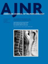
    A, Spinal CE-MRA (1.5 T, sagittal MIP) reveals an extensive pathological arterialization of a ventrolateral epidural venous pouch extending over four vertebral levels (white arrow). B–C, Further reconstructions of the source MRA images (coronal and axial MPR) demonstrate precisely the epidural pouch (white arrow) and show the filling of the intradural radicular drainage vein (white arrowhead). D–E, DSA exams (posteroanterior projections) identify the multisegmental ventrolateral epidural pouch of the SEAVF (white arrow) with multiple left-sided arterial feeders supplied by the thoracic segmental arteries T 10 and T 11. Note the distant origin of the intradural radicular drainage vein (asterisk).

Tables

  • Figures
    • View popup
    Table 1:

    Clinical presentation of patients with spinal epidural arteriovenous fistula

    Patient No.Age (yr)/SexDuration of Symptoms (mo)Symptoms at DiagnosisStatus at Discharge
    178, M6Paraparesis, sphincter dysfunctionImproved
    263, M1Paraparesis, sphincter dysfunction hypesthesia below T10Stable
    377, M1.5Paraparesis, hypesthesia L4, ataxia, sphincter dysfunctionStable
    460, F1.5Paraplegia, hypesthesia disturbances below T10Improved
    568, F24Neurogenic claudication <100 mImproved
    677, M1.5Paraplegia, hypesthesia below L1, sphincter dysfunctionStable
    764, F1Paraparesis, sphincter dysfunctionImproved
    883, M60Paraparesis, hypesthesia, sphincter dysfunctionStable
    972, M0Paraparesis, hypesthesia, sphincter dysfunctionStable
    1080, M12Paraparesis, sphincter dysfunctionStable
    1177, M2Paresis of left foot, ataxiaStable
    1278, M3Paraparesis, hypesthesiaStable
    1359, M5AtaxiaStable
    • View popup
    Table 2:

    Angiomorphologic characteristics of spinal epidural arteriovenous fistulas

    Patient No.Arterial FeederaOrigin of Intradural Radicular Drainage VeinaExtension of Epidural PouchbExtension of Arterialized Perimedullary VeinsbLocation of Arterialized Perimedullary VeinsbNo. of DSAs until Diagnosis
    1L3 RL3 LL3–L4T8–T12D = V2
    2L3 bilateral, L4 LL3 LL2–L4T3–T12D = V2
    3L1 RL1 RL1NDD < V1
    4L3 bilateralL3 RL3T9–L1D > V1
    5Left iliolumbar arteryS1S1T7–T12D > V3
    6T10 L and T11 LL2 LT 10–L3T6–L1D = V3
    7L1 LL2 LL1–L2T3–T12D = V1
    8T12 LT12 LT12 LT11D > V3
    9L3 LL3 LL3 LT6–T12D < V1
    10L4 RL4 RL2–L4T3–T10D = V1
    11L3 LS1 bilateralL3–S1T10–L1D > V1
    12L3 RL3 RL3 RT9–T12D < V1
    13L1 RL1 RL1 RT10–L1D > V1
    • Note:—D indicates dorsal to spinal cord; V, ventral to spinal cord; L, left; R, right; ND, no data.

    • ↵a DSA.

    • ↵b MRA/DSA.

PreviousNext
Back to top

In this issue

American Journal of Neuroradiology: 39 (11)
American Journal of Neuroradiology
Vol. 39, Issue 11
1 Nov 2018
  • Table of Contents
  • Index by author
  • Complete Issue (PDF)
Advertisement
Print
Download PDF
Email Article

Thank you for your interest in spreading the word on American Journal of Neuroradiology.

NOTE: We only request your email address so that the person you are recommending the page to knows that you wanted them to see it, and that it is not junk mail. We do not capture any email address.

Enter multiple addresses on separate lines or separate them with commas.
Spinal Epidural Arteriovenous Fistula with Perimedullary Venous Reflux: Clinical and Neuroradiologic Features of an Underestimated Vascular Disorder
(Your Name) has sent you a message from American Journal of Neuroradiology
(Your Name) thought you would like to see the American Journal of Neuroradiology web site.
CAPTCHA
This question is for testing whether or not you are a human visitor and to prevent automated spam submissions.
Cite this article
M. Mull, A. Othman, M. Dafotakis, F.-J. Hans, G.A. Schubert, F. Jablawi
Spinal Epidural Arteriovenous Fistula with Perimedullary Venous Reflux: Clinical and Neuroradiologic Features of an Underestimated Vascular Disorder
American Journal of Neuroradiology Nov 2018, 39 (11) 2095-2102; DOI: 10.3174/ajnr.A5854

Citation Manager Formats

  • BibTeX
  • Bookends
  • EasyBib
  • EndNote (tagged)
  • EndNote 8 (xml)
  • Medlars
  • Mendeley
  • Papers
  • RefWorks Tagged
  • Ref Manager
  • RIS
  • Zotero
0 Responses
Respond to this article
Share
Bookmark this article
Spinal Epidural Arteriovenous Fistula with Perimedullary Venous Reflux: Clinical and Neuroradiologic Features of an Underestimated Vascular Disorder
M. Mull, A. Othman, M. Dafotakis, F.-J. Hans, G.A. Schubert, F. Jablawi
American Journal of Neuroradiology Nov 2018, 39 (11) 2095-2102; DOI: 10.3174/ajnr.A5854
del.icio.us logo Twitter logo Facebook logo Mendeley logo
  • Tweet Widget
  • Facebook Like
  • Google Plus One
Purchase

Jump to section

  • Article
    • Abstract
    • ABBREVIATIONS:
    • Materials and Methods
    • Results
    • Discussion
    • Conclusions
    • Footnotes
    • References
  • Figures & Data
  • Info & Metrics
  • Responses
  • References
  • PDF

Related Articles

  • No related articles found.
  • PubMed
  • Google Scholar

Cited By...

  • No citing articles found.
  • Crossref (17)
  • Google Scholar

This article has been cited by the following articles in journals that are participating in Crossref Cited-by Linking.

  • Clinical and Imaging Characteristics of Spinal Dural Arteriovenous Fistulas and Spinal Epidural Arteriovenous Fistulas
    Waleed Brinjikji, Elisa Colombo, Harry J Cloft, Giuseppe Lanzino
    Neurosurgery 2021 88 3
  • The clinical value of venous drainage in patients with spinal dural arteriovenous fistula
    F. Jablawi, M. Mull
    Journal of the Neurological Sciences 2019 397
  • Intramedullary Hemorrhage Caused by Lumbosacral Epidural Arteriovenous Fistula with Dual Retrograde Perimedullary Venous Draining Routes: A Case Report and Review of the Literature
    Prasert Iampreechakul, Anusak Liengudom, Korrapakc Wangtanaphat, Porn Narischat, Punjama Lertbutsayanukul, Somkiet Siriwimonmas
    World Neurosurgery 2020 143
  • Unique Angioarchitecture of Sacral Dural Arteriovenous Fistula
    Michael Mull, Gerrit Alexander Schubert, Jens Obersheimer, Fidaa Jablawi
    World Neurosurgery 2020 133
  • Natural History and Clinical Outcomes of Paravertebral Arteriovenous Shunts
    Yueshan Feng, Jiaxing Yu, Jiankun Xu, Chuan He, Lisong Bian, Guilin Li, Ming Ye, Peng Hu, Liyong Sun, Nan Jiang, Feng Ling, Tao Hong, Hongqi Zhang
    Stroke 2021 52 12
  • Treatment strategy and long-term outcome in patients with deep lumbosacral arteriovenous fistulas. A single center analysis in nineteen patients
    F. Jablawi, O. Nikoubashman, M. Dafotakis, G.A. Schubert, F.-J. Hans, M. Mull
    Clinical Neurology and Neurosurgery 2020 188
  • Arteriovenous malformations of the filum terminale: clinical characteristics, angioarchitecture, and management of a rare spinal vascular pathology
    Michael Mull, Manuel Dafotakis, Gerrit Alexander Schubert, Franz-Josef Hans, Fidaa Jablawi
    Neurosurgical Focus 2022 53 1
  • Spinal epidural arteriovenous fistula with intradural venous reflux: The possibility of acquired origin caused by spinal stenosis and/or disc herniation
    Prasert Iampreechakul, Anusak Liengudom, Korrapakc Wangtanaphat, Wuttipong Tirakotai, Yodkhwan Wattanasen, Punjama Lertbutsayanukul, Somkiet Siriwimonmas
    Clinical Neurology and Neurosurgery 2021 207
  • Disorders of the Cauda Equina
    Samantha LoRusso
    CONTINUUM: Lifelong Learning in Neurology 2021 27 1
  • Interactive spinal computed tomography angiography–guided spinal digital subtraction angiography and embolization for thoracolumbar epidural arteriovenous fistulas: illustrative case
    Hirokuni Hashikata, Yoshinori Maki, Ryota Ishibashi, Hiroki Toda
    Journal of Neurosurgery: Case Lessons 2022 4 12

More in this TOC Section

Interventional

  • SAVE vs. Solumbra Techniques for Thrombectomy
  • Delayed Reperfusion Post-Thrombectomy&Thrombolysis
  • Contrast-Induced Encephalopathy after NeuroIR
Show more Interventional

Spine

  • Bern Score Validity for SIH
  • MP2RAGE 7T in MS Lesions of the Cervical Spine
  • Deep Learning for STIR Spine MRI Quality
Show more Spine

Similar Articles

Advertisement

Indexed Content

  • Current Issue
  • Accepted Manuscripts
  • Article Preview
  • Past Issues
  • Editorials
  • Editors Choice
  • Fellow Journal Club
  • Letters to the Editor

Cases

  • Case Collection
  • Archive - Case of the Week
  • Archive - Case of the Month
  • Archive - Classic Case

Special Collections

  • Special Collections

Resources

  • News and Updates
  • Turn around Times
  • Submit a Manuscript
  • Author Policies
  • Manuscript Submission Guidelines
  • Evidence-Based Medicine Level Guide
  • Publishing Checklists
  • Graphical Abstract Preparation
  • Imaging Protocol Submission
  • Submit a Case
  • Become a Reviewer/Academy of Reviewers
  • Get Peer Review Credit from Publons

Multimedia

  • AJNR Podcast
  • AJNR SCANtastic
  • Video Articles

About Us

  • About AJNR
  • Editorial Board
  • Not an AJNR Subscriber? Join Now
  • Alerts
  • Feedback
  • Advertise with us
  • Librarian Resources
  • Permissions
  • Terms and Conditions

American Society of Neuroradiology

  • Not an ASNR Member? Join Now

© 2025 by the American Society of Neuroradiology All rights, including for text and data mining, AI training, and similar technologies, are reserved.
Print ISSN: 0195-6108 Online ISSN: 1936-959X

Powered by HighWire