
Article Figures & Data
Figures
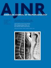
- Fig 1.
Left ear, A, Flat panel CT scan (isotropic voxel, 0.15-mm resolution) of a cadaver temporal bone specimen with a superimposed 3D colored schematic representation of the normal VES on the oblique sagittal plane parallel to the superior semicircular canal. On this plane, the normal saccule (dotted arrow) is more medially and posteriorly located compared with the utricle. The utricle does not protrude into the inferior portion of the vestibule, and the VES does not contact the round (asterisk) and oval (arrowhead) windows. B, MR imaging oblique sagittal reconstruction parallel to the superior semicircular canal of a healthy ear shows superiorly the VES and inferiorly the perilymph filling the inferior third of the vestibule with preservation of the perilymph signal medial to the oval window (arrowhead) and round window (asterisk). C, MR imaging axial reconstruction parallel to the lateral semicircular canal at the inferior third of the vestibule in a healthy subject, showing the vestibule filled by the perilymph (arrow).
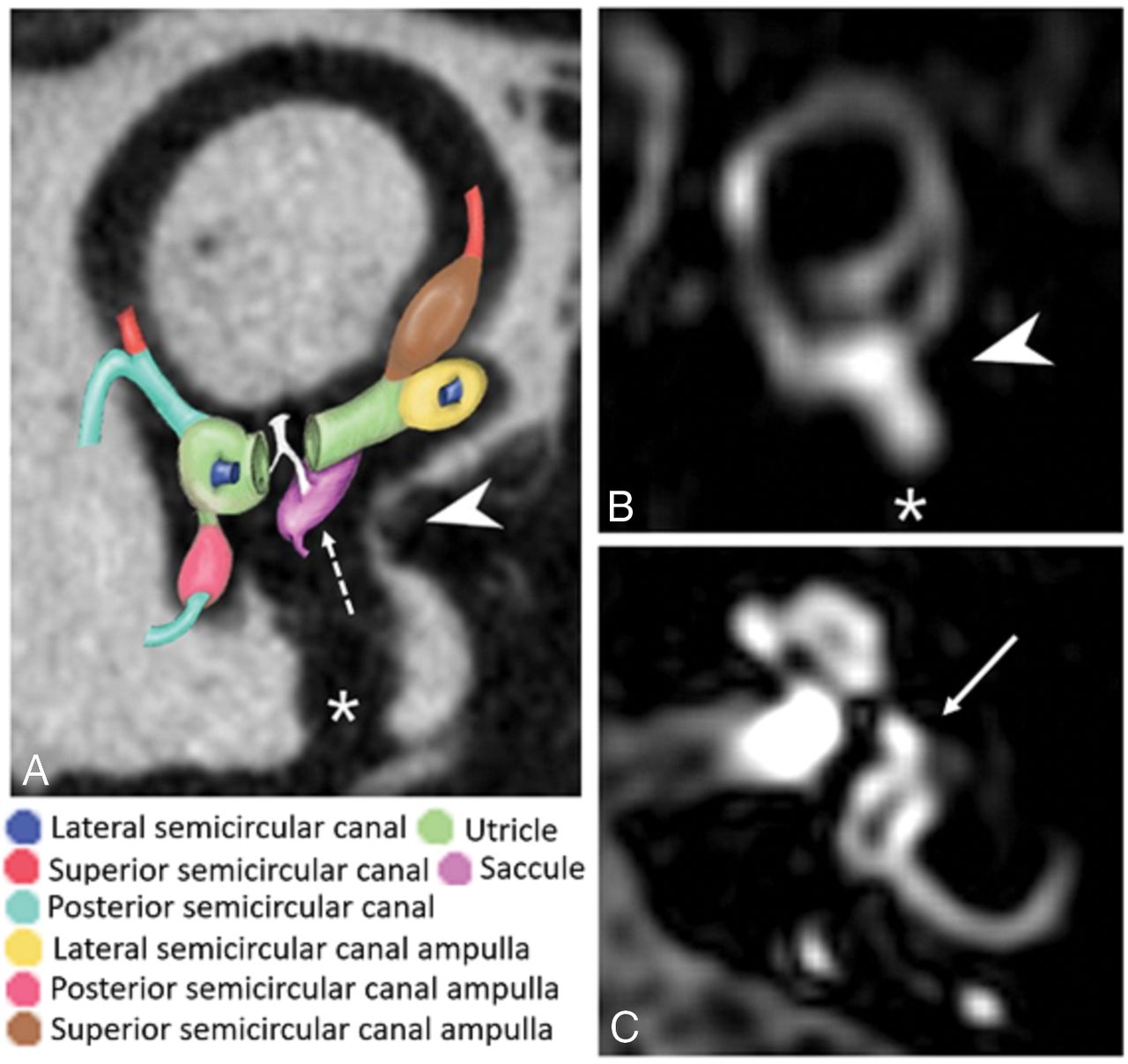
- Fig 2.
Left ear. A, Flat panel CT scan (isotropic voxel, 0.15-mm resolution) of a cadaver temporal bone specimen with superimposed 3D colored schematic representation of the VES on the oblique sagittal plane parallel to the superior semicircular canal, as suggested in patients with MD. The utricle bulges into the inferior third of the vestibule, and the saccule (dotted arrow) bulges more medially; thus, the VES contacts the oval window (arrowhead). The asterisk indicates the round window and the dotted arrow indicates the saccule. B, MR imaging oblique sagittal reconstruction parallel to the superior semicircular canal of an MD ear shows enlargement of the VES bulging into the inferior third of the vestibule and contacting the oval window (arrowhead), with the consequent absence of the normal perilymph signal behind the stapes footplate (asterisk indicates the round window). C, MR imaging axial reconstruction parallel to the lateral semicircular canal at the inferior third of the vestibule in a patient with MD shows the VES contacting the oval window (arrow indicates enlargement of the VES bulging into the inferior third of the vestibule and contacting the oval window).
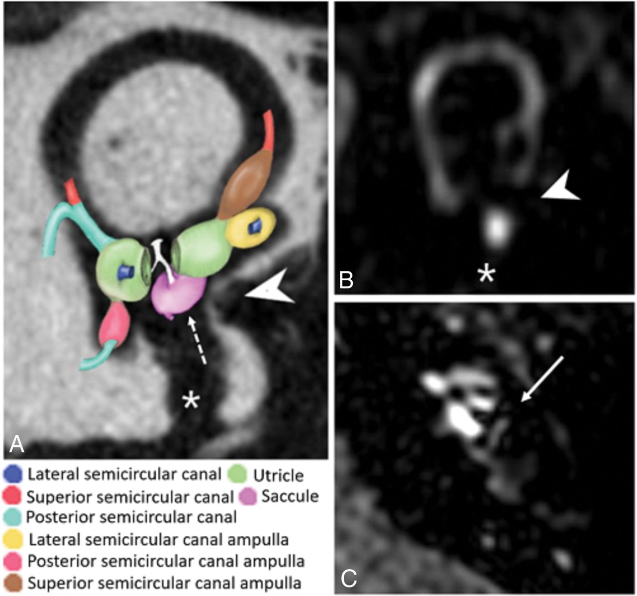
- Fig 3.
Four-hour-delayed postcontrast 3D-FLAIR MR axial image through the basal turns of the cochleae. The right (R) MD ear shows cochlear hydrops (arrowhead) and marked contrast enhancement (arrow) compared with the contralateral (L indicates left) healthy ear (dotted arrow), suggesting BLB breakdown.


Tables
Pre- and Postcontrast T1 FSE Pre- and Postcontrast 3D-FLAIR 3D T2-SSFP Delayed Postcontrast 3D-FLAIR Plane Axial Axial Axial Axial TR (ms) 500 6000 1500 7600 TE (ms) 10 350 194 345 TI (ms) / 2350 / 2100 Fat saturation SPIR SPIR / SPAIR TSE factor 3 182 40 100 Flip angle 90° 90° 90° 90° Slice thickness (mm) 1.5 1 0.6 0.6 Slices (no.) 15 30 22 40 FOV (mm2) 180 × 160 230 × 190 140 × 140 200 × 255 Matrix 256 × 205 232 × 229 264 × 248 250 × 252 Parallel imaging No Yes No Yes Averages 4 6 1 4 Scan time 2 min 51 sec 10 min 6 sec 6 min 35 sec 9 min 15 sec Note:—SSFP indicates steady-state free precession; SPIR, spectral presaturation with inversion recovery; SPAIR, spectral attenuated inversion recovery; 3D FLAIR, Three dimensionally Fluid Attenuated Inversion Recovery; FSE, Fast spin echo; TR, time of repetition; TE, time of echo; TI, time of inversion; TSE, Turbo Spin-Echo; FOV, field of view; /, specific parameter is not available.
- Table 2:
Contact between the oval window and the saccule in sagittal oblique plane (VESCO)
SE (%) (95% CI) SP (%) (95% CI) PPV (%) (95% CI) NPV (%) (95% CI) Symptomatic MD vs asymptomatic MD 81 (61–93) 96 (76–100) 96 (76–100) 81 (61–93) Symptomatic MD vs healthy 81 (61–93) 96 (77–100) 96 (76–100) 82 (62–93) Symptomatic MD vs SSHL 81 (61–93) 96 (77–100) 96 (76–100) 82 (62–93) Symptomatic MD vs other 81 (61–93) 96 (87–99) 88 (68–97) 93 (84–97) Note:—SE indicates sensitivity; SP, specificity; PPV, positive predictive value; NPV, negative predictive value.
SE (%) (95% CI) SP (%) (95% CI) PPV (%) (95% CI) NPV (%) (95% CI) Symptomatic MD vs asymptomatic MD 74 (53–88) 65 (43–83) 71 (51–86) 68 (45–85) Symptomatic MD vs healthy 74 (53–88) 83 (62–95) 83 (62–95) 74 (53–88) Symptomatic MD vs SSHL 74 (53–88) 58 (37–77) 67 (47–82) 67 (43–85) Symptomatic MD vs other 74 (53–88) 69 (57–79) 48 (32–63) 88 (75–94) Note:—SE indicates sensitivity; SP, specificity.
SE (%) (95% CI) SP (%) (95% CI) PPV (%) (95% CI) NPV (%) (95% CI) Symptomatic MD vs asymptomatic MD 74 (53–88) 78 (56–92) 80 (59–92) 72 (50–87) Symptomatic MD vs healthy 74 (53–88) 96 (77–100) 95 (74–100) 77 (57–89) Symptomatic MD vs SSHL 74 (53–88) 83 (62–95) 83 (62–95) 74 (53–88) Symptomatic MD vs other 74 (53–88) 86 (75–93) 67 (47–82) 90 (79–95) Note:—SE indicates sensitivity; SP, specificity.
- Table 5:
Presence of VESCO versus VEH and CH according to the Nakashima criteria in patients with MD
VESCO VEH CH No (n = 7) Mild (n = 11) Severe (n = 9) No (n = 7) Yes (n = 20) No (n = 5) 2 (40%) 3 (60%) 0 (0%) 3 (60%) 2 (40%) Yes (n = 22) 5 (23%) 8 (36%) 9 (41%) 4 (18%) 18 (82%)


