
Article Figures & Data
Figures
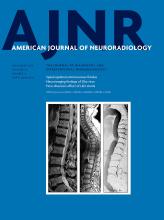
- Fig 1.
Supratentorial abnormalities in patients with the ACTA2 mutation. Upper row: sagittal T2-weighted image (A) and sagittal T1WI (B) show typical hypoplasia and bending of the anterior corpus callosum with associated abnormal radial gyration of the frontal lobes and a deficient anterior cingulate gyrus (white arrows). In patient 7, there is a horizontal orientation of the fornix (black arrow in B), which is also markedly thick. Middle row: axial T1WI (C and D) demonstrates characteristic V-shaped anterior corpus callosum (black circle). Lower row: MR angiography maximum-intensity-projection anteroposterior view (E and F) shows typical neurovascular abnormalities in patients with ACTA2 mutation.
- Fig 2.
Infratentorial malformations in ACTA2 mutation. Upper row: axial T2WI (A and B) at the level of the pons. The twin peaks sign is demonstrated; the pons is flattened with reduction of the anteroposterior diameter and an impression of the basilar artery on the anterior surface with consequent presence of 2 symmetric prominences resembling twin mountains. Middle row: axial T2WI at the level of the cerebral peduncles (C and D) shows mild antero-posterior elongation of the midbrain with reduction of the laterolateral diameter and squeezing of the cerebral peduncles. Right column: normal for comparison.
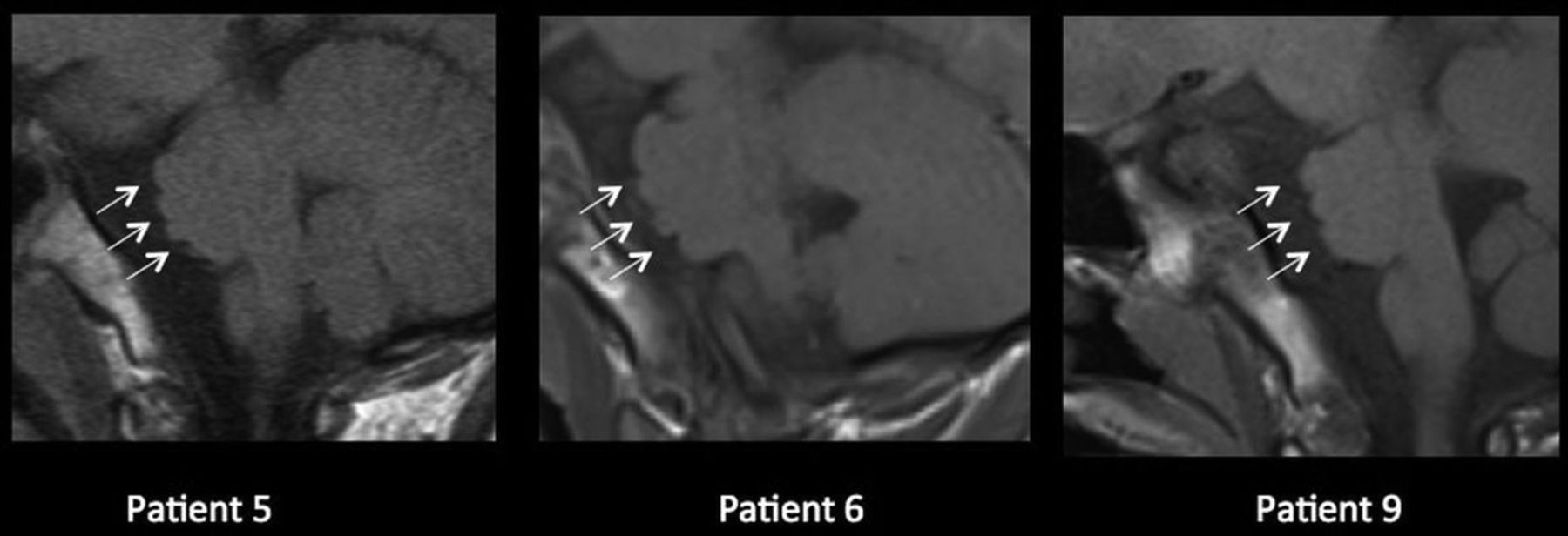
- Fig 3.
In the parasagittal slice, the patients show multiple indentations on the surface of the pons. We speculate that these may be due to stretching of the straightened pontine arterial branches.
- Fig 4.
Neuroradiologic findings in an adult patient without a confirmed ACTA2 mutation (patient 15). Sagittal T2WI (A) and axial T2WI at the level of the pons (B) and the midbrain (C) show marked callosal anterior bending (dotted arrow in A), the twin peaks pontine sign (arrow in B), and reduction of the laterolateral diameter of the midbrain with a squeezed cerebral peduncle. D, Sagittal T2WI demonstrates a marked basilar impression on the pons and anterior bending of the midbrain. The parasagittal right slice (E) demonstrates indentation of the lateral surface of the pons (dotted black arrow) and a straight course of the posterior cerebral arteries (white arrow). Axial T2WI (F) at the level of the proximal segment of the posterior cerebral arteries (pontomesencephalic junction) shows marked compression of the brain stem at that level related to straightening of the arteries (white arrows) and radial gyration of the posterior temporal lobes (black dotted arrows).


