
Article Figures & Data
Figures
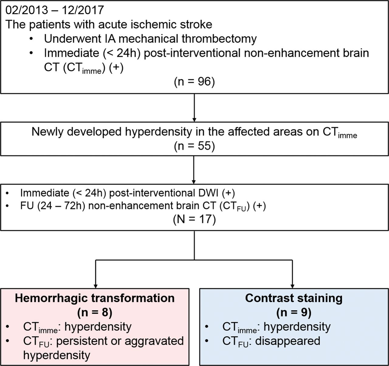
- Fig 1.
Study design.
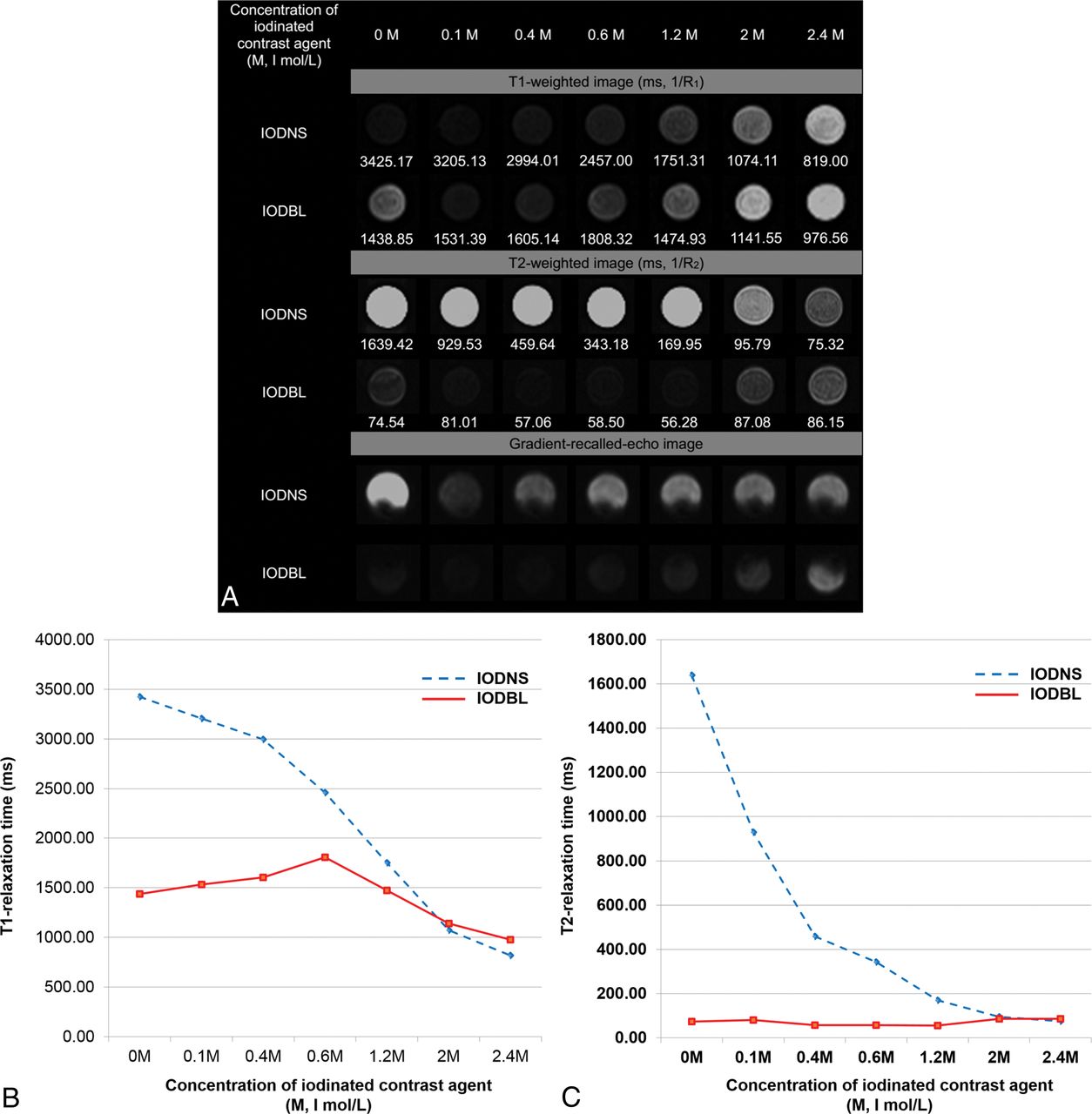
- Fig 2.
MR signal intensity of IODNS and IODBL on T1-weighted, T2-weighted, and gradient recalled-echo images. A, Images from the phantom. B, T1-relaxation times of IODNS and IODBL at different concentrations. C, T2-relaxation times of IODNS and IODBL according to the concentration of iodinated contrast agents. Visipaque (iodixanol, 320 I mg/mL, 2.5 I mol/L, iso-osmolar) was used as the iodinated contrast agent in this phantom.
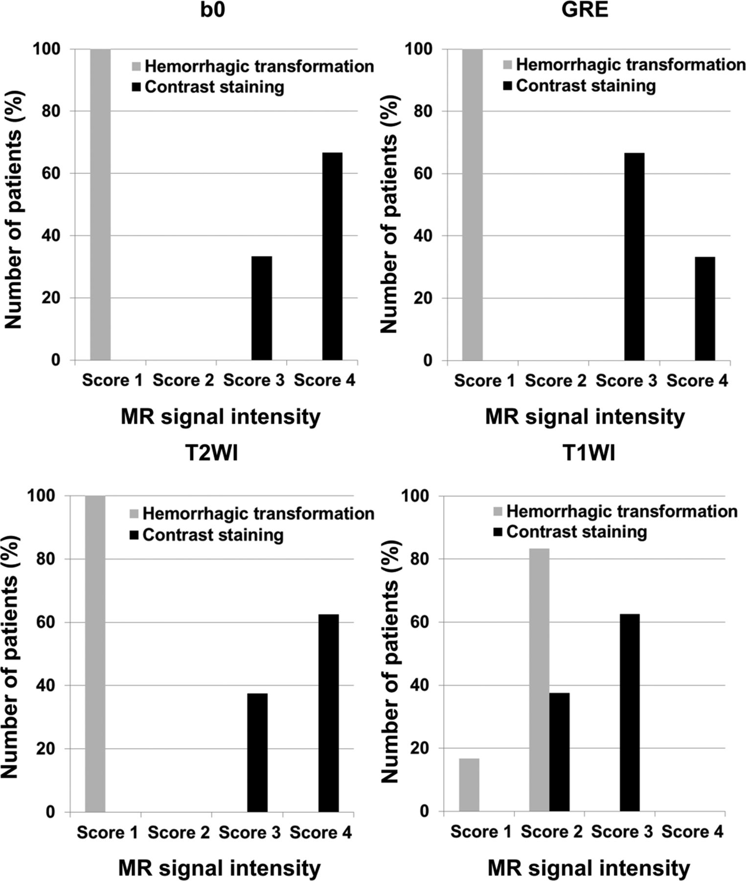
- Fig 3.
Magnetic signal intensity score of hemorrhagic transformation and contrast staining on B0, gradient recalled-echo, T2-weighted, and T1-weighted imaging. Score 1, signal intensity was similar to that of the vessel; score 2, signal intensity was lower than that of the gray matter; score 3, signal intensity was similar to that of the gray matter; and score 4, signal intensity was higher than that of the gray matter.
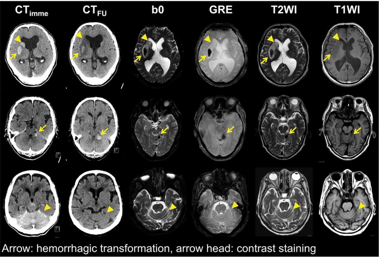
- Fig 4.
Representative images of hemorrhagic transformation and contrast staining in 3 patients with acute ischemic stroke. First row: images of a 66-year-old man with infarction in the territory of the right middle cerebral artery. A persistent hyperdense lesion in the right lentiform nucleus, presumed to be a hemorrhagic transformation (arrow), shows dark signal intensity in the B0, gradient recalled-echo, and T2-weighted images. In contrast, a hyperdense lesion in the right caudate nucleus, which is washed out on the follow-up nonenhanced brain CT and presumed to be contrast staining (arrowhead), demonstrates a high signal intensity in the B0, GRE, and T2-weighted images. This patient was grouped into the hemorrhagic transformation group, and the lesion in the lentiform nucleus was analyzed. Middle row: images of a 68-year-old man with infarction in the region of the right superior cerebellar artery. A persistent hyperdense lesion in the left cerebellum, presumed to be hemorrhagic transformation (arrow), shows dark signal intensity in the B0, GRE, and T2-weighted images. Lower row: images of an 88-year-old woman with bilateral cerebellar infarction. A hyperdense lesion in the bilateral upper cerebellum, which disappeared on CTFU and was presumed to be contrast staining (arrowhead), demonstrates iso-signal intensity in the B0, GRE, and T2-weighted images.


Tables
- Table 1:
MR signal intensities of hemorrhagic transformation and contrast staining in B0 and GRE imagea
Hemorrhagic Transformation (n = 8) Contrast Staining (n = 9) Total (n = 17) P B0 <.001 Score 1 8 (100.00%) 0 (0.00%) 8 (47.06%) Score 2 0 (0.00%) 0 (0.00%) 0 (0.00%) Score 3 0 (0.00%) 3 (33.33%) 3 (17.65%) Score 4 0 (0.00%) 6 (66.67%) 6 (35.29%) GRE <.001 Score 1 8 (100.00%) 0 (0.00%) 8 (47.06%) Score 2 0 (0.00%) 0 (0.00%) 0 (0.00%) Score 3 0 (0.00%) 6 (66.67%) 6 (35.29%) Score 4 0 (0.00%) 3 (33.33%) 3 (17.65%) ↵a Values are numbers of patients with percentages in parentheses. Score 1, signal intensity was similar to that of the vessel; score 2, signal intensity was lower than that of the gray matter; score 3, signal intensity was similar to that of the gray matter; score 4, signal intensity was higher than that of the gray matter.
- Table 2:
MR signal intensities of hemorrhagic transformation and contrast staining in T1WI and T2WIa
Hemorrhagic Transformation (n = 6) Contrast Staining (n = 8) Total (n = 14) P T1WI .042 Score 1 1 (16.67%) 0 (0.00%) 1 (7.14%) Score 2 5 (83.33%) 3 (37.50%) 8 (57.14%) Score 3 0 (0.00%) 5 (62.50%) 5 (35.71%) Score 4 0 (0.00%) 0 (0.00%) 0 (0.00%) T2WI .001 Score 1 6 (100.00%) 0 (0.00%) 6 (42.86%) Score 2 0 (0.00%) 0 (0.00%) 0 (0.00%) Score 3 0 (0.00%) 3 (37.50%) 3 (21.43%) Score 4 0 (0.00%) 5 (62.50%) 5 (35.71%) ↵a Values are numbers of patients with percentages in parentheses. Score 1, signal intensity was similar to that of the vessel; score 2, signal intensity was lower than that of the gray matter; score 3, signal intensity was similar to that of the gray matter; score 4, signal intensity was higher than that of the gray matter.


