Advertisement

Main menu

  • Home
  • Content
    • Current Issue
    • Accepted Manuscripts
    • Article Preview
    • Past Issue Archive
    • AJNR Case Collection
    • Case of the Week Archive
    • Classic Case Archive
    • Case of the Month Archive
  • Special Collections
    • Spinal CSF Leak Articles (Jan 2020-June 2024)
    • 2024 AJNR Journal Awards
    • Most Impactful AJNR Articles
  • Multimedia
    • AJNR Podcast
    • AJNR Scantastics
    • Video Articles
  • For Authors
    • Submit a Manuscript
    • Author Policies
    • Fast publishing of Accepted Manuscripts
    • Graphical Abstract Preparation
    • Manuscript Submission Guidelines
    • Imaging Protocol Submission
    • Submit a Case for the Case Collection
  • About Us
    • About AJNR
    • Editorial Board
  • More
    • Become a Reviewer/Academy of Reviewers
    • Subscribers
    • Permissions
    • Alerts
    • Feedback
    • Advertisers
    • ASNR Home
  • Other Publications
    • ajnr

User menu

  • Alerts
  • Log in

Search

  • Advanced search
American Journal of Neuroradiology

American Journal of Neuroradiology

ASHNR American Society of Functional Neuroradiology ASHNR American Society of Pediatric Neuroradiology ASSR
  • Alerts
  • Log in

Advanced Search

  • Home
  • Content
    • Current Issue
    • Accepted Manuscripts
    • Article Preview
    • Past Issue Archive
    • AJNR Case Collection
    • Case of the Week Archive
    • Classic Case Archive
    • Case of the Month Archive
  • Special Collections
    • Spinal CSF Leak Articles (Jan 2020-June 2024)
    • 2024 AJNR Journal Awards
    • Most Impactful AJNR Articles
  • Multimedia
    • AJNR Podcast
    • AJNR Scantastics
    • Video Articles
  • For Authors
    • Submit a Manuscript
    • Author Policies
    • Fast publishing of Accepted Manuscripts
    • Graphical Abstract Preparation
    • Manuscript Submission Guidelines
    • Imaging Protocol Submission
    • Submit a Case for the Case Collection
  • About Us
    • About AJNR
    • Editorial Board
  • More
    • Become a Reviewer/Academy of Reviewers
    • Subscribers
    • Permissions
    • Alerts
    • Feedback
    • Advertisers
    • ASNR Home
  • Follow AJNR on Twitter
  • Visit AJNR on Facebook
  • Follow AJNR on Instagram
  • Join AJNR on LinkedIn
  • RSS Feeds

Welcome to the new AJNR, Updated Hall of Fame, and more. Read the full announcements.


AJNR is seeking candidates for the position of Associate Section Editor, AJNR Case Collection. Read the full announcement.


Getting new auth cookie, if you see this message a lot, tell someone!
Research ArticleFunctional
Open Access

Interaction of Developmental Venous Anomalies with Resting-State Functional MRI Measures

B. Sundermann, B. Pfleiderer, H. Minnerup, K. Berger and G. Douaud
American Journal of Neuroradiology December 2018, 39 (12) 2326-2331; DOI: https://doi.org/10.3174/ajnr.A5847
B. Sundermann
aFrom the Nuffield Department of Clinical Neurosciences (B.S., G.D.), Oxford Centre for Functional MRI of the Brain (FMRIB), Wellcome Centre for Integrative Neuroimaging, University of Oxford, Oxford, UK
bInstitute of Clinical Radiology (B.S., B.P.), Medical Faculty, University of Münster and University Hospital Münster, Münster, Germany
  • Find this author on Google Scholar
  • Find this author on PubMed
  • Search for this author on this site
  • ORCID record for B. Sundermann
B. Pfleiderer
bInstitute of Clinical Radiology (B.S., B.P.), Medical Faculty, University of Münster and University Hospital Münster, Münster, Germany
  • Find this author on Google Scholar
  • Find this author on PubMed
  • Search for this author on this site
  • ORCID record for B. Pfleiderer
H. Minnerup
cDepartment of Epidemiology and Social Medicine (H.M., K.B.), University of Münster, Münster, Germany.
  • Find this author on Google Scholar
  • Find this author on PubMed
  • Search for this author on this site
  • ORCID record for H. Minnerup
K. Berger
cDepartment of Epidemiology and Social Medicine (H.M., K.B.), University of Münster, Münster, Germany.
  • Find this author on Google Scholar
  • Find this author on PubMed
  • Search for this author on this site
  • ORCID record for K. Berger
G. Douaud
aFrom the Nuffield Department of Clinical Neurosciences (B.S., G.D.), Oxford Centre for Functional MRI of the Brain (FMRIB), Wellcome Centre for Integrative Neuroimaging, University of Oxford, Oxford, UK
  • Find this author on Google Scholar
  • Find this author on PubMed
  • Search for this author on this site
  • ORCID record for G. Douaud
  • Article
  • Figures & Data
  • Supplemental
  • Info & Metrics
  • Responses
  • References
  • PDF
Loading

Abstract

BACKGROUND AND PURPOSE: Functional MR imaging of the brain, used for both clinical and neuroscientific applications, relies on measuring fluctuations in blood oxygenation. Such measurements are susceptible to noise of vascular origin. The purpose of this study was to assess whether developmental venous anomalies, which are frequently observed normal variants, can bias fMRI measures by appearing as true neural signal.

MATERIALS AND METHODS: Large developmental venous anomalies (1 in each of 14 participants) were identified from a large neuroimaging cohort (n = 814). Resting-state fMRI data were decomposed using independent component analysis, a data-driven technique that creates distinct component maps representing aspects of either structured noise or true neural activity. We searched all independent components for maps that exhibited a spatial distribution of their signals following the topography of developmental venous anomalies.

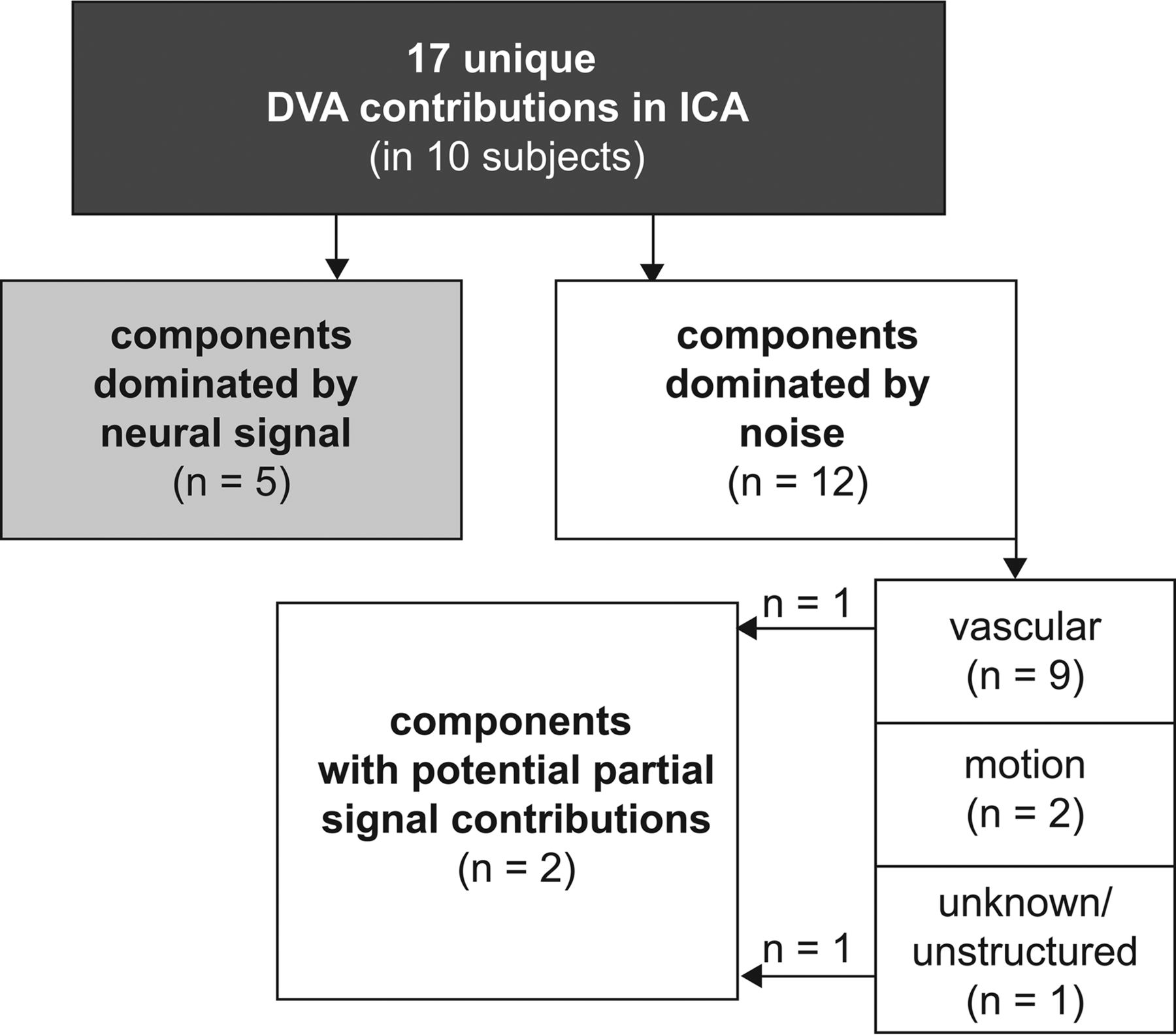
RESULTS: Of the 14 developmental venous anomalies identified, 10 were clearly present in 17 fMRI independent components in total. While 9 (52.9%) of these 17 independent components were dominated by venous contributions and 2 (11.8%) by motion artifacts, 2 independent components (11.8%) showed partial neural signal contributions and 5 independent components (29.4%) unambiguously exhibited typical neural signal patterns.

CONCLUSIONS: Developmental venous anomalies can strongly resemble neural signal as measured by fMRI. They are thus a potential source of bias in fMRI analyses, especially when present in the cortex. This could impede interpretation of local activity in patients, such as in presurgical mapping. In scientific studies with large samples, developmental venous anomaly confounds could be mainly addressed using independent component analysis–based denoising.

ABBREVIATIONS:

BOLD
blood oxygen level–dependent
DVA
developmental venous anomaly
IC
independent component
ICA
independent component analysis
rsfMRI
resting-state fMRI

Functional MR imaging of the brain is extensively used for task-specific presurgical functional mapping1⇓–3 and for task-based group studies.4 Analyses of spontaneous brain activity and connectivity by resting-state fMRI (rsfMRI)5⇓⇓⇓–9 have been more recently introduced as a potential clinical tool, both in presurgical motor10,11 and language mapping,10,12⇓⇓–15 particularly in patients less able to adhere to task instruction, and mapping of epileptogenic foci.8 Additionally, rsfMRI combined with automatic machine learning shows promise for individual diagnosis and prognosis estimation in large datasets, especially in psychiatric and neurodegenerative disorders,16⇓⇓–19 as well as for genome-wide association studies.20 fMRI is based on blood oxygen level–dependent (BOLD) contrast and thus provides indirect measures of neural activity. BOLD changes are attributed predominantly to both extravascular tissue and local capillaries and veins.21 Consequently, there are various sources of potential bias to the BOLD signal measured by fMRI, including global and local perfusion as well as vascular architecture.21

Incidental findings and normal variants are frequently encountered in brain MR imaging.22⇓⇓⇓–26 While debates on the management of incidental findings in scientific studies mostly focus on participant safety,27 little is known about potential biases of particular findings on measures of scientific or clinical interest (putative correlates of neural signals) in fMRI.

Developmental venous anomalies (DVAs)28 are frequent (around 2% prevalence29,30) and are usually clinically irrelevant but can be associated, in rare circumstances, with other vascular lesions, such as cavernous malformations, or with abnormalities of neuronal migration.28,31 DVAs can also exhibit signs of venous congestion.32 The normal venous drainage of the cerebral hemispheres can be divided into 2 systems: 1) The superficial system drains blood from cortical and immediately subcortical capillaries into pial veins; and 2) the deep system drains large parts of the deep white matter and basal ganglia into deep veins (the internal cerebral veins and the basal vein of Rosenthal). The cerebellum features a comparable, 2-system venous angioarchitecture.31 The term DVA describes variants in which a superficial venous territory drains into deep veins or a deep venous territory drains toward either the superficial pial veins or directly into a dural venous sinus.31

The detectability of DVAs depends largely on the imaging techniques used. However, DVAs are characterized by very typical morphologic imaging features. These are large collector veins crossing the brain parenchyma in locations where usually only capillaries and small veins are expected, and radially contributing veins resulting in a typical caput medusae appearance.29,31 These veins drain blood from an atypical territory. The presence of a DVA thus potentially undermines common assumptions about the origin of observed BOLD signal fluctuations in gray matter regions.

The purpose of this study was to assess if and how DVAs bias fMRI measures by assessing the similarity of DVA correlates in rsfMRI to typical patterns of neural activity in a large, community-based imaging population.33 If such similarity exists, then the presence of DVAs might lead to misinterpretations of local activity patterns in presurgical mapping and confound conclusions in group studies and new rsfMRI-based diagnostic approaches.

Materials and Methods

Subjects

This analysis is based on subjects with large DVAs identified during routine radiologic review and quality assessment of brain MR imaging for the BiDirect cohort study.33,34 They were selected from a mixed sample of patients with depression and population-dwelling controls (n = 814). The study was approved by the local ethics committee, and all subjects gave their written informed consent. Demographic characteristics of the BiDirect cohort and the imaging sample used here are presented in On-line Table 1.

MR Imaging Data Acquisition

Structural and functional MR imaging data were acquired using a 3T scanner (Intera with an Achieva upgrade; Philips Healthcare, Best, the Netherlands) and a standard transmit/receive head coil. Full details on the imaging protocol have been published separately.33

The main analyses were based on the following sequences: a 3D T1-weighted gradient-echo sequence with an inversion prepulse—turbo field echo, TR/TE = 7.26/3.56 ms, TI = 404 ms, flip angle = 9°, sagittal orientation, matrix = 256 × 256 mm, field of view = 256 × 256 × 160 mm, voxel size = 1 × 1 × 2 mm reconstructed to 1 × 1 × 1 mm by zero-filling in the k-space; rsfMRI using a T2*-weighted echo-planar imaging technique—fast-field echo, TR/TE = 3000/38 ms, flip angle = 90°, 72 volumes, matrix dimensions = 64 × 64, FOV = 230 × 230 mm, 36 axial slices, thickness = 3.6 mm, pixel size = 3.6 × 3.6 mm.

Additional information about DVA morphology was obtained from the following: FLAIR—TR/TE = 11,000/80 ms, TI = 2600 ms, flip angle = 90°, matrix dimensions = 352 × 206, FOV =230 × 186 mm, 27 axial slices, thickness = 4 mm, gap = 1 mm, reconstructed pixel size = 0.45 × 0.45 mm; and a T2*-weighted fast-field echo sequence—TR/TE = 574/16 ms, flip angle = 18°, matrix dimensions = 256 × 164, FOV = 230 × 183 mm, 27 axial slices, thickness = 4 mm, gap = 1 mm, reconstructed pixel size = 0.45 × 0.45 mm.

DVA Identification

Suspected DVAs were confirmed in a separate step after initial screening during visual data-quality assessment based on T1-weighted images using the following criteria: 1) a large vessel crossing the brain parenchyma, and 2) a typical caput medusae appearance of feeding vessels. Finally, a typical hypointense appearance was confirmed on T2* fast-field echo. We excluded subjects with signs of other associated vascular lesions, for example, cavernous malformations. FLAIR images were inspected for surrounding hyperintensity as a potential sign of venous congestion.32 Demographic information about all subjects selected for the DVA analysis as well as DVA locations is presented in On-line Table 2.

Resting-State fMRI Data Analysis with Independent Component Analysis

Spatial independent component analysis (ICA) refers to a range of data-driven analysis techniques decomposing time-series data into a set of spatially independent components (ICs) characterized by spatial maps and signal time courses. ICA is particularly popular for the analysis of rsfMRI data because it can isolate separate representations of well-known functionally relevant brain networks.35 In addition to these components related to the signal of interest originating from the gray matter, further components represent distinct sources of noise such as arterial or CSF pulsations or movement artifacts.36 ICA is therefore very suitable for identifying and characterizing such biasing influences. ICA can identify general sources of noise independent of specific model assumptions in other fMRI analysis techniques. Thus, findings from ICA generalize to other analysis techniques and to task-fMRI data. Indeed, it can reveal potential biases that might have otherwise not been directly visible despite potentially significantly influencing results and conclusions. ICA is therefore increasingly applied as a preprocessing step for noise clean-up of both rsfMRI and task-fMRI data before performing further analyses (see the Discussion for details).36⇓–38

Two types of components representing venous signal fluctuations are particularly relevant for DVAs: those showing mainly the venous sinuses and large veins and those putatively representing transmedullary and subependymal veins (traditionally referred to as white matter components). Given the partially venous origin of the BOLD signal of interest, venous noise components can exhibit temporal characteristics that greatly resemble signal components.36 Thus, additional spatial features (eg, based on sinus masks) have to be used in automatic noise-classification tools,37 but the underlying spatial assumptions behind these predefined masks could be broken in the presence of a large DVA.

Single-session analyses of the rsfMRI time-series data were conducted using the FMRIB Software Library (FSL, Version 5.0.9; http://www.fmrib.ox.ac.uk/fsl).39,40 Preprocessing included motion correction,41 non-brain tissue masking,42 spatial smoothing using a Gaussian kernel (full width at half maximum = 7 mm), and high-pass temporal filtering (cutoff period = 100 seconds). The resulting image time-series were then decomposed using probabilistic ICA with automatic dimensionality estimation as implemented in MELODIC (Version 3.15; https://fsl.fmrib.ox.ac.uk/fsl/fslwiki/MELODIC).35,43⇓–45

Identification of DVAs in the ICA Decomposition of rsfMRI

To help with the identification of the presence of DVAs in our ICA, we linearly registered ICA spatial maps to the T1-weighted anatomic images using boundary-based registration.46 ICA spatial maps were thresholded at |Z| = 2.3 and overlaid with anatomic images with FSLeyes (Version 0.9.11; https://fsl.fmrib.ox.ac.uk/fsl/fslwiki/FSLeyes). Components were individually assessed for contributions spatially corresponding to the courses of the DVAs (B.S.). Those ICs that were identified as containing DVA rsfMRI signal correlates were subsequently evaluated for whether they exhibited contributions of neural signal of interest or artifacts (eg, motion or arterial pulsations) by taking spatial maps, time courses, and power spectra into account.36

Results

General DVA Characteristics

Sixteen DVAs without an associated second vascular lesion were identified in 16 subjects. None of the DVAs exhibited associated hyperintensities on FLAIR, kinking of the draining vein, or other indirect signs of venous congestion. Two of these findings located in the cerebellum were excluded from further analyses because they were partially cut by the fMRI acquisition volume.

DVA Occurrence in the ICA Decomposition of rsfMRI

All of the 14 DVAs subsequently included were present in at least 1 IC. Ten DVAs (71.4%) had unique local presence, closely following their spatial distribution in at least 1 IC per subject and up to 4 ICs per subject. The remaining 4 DVAs had less specific spatial coverage. In other words, it could not be determined whether alterations in ICs corresponding to these 4 DVAs (at least 1 IC per subject) were directly related to the DVA or whether these alterations represented signal fluctuations of interest in the surrounding tissue or other sources of noise.

In total, 43 ICs thus covered spatial locations specific to DVAs across subjects. Seventeen of these ICs (39.5%) exhibited unique contributions following the spatial distribution of the DVAs, while a further 26 ICs (60.5%) were less specific, with their spatial distribution only partially overlapping that of the DVAs.

Characteristics of rsfMRI ICs Containing DVAs

Among the 17 ICs closely following the spatial distribution of the DVAs, 9 (52.9%) were ICs with predominantly vascular patterns and clear contributions of venous sinuses, deep cerebral veins, or subependymal veins (white matter components), one of which exhibited partial signal contribution. Two ICs (11.8%) were dominated by motion artifacts. Most important, 5 ICs (29.4%) in 3 subjects exhibited dominant patterns typical of true neural signal. Three of these 5 ICs had clean low-frequency power spectra, while contamination in the higher frequency range was present in 2 ICs. One final IC was unstructured in its overall spatial distribution but showed partial overlap with neural signal (see Fig 1 for an overview of these results).

Fig 1.
  • Download figure
  • Open in new tab
  • Download powerpoint
Fig 1.

Overview of dominant effects represented by the independent components which included unique contributions of DVAs.

Illustrative cases are presented in Figs 2 and 3. A full list of the sample including DVA-related findings is presented in On-line Table 2. All 17 ICs distinctly following the spatial distribution of the DVAs are presented in On-line Figs 1–10.

Fig 2.
  • Download figure
  • Open in new tab
  • Download powerpoint
Fig 2.

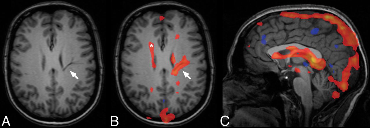
An example of a unique contribution of a DVA with deep drainage in an IC dominated by venous pulsations (subject 5). Typical anatomic features of the DVA (arrow, A) with a caput medusae appearance, unique contribution following the course of the DVA through the parenchyma (arrow, B) and contributions attributable to normal subependymal veins in the periventricular white matter (asterisk, B). C, Midsagittal view representing the typical venous character of this component, including venous sinuses and deep veins.

Fig 3.
  • Download figure
  • Open in new tab
  • Download powerpoint
Fig 3.

An example of a unique contribution of a DVA (arrows) in an IC exhibiting typical characteristics of a neural signal component (subject 3).

Discussion

In summary, most DVAs clearly appeared in ≥1 component on the basis of ICA decomposition of the rsfMRI data. While some of these ICs demonstrated unambiguous venous ICs including the venous sinuses, some of the DVAs were present in ICs that mostly exhibited typical features of neural signal ICs or that could not be unambiguously classified as noise. These findings show that signal fluctuations in DVAs can contribute to the fMRI signal in the brain and thus have the potential to bias conclusions in both clinical and scientific fMRI analyses if not appropriately considered, especially in a superficial/cortical location. DVAs could thus lead to pseudoactivations in presurgical mapping, biasing the possible extent of subsequent tumor resection if not recognized. Local pseudoactivity might also bias conclusions in the rsfMRI group studies, focusing on disease mechanisms, particularly in small samples. Finally, altered activity patterns may lead to false individual diagnostic decisions in highly automated diagnostic modeling, for example, when a classifier (supervised learning) is applied to rsfMRI data biased by a DVA in a single subject.

Potentially strong biases of vascular abnormalities on fMRI measures are well-known from imaging in patients with arteriovenous malformations, which are high-flow lesions.1,47 Our findings demonstrate that such biases can also be caused by usually clinically irrelevant low-flow lesions such as DVAs. Perfusion imaging in DVAs has revealed a diverse pattern: The large collector veins typically present as strictly local hyperperfusion compared with the surrounding brain parenchyma. Surrounding brain tissue can either exhibit normal perfusion patterns48 or show signs of venous congestion with delayed perfusion and increased cerebral blood volume extending beyond the visible transparenchymal vessels.32,48⇓⇓⇓–52 Such venous congestion may be due to a rare stenosis of the draining vein31 and could further invalidate assumptions of fMRI in the affected area. We did not observe indirect signs of such venous congestion in our sample. Evidence of a more widespread influence of DVAs on adjacent brain tissue is provided by findings of reduced uptake of fluorodeoxyglucose in a subset of subjects with DVAs, suggesting hypometabolism.53

In addition to identification of interpretable signal components,36 ICA has a practical application for reduction of such biases. Indeed, it can be used to separate signals of interest from noise in fMRI data based on either hand classification of ICs36,54 or by using automated IC classification tools.36,37,55⇓⇓⇓⇓⇓⇓⇓⇓⇓–65 Our results suggest that biasing signal fluctuations in DVAs can not only be identified but also addressed by ICA-based denoising in most cases. However, our results also demonstrate that a non-negligible proportion of DVAs cannot be reasonably separated from the neural signal of interest and may contaminate clear signal ICs, as well as unclear ICs (which are typically not removed from the dataset with these cleaning methods). The co-occurrence of DVA signal and typical neural signal patterns might be mainly based on similarly low (and potentially aliased) frequencies of the BOLD signal fluctuations in the temporal domain. We therefore believe that this observation points to the typical problem that the indirect measurement of neural activity by BOLD fMRI can be confounded by vascular sources of noise.55 In our opinion, this aspect is underappreciated in many fMRI and particularly rsfMRI studies, and both researchers and clinical personnel should be more aware of this general issue.

Another practically important finding is that the significant signal alterations related to DVAs strictly followed the spatial distribution of the collecting vein and large tributaries. This finding supports the idea that a potential bias is relatively local, though subthreshold alterations in the immediate vicinity cannot be excluded.

An important-but-controversial differential diagnostic aspect is the rare observation of arteriovenous malformations draining into DVAs or microshunts.66,67 While the latter is primarily an angiographic finding, whether the observation of DVA signal fluctuations with arteries, rather than with veins, might add to the diagnostic information in such cases remains to be evaluated. However, current temporal resolution typically below the frequency of arterial pulsations does not facilitate reliable diagnostic assessment of this issue due to aliasing.38,68

Limitations

This analysis focused on large DVAs clearly identifiable by their typical branching morphology (ie, no hard size criterion). Because 3D susceptibility-weighted imaging or contrast-enhanced T1-weighted data were not available in this sample, the true DVA prevalence is probably underestimated. Thus, the sample size of 14 subjects with clear DVAs in this study is comparatively small. In particular, DVAs with a superficial drainage pattern were potentially underrepresented because they can be more difficult to identify by nonenhanced MR imaging. We expect superficial DVAs to be more problematic, even if smaller, because the spatial interpretation of the signal origin (cortex versus DVA) is more difficult in these cases. Because we did not observe rare DVAs with signs of venous congestion in our substantial cohort of 814 participants, we unfortunately could not assess how rsfMRI captures this clinically relevant information. While focusing on ICA outputs might be perceived at first as a limitation, this analysis method actually provides an unbiased way to assess the true characteristics of the DVA signal and therefore its potential bias.

Conclusions

This work provides a proof of concept that DVAs can have features very similar to those of neural signal patterns and can thus potentially be a source of bias in fMRI analyses, probably especially when present in a superficial location involving the cortex. Thus, our study raises awareness of a potential issue that has been neglected so far. Although most effects of DVAs on fMRI signal were local and potentially amenable to dedicated noise-correction methods, there is evidence of more widespread alterations and a contamination of putative neural signal. In the clinical setting, physicians should be aware of potential “pseudoactivations” caused by DVAs, especially in the context of presurgical mapping, as well as potential biases these could cause in highly automated diagnostic approaches using supervised learning16⇓⇓–19 in big data and genome-wide association studies.20 DVAs in brain regions of interest could bias conclusions in small-group studies and thus warrant exclusion on a case-by-case basis.

Even though DVAs are usually not a clinically relevant finding, they should thus be reported to researchers by radiologists or neuroradiologists involved in the routine evaluation of scientific MR images of the brain. Awareness of potential biases caused by these frequent normal variants is important not only for neuroscientists but also for correct interpretation of clinical fMRI data.

Acknowledgments

The authors thank all study participants and the entire team of the BiDirect study, including collaborators in associated institutions.

Footnotes

  • Disclosures: Benedikt Sundermann—RELATED: Grant: Deutsche Forschungsgemeinschaft, Comments: research fellowship SU 917/1–1. Heike Minnerup—RELATED: Grant: German Federal Ministry of Education and Research, Comments: grants 01ER1205, 01ER0816, and 01ER1506.* Klaus Berger—RELATED: Grant: German Ministry of Research and Education.* *Money paid to the institution.

  • B.S. was funded by a research fellowship from the Deutsche Forschungsgemeinschaft (SU 917/1-1). BiDirect is funded by a research grant (01ER0816 and 01ER1506) from the German Federal Ministry of Education and Research. H.M. was funded by a German Federal Ministry of Education and Research grant (01ER1205). G.D. was supported by the UK Medical Research Council (MR/K006673/1).

  • Preliminary results of this work previously presented at: Annual Meeting of the European Society of Neuroradiology, September 13–17, 2017; Malmö, Sweden.

Indicates open access to non-subscribers at www.ajnr.org

References

  1. 1.↵
    1. Bartsch AJ,
    2. Homola G,
    3. Biller A, et al
    . Diagnostic functional MRI: illustrated clinical applications and decision-making. J Magn Reson Imaging 2006;23:921–32 doi:10.1002/jmri.20579 pmid:16649199
    CrossRefPubMed
  2. 2.↵
    1. Leuthardt EC,
    2. Allen M,
    3. Kamran M, et al
    . Resting-state blood oxygen level-dependent functional MRI: a paradigm shift in preoperative brain mapping. Stereotact Funct Neurosurg 2015;93:427–39 doi:10.1159/000442424 pmid:26784290
    CrossRefPubMed
  3. 3.↵
    1. Gabriel M,
    2. Brennan NP,
    3. Peck KK, et al
    . Blood oxygen level dependent functional magnetic resonance imaging for presurgical planning. Neuroimaging Clin N Am 2014;24:557–71 doi:10.1016/j.nic.2014.07.003 pmid:25441500
    CrossRefPubMed
  4. 4.↵
    1. Bandettini PA
    . Twenty years of functional MRI: the science and the stories. Neuroimage 2012;62:575–88 doi:10.1016/j.neuroimage.2012.04.026 pmid:22542637
    CrossRefPubMed
  5. 5.↵
    1. Zhang D,
    2. Raichle ME
    . Disease and the brain's dark energy. Nat Rev Neurol 2010;6:15–28 doi:10.1038/nrneurol.2009.198 pmid:20057496
    CrossRefPubMed
  6. 6.↵
    1. van den Heuvel MP,
    2. Hulshoff Pol HE
    . Exploring the brain network: a review on resting-state fMRI functional connectivity. Eur Neuropsychopharmacol 2010;20:519–34 doi:10.1016/j.euroneuro.2010.03.008 pmid:20471808
    CrossRefPubMed
  7. 7.↵
    1. Barkhof F,
    2. Haller S,
    3. Rombouts SA
    . Resting-state functional MR imaging: a new window to the brain. Radiology 2014;272:29–49 doi:10.1148/radiol.14132388 pmid:24956047
    CrossRefPubMed
  8. 8.↵
    1. Lee MH,
    2. Smyser CD,
    3. Shimony JS
    . Resting-state fMRI: a review of methods and clinical applications. AJNR Am J Neuroradiol 2013;34:1866–72 doi:10.3174/ajnr.A3263 pmid:22936095
    Abstract/FREE Full Text
  9. 9.↵
    1. Lv H,
    2. Wang Z,
    3. Tong E, et al
    . Resting-state functional MRI: everything that nonexperts have always wanted to know. AJNR Am J Neuroradiol 2018;39:1390–99 doi:10.3174/ajnr.A5527 pmid:29348136
    Abstract/FREE Full Text
  10. 10.↵
    1. Leuthardt EC,
    2. Guzman G,
    3. Bandt SK, et al
    . Integration of resting state functional MRI into clinical practice: a large single institution experience. PLoS One 2018;13:e0198349 doi:10.1371/journal.pone.0198349 pmid:29933375
    CrossRefPubMed
  11. 11.↵
    1. Hou BL,
    2. Bhatia S,
    3. Carpenter JS
    . Quantitative comparisons on hand motor functional areas determined by resting state and task BOLD fMRI and anatomical MRI for pre-surgical planning of patients with brain tumors. Neuroimage Clin 2016;11:378–87 doi:10.1016/j.nicl.2016.03.003 pmid:27069871
    CrossRefPubMed
  12. 12.↵
    1. Parker Jones O,
    2. Voets NL,
    3. Adcock JE, et al
    . Resting connectivity predicts task activation in pre-surgical populations. Neuroimage Clin 2016;13:378–85 doi:10.1016/j.nicl.2016.12.028 pmid:28123949
    CrossRefPubMed
  13. 13.↵
    1. Lu J,
    2. Zhang H,
    3. Hameed NU, et al
    . An automated method for identifying an independent component analysis-based language-related resting-state network in brain tumor subjects for surgical planning. Sci Rep 2017;7:13769 doi:10.1038/s41598-017-14248-5 pmid:29062010
    CrossRefPubMed
  14. 14.↵
    1. Branco P,
    2. Seixas D,
    3. Deprez S, et al
    . Resting-state functional magnetic resonance imaging for language preoperative planning. Front Hum Neurosci 2016;10:11 doi:10.3389/fnhum.2016.00011 pmid:26869899
    CrossRefPubMed
  15. 15.↵
    1. Kollndorfer K,
    2. Fischmeister FP,
    3. Kasprian G, et al
    . A systematic investigation of the invariance of resting-state network patterns: is resting-state fMRI ready for pre-surgical planning? Front Hum Neurosci 2013;7:95 doi:10.3389/fnhum.2013.00095 pmid:23532457
    CrossRefPubMed
  16. 16.↵
    1. Sundermann B,
    2. Herr D,
    3. Schwindt W, et al
    . Multivariate classification of blood oxygen level-dependent fMRI data with diagnostic intention: a clinical perspective. AJNR Am J Neuroradiol 2014;35:848–55 doi:10.3174/ajnr.A3713 pmid:24029388
    Abstract/FREE Full Text
  17. 17.↵
    1. Wolfers T,
    2. Buitelaar JK,
    3. Beckmann CF, et al
    . From estimating activation locality to predicting disorder: a review of pattern recognition for neuroimaging-based psychiatric diagnostics. Neurosci Biobehav Rev 2015;57:328–49 doi:10.1016/j.neubiorev.2015.08.001 pmid:26254595
    CrossRefPubMed
  18. 18.↵
    1. Rathore S,
    2. Habes M,
    3. Iftikhar MA, et al
    . A review on neuroimaging-based classification studies and associated feature extraction methods for Alzheimer's disease and its prodromal stages. Neuroimage 2017;155:530–48 doi:10.1016/j.neuroimage.2017.03.057 pmid:28414186
    CrossRefPubMed
  19. 19.↵
    1. de Vos F,
    2. Koini M,
    3. Schouten TM, et al
    . A comprehensive analysis of resting state fMRI measures to classify individual patients with Alzheimer's disease. Neuroimage 2018;167:62–72 doi:10.1016/j.neuroimage.2017.11.025 pmid:29155080
    CrossRefPubMed
  20. 20.↵
    1. Elliott L,
    2. Sharp K,
    3. Alfaro-Almagro F, et al
    . Genome-wide association studies of brain structure and function in the UK biobank. bioRxiv 2018:178806 doi:10.1101/178806
    Abstract/FREE Full Text
  21. 21.↵
    1. Kim SG,
    2. Ogawa S
    . Biophysical and physiological origins of blood oxygenation level-dependent fMRI signals. J Cereb Blood Flow Metab 2012;32:1188–206 doi:10.1038/jcbfm.2012.23 pmid:22395207
    CrossRefPubMed
  22. 22.↵
    1. Langner S,
    2. Buelow R,
    3. Fleck S, et al
    . Management of intracranial incidental findings on brain MRI. Rofo 2016;188:1123–33 doi:10.1055/s-0042-111075 pmid:27433969
    CrossRefPubMed
  23. 23.↵
    1. Sandeman EM,
    2. Hernandez Mdel C,
    3. Morris Z, et al
    . Incidental findings on brain MR imaging in older community-dwelling subjects are common but serious medical consequences are rare: a cohort study. PLoS One 2013;8:e71467 doi:10.1371/journal.pone.0071467 pmid:23967214
    CrossRefPubMed
  24. 24.↵
    1. Bos D,
    2. Poels MM,
    3. Adams HH, et al
    . Prevalence, clinical management, and natural course of incidental findings on brain MR images: the population-based Rotterdam scan study. Radiology 2016;281:507–15 doi:10.1148/radiol.2016160218 pmid:27337027
    CrossRefPubMed
  25. 25.↵
    1. Morris Z,
    2. Whiteley WN,
    3. Longstreth WT Jr., et al
    . Incidental findings on brain magnetic resonance imaging: systematic review and meta-analysis. BMJ 2009;339:b3016 doi:10.1136/bmj.b3016 pmid:19687093
    Abstract/FREE Full Text
  26. 26.↵
    1. Vernooij MW,
    2. Ikram MA,
    3. Tanghe HL, et al
    . Incidental findings on brain MRI in the general population. N Engl J Med 2007;357:1821–28 doi:10.1056/NEJMoa070972 pmid:17978290
    CrossRefPubMed
  27. 27.↵
    1. Borra RJ,
    2. Sorensen AG
    . Incidental findings in brain MRI research: what do we owe our subjects? J Am Coll Radiol 2011;8:848–52 doi:10.1016/j.jacr.2011.08.009 pmid:22137002
    CrossRefPubMed
  28. 28.↵
    1. Lasjaunias P,
    2. Burrows P,
    3. Planet C
    . Developmental venous anomalies (DVA): the so-called venous angioma. Neurosurg Rev 1986;9:233–42 doi:10.1007/BF01743138 pmid:3550523
    CrossRefPubMed
  29. 29.↵
    1. Lee M,
    2. Kim MS
    . Image findings in brain developmental venous anomalies. J Cerebrovasc Endovasc Neurosurg 2012;14:37–43 doi:10.7461/jcen.2012.14.1.37 pmid:23210028
    CrossRefPubMed
  30. 30.↵
    1. Sarwar M,
    2. McCormick WF
    . Intracerebral venous angioma: case report and review. Arch Neurol 1978;35:323–25 doi:10.1001/archneur.1978.00500290069012 pmid:646686
    CrossRefPubMed
  31. 31.↵
    1. Ruíz DS,
    2. Yilmaz H,
    3. Gailloud P
    . Cerebral developmental venous anomalies: current concepts. Ann Neurol 2009;66:271–83 doi:10.1002/ana.21754 pmid:19798638
    CrossRefPubMed
  32. 32.↵
    1. Jung HN,
    2. Kim ST,
    3. Cha J, et al
    . Diffusion and perfusion MRI findings of the signal-intensity abnormalities of brain associated with developmental venous anomaly. AJNR Am J Neuroradiol 2014;35:1539–42 doi:10.3174/ajnr.A3900 pmid:24651815
    Abstract/FREE Full Text
  33. 33.↵
    1. Teuber A,
    2. Sundermann B,
    3. Kugel H, et al
    . MR imaging of the brain in large cohort studies: feasibility report of the population- and patient-based BiDirect study. Eur Radiol 2017;27:231–38 doi:10.1007/s00330-016-4303-9 pmid:27059857
    CrossRefPubMed
  34. 34.↵
    1. Teismann H,
    2. Wersching H,
    3. Nagel M, et al
    . Establishing the bidirectional relationship between depression and subclinical arteriosclerosis: rationale, design, and characteristics of the BiDirect study. BMC Psychiatry 2014;14:174 doi:10.1186/1471-244X-14-174 pmid:24924233
    CrossRefPubMed
  35. 35.↵
    1. Beckmann CF,
    2. DeLuca M,
    3. Devlin JT, et al
    . Investigations into resting-state connectivity using independent component analysis. Philos Trans R Soc Lond B Biol Sci 2005;360:1001–13 doi:10.1098/rstb.2005.1634 pmid:16087444
    CrossRefPubMed
  36. 36.↵
    1. Griffanti L,
    2. Douaud G,
    3. Bijsterbosch J, et al
    . Hand classification of fMRI ICA noise components. Neuroimage 2017;154:188–205 doi:10.1016/j.neuroimage.2016.12.036 pmid:27989777
    CrossRefPubMed
  37. 37.↵
    1. Salimi-Khorshidi G,
    2. Douaud G,
    3. Beckmann CF, et al
    . Automatic denoising of functional MRI data: combining independent component analysis and hierarchical fusion of classifiers. Neuroimage 2014;90:449–68 doi:10.1016/j.neuroimage.2013.11.046 pmid:24389422
    CrossRefPubMed
  38. 38.↵
    1. Griffanti L,
    2. Salimi-Khorshidi G,
    3. Beckmann CF, et al
    . ICA-based artefact removal and accelerated fMRI acquisition for improved resting state network imaging. Neuroimage 2014;95:232–47 doi:10.1016/j.neuroimage.2014.03.034 pmid:24657355
    CrossRefPubMed
  39. 39.↵
    1. Smith SM,
    2. Jenkinson M,
    3. Woolrich MW, et al
    . Advances in functional and structural MR image analysis and implementation as FSL. Neuroimage 2004;23(Suppl 1):S208–19 doi:10.1016/j.neuroimage.2004.07.051 pmid:15501092
    CrossRefPubMed
  40. 40.↵
    1. Jenkinson M,
    2. Beckmann CF,
    3. Behrens TE, et al
    . FSL. Neuroimage 2012;62:782–90 doi:10.1016/j.neuroimage.2011.09.015 pmid:21979382
    CrossRefPubMed
  41. 41.↵
    1. Jenkinson M,
    2. Bannister P,
    3. Brady M, et al
    . Improved optimization for the robust and accurate linear registration and motion correction of brain images. Neuroimage 2002;17:825–41 doi:10.1006/nimg.2002.1132 pmid:12377157
    CrossRefPubMed
  42. 42.↵
    1. Smith SM
    . Fast robust automated brain extraction. Hum Brain Mapp 2002;17:143–55 doi:10.1002/hbm.10062 pmid:12391568
    CrossRefPubMed
  43. 43.↵
    1. Beckmann CF,
    2. Smith SM
    . Probabilistic independent component analysis for functional magnetic resonance imaging. IEEE Trans Med Imaging 2004;23:137–52 doi:10.1109/TMI.2003.822821 pmid:14964560
    CrossRefPubMed
  44. 44.↵
    1. Minka T
    . Automatic choice of dimensionality for PCA: technical report 514. MIT Media Lab Vision and Modeling Group. 2000. https://tminka.github.io/papers/pca/minka-pca.pdf. Accessed April 12, 2017.
  45. 45.↵
    1. Hyvärinen A
    . Fast and robust fixed-point algorithms for independent component analysis. IEEE Trans Neural Netw 1999;10:626–34 doi:10.1109/72.761722 pmid:18252563
    CrossRefPubMed
  46. 46.↵
    1. Greve DN,
    2. Fischl B
    . Accurate and robust brain image alignment using boundary-based registration. Neuroimage 2009;48:63–72 doi:10.1016/j.neuroimage.2009.06.060 pmid:19573611
    CrossRefPubMed
  47. 47.↵
    1. Lehéricy S,
    2. Biondi A,
    3. Sourour N, et al
    . Arteriovenous brain malformations: is functional MR imaging reliable for studying language reorganization in patients? Initial observations. Radiology 2002;223:672–82 doi:10.1148/radiol.2233010792 pmid:12034934
    CrossRefPubMed
  48. 48.↵
    1. Kroll H,
    2. Soares BP,
    3. Saloner D, et al
    . Perfusion-CT of developmental venous anomalies: typical and atypical hemodynamic patterns. J Neuroradiol 2010;37:239–42 doi:10.1016/j.neurad.2009.09.002 pmid:19959233
    CrossRefPubMed
  49. 49.↵
    1. Camacho DL,
    2. Smith JK,
    3. Grimme JD, et al
    . Atypical MR imaging perfusion in developmental venous anomalies. AJNR Am J Neuroradiol 2004;25:1549–52 pmid:15502136
    Abstract/FREE Full Text
  50. 50.↵
    1. Hanson EH,
    2. Roach CJ,
    3. Ringdahl EN, et al
    . Developmental venous anomalies: appearance on whole-brain CT digital subtraction angiography and CT perfusion. Neuroradiology 2011;53:331–41 doi:10.1007/s00234-010-0739-9 pmid:20652805
    CrossRefPubMed
  51. 51.↵
    1. Sharma A,
    2. Zipfel GJ,
    3. Hildebolt C, et al
    . Hemodynamic effects of developmental venous anomalies with and without cavernous malformations. AJNR Am J Neuroradiol 2013;34:1746–51 doi:10.3174/ajnr.A3516 pmid:23598827
    Abstract/FREE Full Text
  52. 52.↵
    1. Iv M,
    2. Fischbein NJ,
    3. Zaharchuk G
    . Association of developmental venous anomalies with perfusion abnormalities on arterial spin labeling and bolus perfusion-weighted imaging. J Neuroimaging 2015;25:243–50 doi:10.1111/jon.12119 pmid:24717021
    CrossRefPubMed
  53. 53.↵
    1. Larvie M,
    2. Timerman D,
    3. Thum JA
    . Brain metabolic abnormalities associated with developmental venous anomalies. AJNR Am J Neuroradiol 2015;36:475–80 doi:10.3174/ajnr.A4172 pmid:25477358
    Abstract/FREE Full Text
  54. 54.↵
    1. Kelly RE Jr.,
    2. Alexopoulos GS,
    3. Wang Z, et al
    . Visual inspection of independent components: defining a procedure for artifact removal from fMRI data. J Neurosci Methods 2010;189:233–45 doi:10.1016/j.jneumeth.2010.03.028 pmid:20381530
    CrossRefPubMed
  55. 55.↵
    1. Murphy K,
    2. Birn RM,
    3. Bandettini PA
    . Resting-state fMRI confounds and cleanup. Neuroimage 2013;80:349–59 doi:10.1016/j.neuroimage.2013.04.001 pmid:23571418
    CrossRefPubMed
  56. 56.↵
    1. Thomas CG,
    2. Harshman RA,
    3. Menon RS
    . Noise reduction in BOLD-based fMRI using component analysis. Neuroimage 2002;17:1521–37 doi:10.1006/nimg.2002.1200 pmid:12414291
    CrossRefPubMed
  57. 57.↵
    1. Beall EB,
    2. Lowe MJ
    . Isolating physiologic noise sources with independently determined spatial measures. Neuroimage 2007;37:1286–300 doi:10.1016/j.neuroimage.2007.07.004 pmid:17689982
    CrossRefPubMed
  58. 58.↵
    1. Perlbarg V,
    2. Bellec P,
    3. Anton JL, et al
    . CORSICA: correction of structured noise in fMRI by automatic identification of ICA components. Magn Reson Imaging 2007;25:35–46 doi:10.1016/j.mri.2006.09.042 pmid:17222713
    CrossRefPubMed
  59. 59.↵
    1. Sui J,
    2. Adali T,
    3. Pearlson GD, et al
    . An ICA-based method for the identification of optimal FMRI features and components using combined group-discriminative techniques. Neuroimage 2009;46:73–86 doi:10.1016/j.neuroimage.2009.01.026 pmid:19457398
    CrossRefPubMed
  60. 60.↵
    1. Storti SF,
    2. Formaggio E,
    3. Nordio R, et al
    . Automatic selection of resting-state networks with functional magnetic resonance imaging. Front Neurosci 2013;7:72 doi:10.3389/fnins.2013.00072 pmid:23730268
    CrossRefPubMed
  61. 61.↵
    1. Bhaganagarapu K,
    2. Jackson GD,
    3. Abbott DF
    . An automated method for identifying artifact in independent component analysis of resting-state FMRI. Front Hum Neurosci 2013;7:343 doi:10.3389/fnhum.2013.00343 pmid:23847511
    CrossRefPubMed
  62. 62.↵
    1. Tohka J,
    2. Foerde K,
    3. Aron AR, et al
    . Automatic independent component labeling for artifact removal in fMRI. Neuroimage 2008;39:1227–45 doi:10.1016/j.neuroimage.2007.10.013 pmid:18042495
    CrossRefPubMed
  63. 63.↵
    1. De Martino F,
    2. Gentile F,
    3. Esposito F, et al
    . Classification of fMRI independent components using IC-fingerprints and support vector machine classifiers. Neuroimage 2007;34:177–94 doi:10.1016/j.neuroimage.2006.08.041 pmid:17070708
    CrossRefPubMed
  64. 64.↵
    1. Sochat V,
    2. Supekar K,
    3. Bustillo J, et al
    . A robust classifier to distinguish noise from fMRI independent components. PLoS One 2014;9:e95493 doi:10.1371/journal.pone.0095493 pmid:24748378
    CrossRefPubMed
  65. 65.↵
    1. Caballero-Gaudes C,
    2. Reynolds RC
    . Methods for cleaning the BOLD fMRI signal. Neuroimage 2017;154:128–49 doi:10.1016/j.neuroimage.2016.12.018 pmid:27956209
    CrossRefPubMed
  66. 66.↵
    1. Pereira VM,
    2. Geibprasert S,
    3. Krings T, et al
    . Pathomechanisms of symptomatic developmental venous anomalies. Stroke 2008;39:3201–15 doi:10.1161/STROKEAHA.108.521799 pmid:18988912
    Abstract/FREE Full Text
  67. 67.↵
    1. Tomycz ND,
    2. Vora NA,
    3. Kanal E, et al
    . Intracranial arterialized venous angioma: case report with new insights from functional brain MRI. Diagn Interv Radiol 2010;16:13–15 doi:10.4261/1305-3825.DIR.1627-08.1 pmid:19813172
    CrossRefPubMed
  68. 68.↵
    1. Lowe MJ,
    2. Mock BJ,
    3. Sorenson JA
    . Functional connectivity in single and multislice echoplanar imaging using resting-state fluctuations. Neuroimage 1998;7:119–32 doi:10.1006/nimg.1997.0315 pmid:9558644
    CrossRefPubMed
  • Received May 10, 2018.
  • Accepted after revision August 25, 2018.
  • © 2018 by American Journal of Neuroradiology
View Abstract
PreviousNext
Back to top

In this issue

American Journal of Neuroradiology: 39 (12)
American Journal of Neuroradiology
Vol. 39, Issue 12
1 Dec 2018
  • Table of Contents
  • Index by author
  • Complete Issue (PDF)
Advertisement
Print
Download PDF
Email Article

Thank you for your interest in spreading the word on American Journal of Neuroradiology.

NOTE: We only request your email address so that the person you are recommending the page to knows that you wanted them to see it, and that it is not junk mail. We do not capture any email address.

Enter multiple addresses on separate lines or separate them with commas.
Interaction of Developmental Venous Anomalies with Resting-State Functional MRI Measures
(Your Name) has sent you a message from American Journal of Neuroradiology
(Your Name) thought you would like to see the American Journal of Neuroradiology web site.
CAPTCHA
This question is for testing whether or not you are a human visitor and to prevent automated spam submissions.
Cite this article
B. Sundermann, B. Pfleiderer, H. Minnerup, K. Berger, G. Douaud
Interaction of Developmental Venous Anomalies with Resting-State Functional MRI Measures
American Journal of Neuroradiology Dec 2018, 39 (12) 2326-2331; DOI: 10.3174/ajnr.A5847

Citation Manager Formats

  • BibTeX
  • Bookends
  • EasyBib
  • EndNote (tagged)
  • EndNote 8 (xml)
  • Medlars
  • Mendeley
  • Papers
  • RefWorks Tagged
  • Ref Manager
  • RIS
  • Zotero
0 Responses
Respond to this article
Share
Bookmark this article
Interaction of Developmental Venous Anomalies with Resting-State Functional MRI Measures
B. Sundermann, B. Pfleiderer, H. Minnerup, K. Berger, G. Douaud
American Journal of Neuroradiology Dec 2018, 39 (12) 2326-2331; DOI: 10.3174/ajnr.A5847
del.icio.us logo Twitter logo Facebook logo Mendeley logo
  • Tweet Widget
  • Facebook Like
  • Google Plus One
Purchase

Jump to section

  • Article
    • Abstract
    • ABBREVIATIONS:
    • Materials and Methods
    • Results
    • Discussion
    • Conclusions
    • Acknowledgments
    • Footnotes
    • References
  • Figures & Data
  • Supplemental
  • Info & Metrics
  • Responses
  • References
  • PDF

Related Articles

  • PubMed
  • Google Scholar

Cited By...

  • Correlates of asymmetric venous drainage in resting state functional magnetic resonance imaging data
  • Functional connectivity of cognition-related brain networks in adults with fetal alcohol syndrome
  • Crossref (4)
  • Google Scholar

This article has been cited by the following articles in journals that are participating in Crossref Cited-by Linking.

  • Approaching altered inhibitory control in phenylketonuria: A functional MRI study with a Go‐NoGo task in young female adults
    Benedikt Sundermann, Stefan Garde, Mahboobeh Dehghan Nayyeri, Josef Weglage, Johanna Rau, Bettina Pfleiderer, Reinhold Feldmann
    European Journal of Neuroscience 2020 52 8
  • Functional MRI in Neuro-Oncology: State of the Art and Future Directions
    Luca Pasquini, Kyung K. Peck, Mehrnaz Jenabi, Andrei Holodny
    Radiology 2023 308 3
  • Prevalence and anatomical characteristics of developmental venous anomalies: an MRI study
    Karolina Brzegowy, Natalia Kowalska, Bernard Solewski, Agata Musiał, Tomasz Kasprzycki, Izabela Herman-Sucharska, Jerzy A. Walocha
    Neuroradiology 2021 63 7
  • Functional connectivity of cognition-related brain networks in adults with fetal alcohol syndrome
    Benedikt Sundermann, Reinhold Feldmann, Christian Mathys, Johanna M. H. Rau, Stefan Garde, Anna Braje, Josef Weglage, Bettina Pfleiderer
    BMC Medicine 2023 21 1

More in this TOC Section

  • Glutaric Aciduria Type 1: DK vs. Conventional MRI
  • Kurtosis and Epileptogenic Tubers: A Pilot Study
  • Brain Iron in Niemann-Pick Type C: 7T Study
Show more Functional

Similar Articles

Advertisement

Indexed Content

  • Current Issue
  • Accepted Manuscripts
  • Article Preview
  • Past Issues
  • Editorials
  • Editors Choice
  • Fellow Journal Club
  • Letters to the Editor

Cases

  • Case Collection
  • Archive - Case of the Week
  • Archive - Case of the Month
  • Archive - Classic Case

Special Collections

  • Special Collections

Resources

  • News and Updates
  • Turn around Times
  • Submit a Manuscript
  • Author Policies
  • Manuscript Submission Guidelines
  • Evidence-Based Medicine Level Guide
  • Publishing Checklists
  • Graphical Abstract Preparation
  • Imaging Protocol Submission
  • Submit a Case
  • Become a Reviewer/Academy of Reviewers
  • Get Peer Review Credit from Publons

Multimedia

  • AJNR Podcast
  • AJNR SCANtastic
  • Video Articles

About Us

  • About AJNR
  • Editorial Board
  • Not an AJNR Subscriber? Join Now
  • Alerts
  • Feedback
  • Advertise with us
  • Librarian Resources
  • Permissions
  • Terms and Conditions

American Society of Neuroradiology

  • Not an ASNR Member? Join Now

© 2025 by the American Society of Neuroradiology All rights, including for text and data mining, AI training, and similar technologies, are reserved.
Print ISSN: 0195-6108 Online ISSN: 1936-959X

Powered by HighWire