Advertisement

Main menu

  • Home
  • Content
    • Current Issue
    • Accepted Manuscripts
    • Article Preview
    • Past Issue Archive
    • AJNR Case Collection
    • Case of the Week Archive
    • Classic Case Archive
    • Case of the Month Archive
  • Special Collections
    • Spinal CSF Leak Articles (Jan 2020-June 2024)
    • 2024 AJNR Journal Awards
    • Most Impactful AJNR Articles
  • Multimedia
    • AJNR Podcast
    • AJNR Scantastics
    • Video Articles
  • For Authors
    • Submit a Manuscript
    • Author Policies
    • Fast publishing of Accepted Manuscripts
    • Graphical Abstract Preparation
    • Manuscript Submission Guidelines
    • Imaging Protocol Submission
    • Submit a Case for the Case Collection
  • About Us
    • About AJNR
    • Editorial Board
  • More
    • Become a Reviewer/Academy of Reviewers
    • Subscribers
    • Permissions
    • Alerts
    • Feedback
    • Advertisers
    • ASNR Home
  • Other Publications
    • ajnr

User menu

  • Alerts
  • Log in

Search

  • Advanced search
American Journal of Neuroradiology

American Journal of Neuroradiology

ASHNR American Society of Functional Neuroradiology ASHNR American Society of Pediatric Neuroradiology ASSR
  • Alerts
  • Log in

Advanced Search

  • Home
  • Content
    • Current Issue
    • Accepted Manuscripts
    • Article Preview
    • Past Issue Archive
    • AJNR Case Collection
    • Case of the Week Archive
    • Classic Case Archive
    • Case of the Month Archive
  • Special Collections
    • Spinal CSF Leak Articles (Jan 2020-June 2024)
    • 2024 AJNR Journal Awards
    • Most Impactful AJNR Articles
  • Multimedia
    • AJNR Podcast
    • AJNR Scantastics
    • Video Articles
  • For Authors
    • Submit a Manuscript
    • Author Policies
    • Fast publishing of Accepted Manuscripts
    • Graphical Abstract Preparation
    • Manuscript Submission Guidelines
    • Imaging Protocol Submission
    • Submit a Case for the Case Collection
  • About Us
    • About AJNR
    • Editorial Board
  • More
    • Become a Reviewer/Academy of Reviewers
    • Subscribers
    • Permissions
    • Alerts
    • Feedback
    • Advertisers
    • ASNR Home
  • Follow AJNR on Twitter
  • Visit AJNR on Facebook
  • Follow AJNR on Instagram
  • Join AJNR on LinkedIn
  • RSS Feeds

Welcome to the new AJNR, Updated Hall of Fame, and more. Read the full announcements.


AJNR is seeking candidates for the position of Associate Section Editor, AJNR Case Collection. Read the full announcement.


LetterLETTER

Vessel Wall Enhancement in Treated Unruptured Aneurysms

D.G. Corrêa, R.A. Cadete and L.C. Hygino da Cruz
American Journal of Neuroradiology December 2018, 39 (12) E129-E130; DOI: https://doi.org/10.3174/ajnr.A5843
D.G. Corrêa
aClínica de Diagnóstico por Imagem
bRio de Janeiro, Brazil
  • Find this author on Google Scholar
  • Find this author on PubMed
  • Search for this author on this site
  • ORCID record for D.G. Corrêa
R.A. Cadete
aClínica de Diagnóstico por Imagem
bRio de Janeiro, Brazil
  • Find this author on Google Scholar
  • Find this author on PubMed
  • Search for this author on this site
  • ORCID record for R.A. Cadete
L.C. Hygino da Cruz Jr.
aClínica de Diagnóstico por Imagem
bRio de Janeiro, Brazil
  • Find this author on Google Scholar
  • Find this author on PubMed
  • Search for this author on this site
  • ORCID record for L.C. Hygino da Cruz Jr.
  • Article
  • Figures & Data
  • Info & Metrics
  • Responses
  • References
  • PDF
Loading

We read with great interest the article “Vessel Wall Enhancement in Unruptured Intracranial Aneurysms: An Indicator for Higher Risk of Rupture? High-Resolution MR Imaging and Correlated Histologic Findings,” published in the American Journal of Neuroradiology by Larsen et al.1 The authors found that wall enhancement in vessel wall MR imaging is associated with inflammatory cell invasion, neovascularization, and the presence of vasa vasorum, factors that provide valuable information for unruptured aneurysm risk stratification.1 Previous studies have suggested that aneurysm wall inflammation may precede, rather than result from, rupture. In as much as inflammation affects aneurysm growth and rupture, vessel wall enhancement may be a marker of rupture risk.2

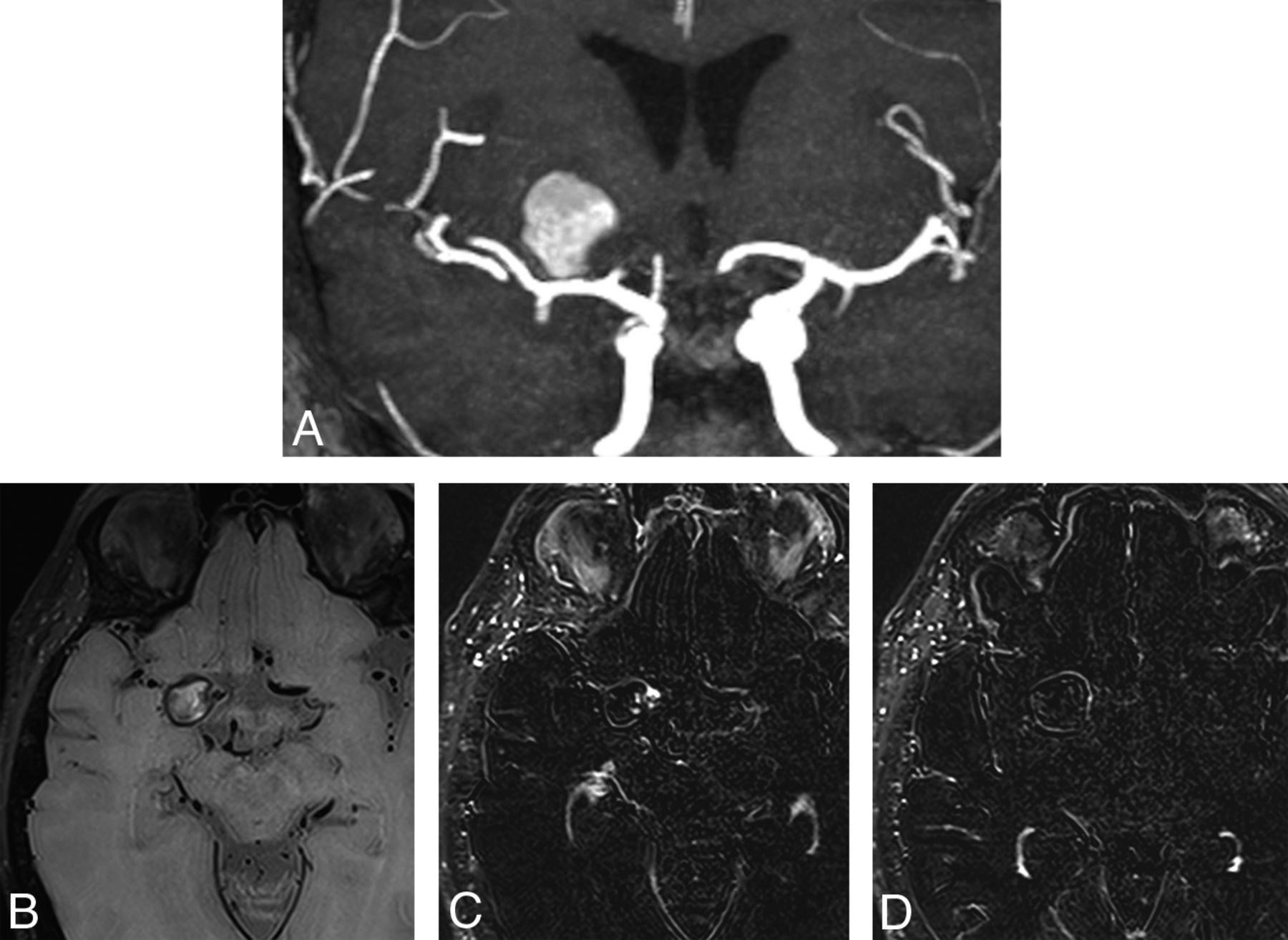
However, we would like to discuss the clinical importance of the presence of aneurysm wall enhancement, detected by high-resolution MR imaging after treatment. We present the case of a 68-year-old woman with an unruptured anterior cerebral artery aneurysm treated with a flow diverter. Follow-up 3T MR angiography 1 month after flow-diverter placement showed magnetic susceptibility artifacts in the A1 portion of the right anterior cerebral artery, secondary to the flow diverter. The aneurysm, which measured 1.7 × 1.5 cm, had a T1-hyperintense signal within it due to thrombosis or slow flow induced by the treatment. Subtraction of the precontrast from the postcontrast enhanced vessel wall images revealed a thin rim of enhancement around the aneurysm wall, without edema in the surrounding parenchyma (Figure). The patient was asymptomatic at the time of the examination and is being monitored as an outpatient.

FIGURE.
  • Download figure
  • Open in new tab
  • Download powerpoint
FIGURE.

A, Maximum intensity projection of TOF-MR angiography shows an aneurysm adjacent to the A1 portion of the right anterior cerebral artery. B, T1 black-blood vessel wall imaging without contrast shows a hyperintense signal area inside the aneurysm due to treatment-induced thrombosis or slow flow. C and D, Subtraction of precontrast from postcontrast enhanced vessel wall images shows a thin rim of enhancement around the aneurysm wall and the flow diverter.

A thin rim of circumferential contrast enhancement around a treated aneurysm is commonly seen in contrast-enhanced TOF-MR angiography following treatment and should be an expected finding. This contrast enhancement is thought to be attributed to a combination of organized peripherally distributed intra-aneurysmal thrombosis, vasa vasorum within the adventitial layer of the aneurysm wall, and/or growth of vascularized tissue into an implanted coil mass or flow diverter due to inflammation or healing.3

Guan et al4 described 3 patients with intracranial aneurysms treated with flow diversion. Post treatment vessel wall high-resolution MR imaging to evaluate aneurysm obliteration revealed intra-aneurysm thrombus development, reduced aneurysm filling, and vessel wall enhancement of the aneurysm sac in all 3 patients. Although the authors did not discuss the significance of the wall enhancement detected by vessel wall imaging, all 3 patients were clinically well and in outpatient follow-up, like the case presented here. In such post treatment scenarios, vessel wall enhancement detected by high-resolution vessel wall MR imaging may be of no clinical significance and not a sign of elevated rupture risk, though this technique is more sensitive to inflammatory changes, atherosclerotic plaques, arterial dissection, and other causes of intracranial arterial narrowing than TOF-MR angiography.2

Hence, whereas vessel wall enhancement of nontreated intracranial aneurysms seen in high-resolution vessel wall imaging may be a marker of rupture risk,1,2 wall enhancement of a treated aneurysm may not necessarily be pathologic, though there is a lack of studies addressing the utility of vessel wall imaging in treated aneurysms in the literature.

References

  1. 1.↵
    1. Larsen N,
    2. von der Brelie C,
    3. Trick D, et al
    . Vessel wall enhancement in unruptured intracranial aneurysms: an indicator for higher risk of rupture? High-resolution MR imaging and correlated histologic findings. AJNR Am J Neuroradiol 2018;39:1617–21 doi:10.3174/ajnr.A5731 pmid:30026386
    Abstract/FREE Full Text
  2. 2.↵
    1. Mandell DM,
    2. Mossa-Basha M,
    3. Qiao Y, et al
    ; Vessel Wall Imaging Study Group of the American Society of Neuroradiology. Intracranial vessel wall MRI: principles and expert consensus recommendations of the American Society of Neuroradiology. AJNR Am J Neuroradiol 2017;38:218–29 doi:10.3174/ajnr.A4893 pmid:27469212
    Abstract/FREE Full Text
  3. 3.↵
    1. Wallace RC,
    2. Karis JP,
    3. Partovi S, et al
    . Noninvasive imaging of treated cerebral aneurysms, Part I: MR angiographic follow-up of coiled aneurysms. AJNR Am J Neuroradiol 2007;28:1001–08 doi:10.3174/ajnr.A0662 pmid:17569946
    Abstract/FREE Full Text
  4. 4.↵
    1. Guan J,
    2. Karsy M,
    3. McNally S, et al
    . High-resolution magnetic resonance imaging of intracranial aneurysms treated by flow diversion. Interdisciplinary Neurosurgery 2017;10:69–74 doi:10.1016/j.inat.2017.07.004
    CrossRef
  • © 2018 by American Journal of Neuroradiology
PreviousNext
Back to top

In this issue

American Journal of Neuroradiology: 39 (12)
American Journal of Neuroradiology
Vol. 39, Issue 12
1 Dec 2018
  • Table of Contents
  • Index by author
  • Complete Issue (PDF)
Advertisement
Print
Download PDF
Email Article

Thank you for your interest in spreading the word on American Journal of Neuroradiology.

NOTE: We only request your email address so that the person you are recommending the page to knows that you wanted them to see it, and that it is not junk mail. We do not capture any email address.

Enter multiple addresses on separate lines or separate them with commas.
Vessel Wall Enhancement in Treated Unruptured Aneurysms
(Your Name) has sent you a message from American Journal of Neuroradiology
(Your Name) thought you would like to see the American Journal of Neuroradiology web site.
CAPTCHA
This question is for testing whether or not you are a human visitor and to prevent automated spam submissions.
Cite this article
D.G. Corrêa, R.A. Cadete, L.C. Hygino da Cruz
Vessel Wall Enhancement in Treated Unruptured Aneurysms
American Journal of Neuroradiology Dec 2018, 39 (12) E129-E130; DOI: 10.3174/ajnr.A5843

Citation Manager Formats

  • BibTeX
  • Bookends
  • EasyBib
  • EndNote (tagged)
  • EndNote 8 (xml)
  • Medlars
  • Mendeley
  • Papers
  • RefWorks Tagged
  • Ref Manager
  • RIS
  • Zotero
0 Responses
Respond to this article
Share
Bookmark this article
Vessel Wall Enhancement in Treated Unruptured Aneurysms
D.G. Corrêa, R.A. Cadete, L.C. Hygino da Cruz
American Journal of Neuroradiology Dec 2018, 39 (12) E129-E130; DOI: 10.3174/ajnr.A5843
del.icio.us logo Twitter logo Facebook logo Mendeley logo
  • Tweet Widget
  • Facebook Like
  • Google Plus One
Purchase

Jump to section

  • Article
    • References
  • Figures & Data
  • Info & Metrics
  • Responses
  • References
  • PDF

Related Articles

  • Reply:
  • PubMed
  • Google Scholar

Cited By...

  • No citing articles found.
  • Crossref (1)
  • Google Scholar

This article has been cited by the following articles in journals that are participating in Crossref Cited-by Linking.

  • High-Resolution Vessel Wall MRI of Endovascularly Treated Intracranial Aneurysms
    Łukasz Zwarzany, Mateusz Owsiak, Ernest Tyburski, Wojciech Poncyljusz
    Tomography 2022 8 1

More in this TOC Section

  • Brain AVM’s Nidus: What if We Hadn’t Understood Anything?
  • Letter to the Editor regarding “Automated Volumetric Software in Dementia: Help or Hindrance to the Neuroradiologist?”
  • Reply:
Show more Letters

Similar Articles

Advertisement

Indexed Content

  • Current Issue
  • Accepted Manuscripts
  • Article Preview
  • Past Issues
  • Editorials
  • Editors Choice
  • Fellow Journal Club
  • Letters to the Editor

Cases

  • Case Collection
  • Archive - Case of the Week
  • Archive - Case of the Month
  • Archive - Classic Case

Special Collections

  • Special Collections

Resources

  • News and Updates
  • Turn around Times
  • Submit a Manuscript
  • Author Policies
  • Manuscript Submission Guidelines
  • Evidence-Based Medicine Level Guide
  • Publishing Checklists
  • Graphical Abstract Preparation
  • Imaging Protocol Submission
  • Submit a Case
  • Become a Reviewer/Academy of Reviewers
  • Get Peer Review Credit from Publons

Multimedia

  • AJNR Podcast
  • AJNR SCANtastic
  • Video Articles

About Us

  • About AJNR
  • Editorial Board
  • Not an AJNR Subscriber? Join Now
  • Alerts
  • Feedback
  • Advertise with us
  • Librarian Resources
  • Permissions
  • Terms and Conditions

American Society of Neuroradiology

  • Not an ASNR Member? Join Now

© 2025 by the American Society of Neuroradiology All rights, including for text and data mining, AI training, and similar technologies, are reserved.
Print ISSN: 0195-6108 Online ISSN: 1936-959X

Powered by HighWire