
Article Figures & Data
Figures
- Fig 1.
ROIs used in the magnitude images for calculating the signal-to-noise ratios. A, The circumferential of both carotid arteries (I) was used as a marker for the carotid vessel wall that runs through the suprasellar cistern, which was used as marker for CSF (II). B, The circumferential of the basilar artery (I) was used as marker for the basilar vessel wall that runs through the pontine cistern, which was used as second marker for CSF (II). For the pituitary stalk, a homogeneous hyperintense part of the center was used (III). In the lumen of the middle cerebral artery, an ROI was drawn as a marker for blood (IV), and in the left orbital gyrus, an ROI was drawn as marker for brain tissue (V). The ROIs are drawn for illustrative purposes, and the exact contours may differ in the real measurements, depending on patients' specific anatomies.
- Fig 2.
Sample images in the axial plane of the 7 different scan variants performed at 3T (precontrast, in order of decreasing scan duration). Both distal intracranial internal carotid arteries (white arrowheads) with the bifurcation of the posterior communicating artery are depicted, surrounded by CSF. 1, T1WI VISTA variant 1 (8:24 minutes). 2, Proton density–weighted VISTA variant 2 (7:50 minutes), adjusted from Qiao et al.19 3, T1WI VIRTA variant 3 (6:42 minutes). 4, T1WI VIRTA variant 4 (6:01 minutes). 5, T1WI VISTA variant 5 (5:52 minutes), adjusted from Qiao et al19 with a shorter TR. 6, T1WI VISTA variant 6 (5:49 minutes). 7, T1WI VIRTA variant 7 (4:39 minutes).
- Fig 3.
Sample images in the sagittal plane of the 7 different scan variants performed at 3T (precontrast, in the order of decreasing scan duration). The right MCA (black arrowheads) is depicted in all images, surrounded by CSF and brain parenchyma. 1, T1WI VISTA variant 1 (8:24 minutes). 2, Proton density–weighted VISTA variant 2 (7:50 minutes), adjusted from Qiao et al.19 3, T1WI VIRTA variant 3 (6:42 minutes). 4, T1WI VIRTA variant 4 (6:01 minutes). 5, T1WI VISTA variant 5 (5:52 minutes), adjusted from Qiao et al19 with a shorter TR. 6, T1WI VISTA variant 6 (5:49 minutes). 7, T1WI VIRTA variant 7 (4:39 minutes).
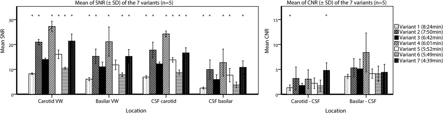
- Fig 4.
Barplots showing the mean and SD of the clinically relevant SNRs and CNRs of all 7 vessel wall imaging variants. The means and SDs are calculated for 5 subjects The mean of the left and right intracranial internal carotid arteries was used for the carotid vessel wall. The asterisks indicate a significant difference compared with reference variant 3 (corrected P value for multiple comparisons, P < .008) using generalized estimating equations to account for repeated measures on the same subjects. VW indicates vessel wall.
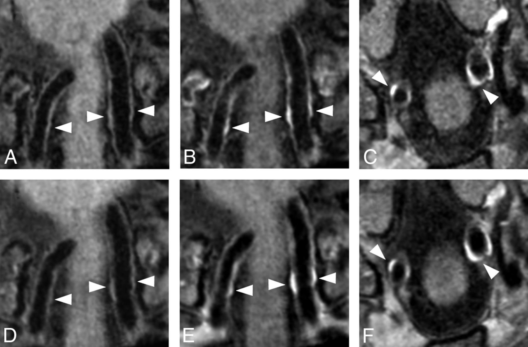
- Fig 5.
Matching vessel wall lesions of the distal vertebral arteries (white arrowheads) seen before and after contrast with variant 3 (6:42 minutes; A–C) and variant 7 (4:39 minutes; D–F) in a 59-year-old man with multiple cardiovascular risk factors. Subtle (concentric) wall thickening is seen in both the left and right vertebral arteries before contrast (A and D), with clear contrast enhancement on the postcontrast image (B and E). Variant 3 (6:42 minutes, B and C) was acquired approximately 2 minutes after contrast injection, and variant 7 (4:39 minutes, E and F), approximately 9 minutes after contrast injection. Some patient motion was seen in the postcontrast series of variant 7. Postcontrast transverse images of variants 3 (C) and 7 (F) show clear vessel wall enhancement in both vertebral arteries (white arrowheads).


Tables
T1WI VISTAd Variant 1 PDW VISTA Variant 2a T1WI VIRTAd Variant 3b T1WI VIRTA Variant 4 T1WI VISTA Variant 5c T1WI VISTA Variant 6 T1WI VIRTA Variant 7 Scan duration (min) 8:24 7:50 6:42 6:01 5:52 5:49 4:39 TR/TE (ms) 1500/38 2000/40 1500/37 1500/40 1500/40 1500/38 1500/40 FOV (mm3) 200 × 166 × 45 200 × 166 × 45 200 × 166 × 45 200 × 166 × 45 200 × 166 × 45 200 × 166 × 45 200 × 166 × 45 Acquired voxel (mm3) 0.5 × 0.5 × 0.5 0.5 × 0.5 × 0.5 0.6 × 0.6 × 1.0 0.5 × 0.5 × 1.0 0.5 × 0.5 × 0.5 0.6 × 0.6 × 0.6 0.5 × 0.5 × 1.0 Reconstructed voxel (mm3) 0.5 × 0.5 × 0.5 0.5 × 0.5 × 0.5 0.5 × 0.5 × 0.5 0.5 × 0.5 × 0.5 0.5 × 0.5 × 0.5 0.6 × 0.6 × 0.6 0.5 × 0.5 × 0.5 Oversample factor 1.8 1.2 1.8 1.8 1.2 1.8 1.8 SENSE reduction factor 1.5 2 1.5 2 2 1.5 2 Overcontiguous slices No (90) No (90) Yes (90) Yes (90) No (90) No (75) Yes (90) TSE + startup echoes 56 + 3 60 + 5 56 + 5 56 + 5 60 + 5 56 + 4 56 + 5 Turbo direction Radial Radial Y-axis Radial Radial Radial Radial Refocusing control (αmin/αmax) 30/120 50/120 25/120 50/120 50/120 30/120 50/120 Readout bandwidth (Hz) 607.6 358.6 732.1 360.1 358.6 753 360.1 Reference tissue T1/T2 (ms) 1200/80 1200/100 1200/100 1200/100 1200/100 1200/80 1200/100 Anti-DRIVE Yes No Yes No No Yes Yes Elliptic k-space shutter Yes Yes No No Yes Yes Yes Precontrast P Value Postcontrast P Value T1WI VIRTA (Variant 3)f T1WI VIRTA (Variant 7) T1WI VIRTA (Variant 3)f T1WI VIRTA (Variant 7) SNRtissueb 28.0 ± 1.8 40.5 ± 3.4 .005g 27.9 ± 1.3 42.4 ± 2.8 .005g SNRcarotid vessel wallc 15.3 ± 1.4 20.8 ± 2.7 .005g 17.2 ± 0.9 26.4 ± 4.0 .005g SNRbasilar vessel wall 13.0 ± 1.4 15.9 ± 1.7 .005g 13.4 ± 2.4 19.2 ± 1.8 .005g SNRsuprasellar CSF 9.1 ± 2.0 11.9 ± 2.2 .007g 11.3 ± 0.7 14.1 ± 2.4 .013 SNRpontine CSF 5.9 ± 2.1 8.0 ± 2.0 .007g 6.5 ± 1.8 11.2 ± 2.6 .007g SNRblood 3.6 ± 0.6 5.4 ± 1.2 .005g 3.8 ± 0.5 6.3 ± 1.4 .007g SNRpituitary gland 23.0 ± 3.4 33.4 ± 4.7 .005g 33.3 ± 3.5 49.5 ± 6.7 .005g CNRcarotid vessel wall–CSFd 6.1 ± 1.6 9.0 ± 1.5 .005g 6.0 ± 0.7 12.3 ± 3.6 .007g CNRbasilar vessel wall–CSFe 7.2 ± 1.4 7.9 ± 1.4 .059 7.0 ± 2.0 8.0 ± 1.9 .285 CNRcarotid vessel wall–bloodc 11.6 ± 1.3 15.5 ± 2.9 .013 13.5 ± 1.0 20.1 ± 3.8 .007g ↵a Data are mean and SD calculated in 10 subjects. A Wilcoxon signed rank test was used to compare the differences between precontrast variants 3 and 7 and postcontrast variants 3 and 7 (additional statistical comparisons are shown in On-line Table 2).
↵b Tissue ROI is located at the left orbital gyri.
↵c The mean of the left and right distal intracranial internal carotid arteries.
↵d Suprasellar CSF is used as reference.
↵e Pontine CSF is used as reference.
↵f Currently used in our clinic.
↵g Statistically significant (P < .008).


Jump to section
Related Articles
Cited By...
- No citing articles found.