
Article Figures & Data
Figures
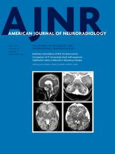
- Fig 1.
Measurement techniques used in the study. A, Preterm neonate, gestational age 35 weeks 6 days. T1-weighted MR image shows measurement of the vermis height from the culmen to the uvular lobule on the midsagittal image. B and C, Preterm neonate, gestational age 30 weeks 6 days. T2-weighted MR images show the cerebellar diameter measurement at the widest points of the cerebellum on axial and coronal images, respectively. D, Preterm neonate, gestational age 33 weeks 4 days. T1-weighted image shows cranial base angle measurement with the fonticulus frontalis used as the anterior landmark; midsella, as the vertex point; and the distal tip of the ossified clivus, as the terminal point. E, Measurement of the tentorial angle between the Twining line and a line drawn through tentorium cerebelli with tuberculum sella used as the originating point. F, T1-weighted MR image shows measurement of the infratentorial angle, using a line originating from the midsella drawn through the midpoint of the dural reflection of the torcula herophili to the inner cortex, and a line drawn between the midsella and basion.
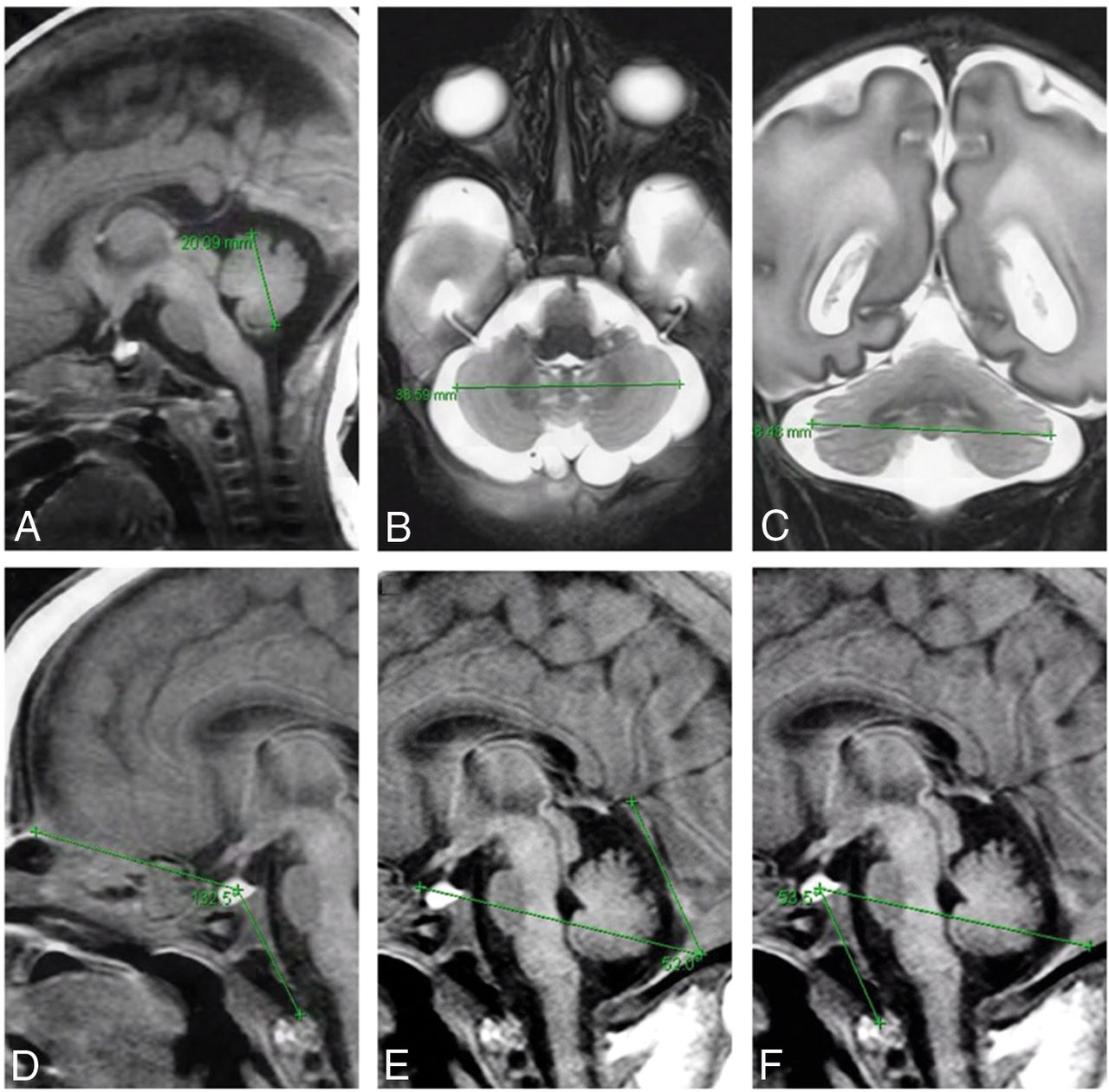
- Fig 2.
Characteristic MR imaging findings of neonates with a RASopathy. A, Preterm neonate, gestational age 28 weeks 6 days, MR imaging performed at postmenstrual age 30 weeks 5 days. T2-weighted midsagittal MR image shows a vertical tentorium and splenium of the corpus callosum. B, Preterm neonate, gestational age 34 weeks 2 days. T2-weighted axial MR image demonstrates the presence of hemorrhagic and cystic lesions in the peripheral regions of the cerebellum. C, Preterm neonate, gestational age 34 weeks 2 days. T2-weighted axial MR imaging shows a mildly enlarged extracerebral space with severe white matter injury, which evolved into extensive cysts. D, Preterm neonate, gestational age 34 weeks. Axial T2-weighted MR image shows a severely enlarged extracerebral space, punctate white matter lesions, and a small amount of blood in the lateral ventricles.
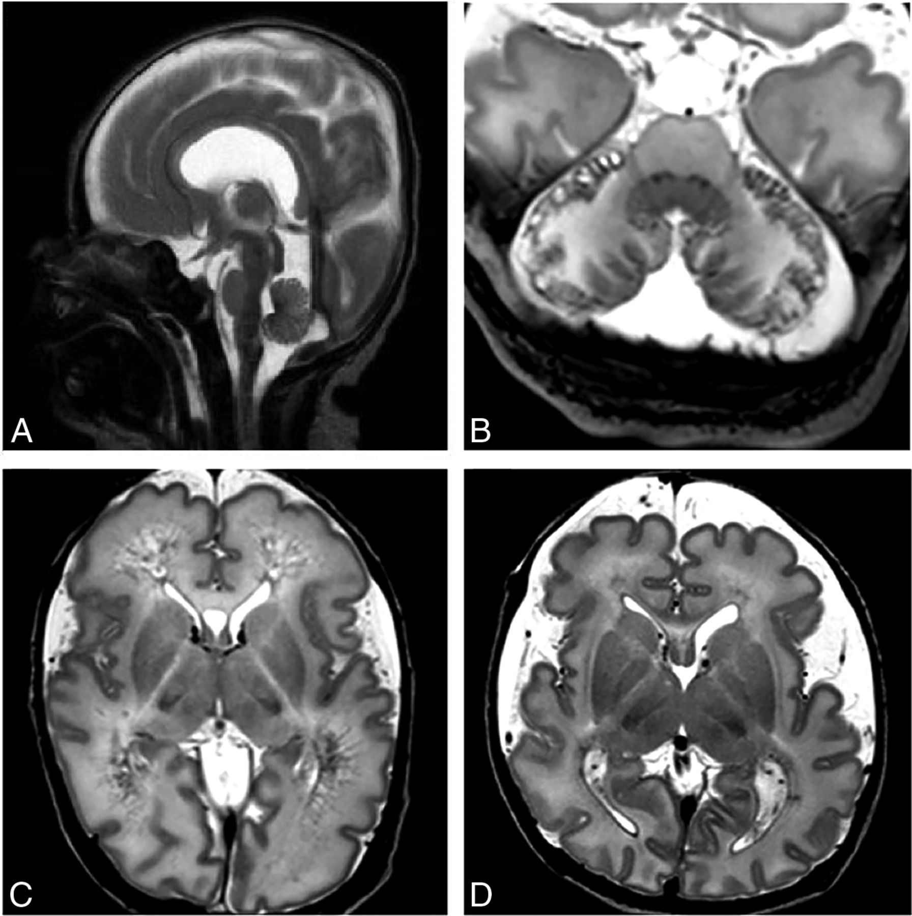
- Fig 3.
Boxplot graphs show the comparison of the vermis height (top left), cranial base angle (top right), tentorial angle (bottom left), and infratentorial angle (bottom right) between infants with a RASopathy and the control group.


Tables
Clinical characteristics and MRI angle measurements and assessments
Group 1 (n = 16) Group 2 (n = 32) P Valuea Gestational age (mean) (wk) 35.6 ± 4.2 37.3 ± 4.5 .1b Postmenstrual age at MRI (mean) (wk) 37.6 ± 4.8 39.8 ± 6.5 .2b Vermis height (mean) (mm) 20.5 ± 4.4 23.7 ± 3.9 .01be Transcerebellar diameter (mean) Axial 48.6 ± 9.1 52.3 ± 9.8 .2b Coronal 48.8 ± 9.6 52.7 ± 9.7 .2b Cranial base angle (mean) 130.3° ± 4.0° 134.8° ± 5.3° .005ce Tentorial angle (mean) 55.4° ± 3.4° 46.6° ± 5.8° <.001ce Infratentorial angle (mean) 52.2° ± 6.5° 47.7° ± 4.0° .001be Intraventricular hemorrhage (No.) (%) 8 (47) 0 (0) <.001de Cerebellar abnormality (No.) (%) 9 (56) 0 (0) <.001de Hemorrhagic lesion 6 (67) Hemorrhagic-cystic lesion 3 (33) Enlarged extracerebral space (No.) (%) 13 (81) 0 (0) <.001de Mild 10 (77) Severe 3 (23) Delayed cortical gyrification (No.) (%) 12 (75) 0 (0) <.001de WMI after cerebral hemorrhage (No.) (%) 9 (56) 0 (0) <.001de Punctate white matter lesion 7 (78) Cystic lesion 2 (22) Abnormality in DWI (n = 12) (No.) (%) 7 (58) - Focal restriction 6 (86) Extensive restriction 1 (14) Corpus callosum abnormality (No.) (%) 4 (25) 0 (0) <.001de Vertical appearance 3 (75) Hypoplasia 1 (25)


Jump to section
Related Articles
Cited By...
- No citing articles found.