
Article Figures & Data
Figures
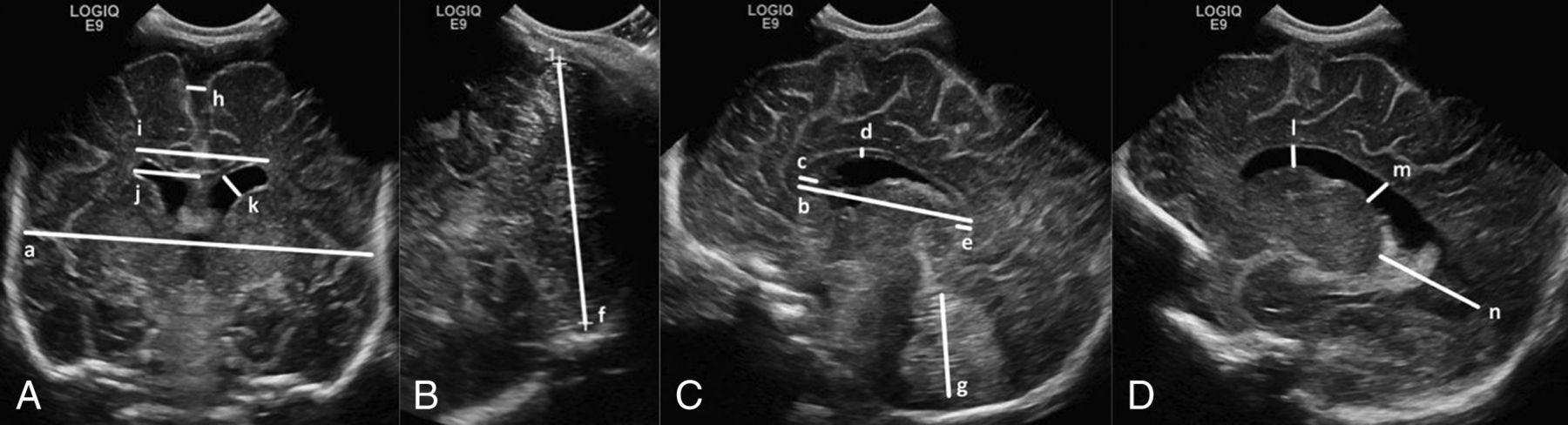
- Fig 1.
Cranial ultrasonography linear measures: images through the anterior fontanel in the coronal plane at the level of foramina of Monro (A) and the sagittal (C) and parasagittal (D) planes, and an image through the mastoid fontanel in the coronal plane posterior to the fourth ventricle (B). Brain tissue: biparietal diameter (a), corpus callosum length (b), corpus callosum genu width (c), corpus callosum body height (d), corpus callosum splenium width (e), transcerebellar diameter (f), and vermis height (g). Fluid spaces: interhemispheric distance (h), ventricular width (i), ventricular index (j), anterior horn width (k), anterior horn height (l), ventricular midbody width (m), and thalamo-occipital distance (n).
- Fig 2.
Cranial ultrasonography linear measures with respect to postnatal age. A, Brain tissue. B, Fluid spaces.


Tables
Perinatal Variable Summary (N = 144 Infants) Gestational age (mean) (SD) (wk) 27.7 (1.5) Birth weight (mean) (SD) (g) 1017 (259) Small for gestational age, birth weight <−2 SDs (No.) (%) 16 (11) Female (No.) (%) 75 (52) Multiple gestations (No.) (%) 64 (44) Cesarean delivery (No.) (%) 106 (74) Antenatal corticosteroids (No.) (%) 134 (93) Antenatal magnesium sulfate (No.) (%) 103 (72) Respiratory distress from birth (No.) (%) 141 (98) Surfactant (No.) (%) 91 (63) Duration of positive pressure ventilation (median) (25th–75th centile) (days) 19 (0–96) Duration of supplemental oxygen (median) (25th–75th centile) (days) 19 (4–56) Bronchopulmonary dysplasiaa (No.) (%) 46 (32) Postnatal corticosteroids (No.) (%) 18 (13) Intraventricular hemorrhage, grades I or II (No.) (%) 25 (17) Retinopathy of prematurity (No.) (%) 32 (22) Sepsis confirmedb (No.) (%) 20 (14) NEC (No.) (%) 17 (12) Sepsis confirmed and/or NEC (No.) (%) 31 (22) Survived to discharge home (No.) (%) 139 (97) Linear Measure Intraobserver Interobserver ICC 95% CI ICC 95% CI Brain tissue Biparietal diameter 0.98 0.95–0.99 0.97 0.94–0.99 Corpus callosum length 0.99 0.97–0.99 0.98 0.95–0.99 Corpus callosum genu width 0.78 0.53–0.90 0.38 −0.04–0.69 Corpus callosum body height 0.74 0.52–0.87 0.58 0.27–0.78 Corpus callosum splenium width 0.80 0.56–0.92 0.21 −0.22–0.58 Transcerebellar diameter 0.99 0.98–0.99 0.98 0.96–0.99 Vermis height 0.91 0.82–0.96 0.81 0.62–0.91 Fluid spaces Interhemispheric distance 0.98 0.96–0.99 0.98 0.95–0.99 Anterior horn width, left 0.94 0.87–0.97 0.91 0.81–0.96 Anterior horn width, right 0.96 0.90–0.98 0.95 0.89–0.98 Ventricular index, left 0.85 0.70–0.93 0.85 0.69–0.93 Ventricular index, right 0.63 0.32–0.82 0.59 0.26–0.80 Ventricular width 0.87 0.72–0.94 0.89 0.76–0.95 Anterior horn height, left 0.86 0.72–0.94 0.76 0.53–0.88 Anterior horn height, right 0.92 0.82–0.96 0.78 0.55–0.90 Ventricular midbody height, left 0.73 0.49–0.87 0.47 0.11–0.72 Ventricular midbody height, right 0.81 0.61–0.91 0.52 0.18–0.75 Thalamo-occipital distance, left 0.93 0.81–0.97 0.80 0.52–0.93 Thalamo-occipital distance, right 0.92 0.80–0.97 0.62 0.22–0.84 Note:—ICC indicates intraclass correlation coefficient.
Perinatal Variable Adjusteda Growth (mm/week PNA) Interaction Termb (P Value) β 95% CI Biparietal diameter GA <28 weeks 1.88 1.71–2.05 .002 ≥28 weeks 1.29 0.96–1.62 Corpus callosum length BW z score ≥−2 SD 0.91 0.84–0.98 .045 <−2 SD 0.73 0.57–0.89 Postnatal corticosteroids No 0.92 0.85–1.00 .046 Yes 0.77 0.63–0.90 Bronchopulmonary dysplasia No 0.98 0.88–1.07 .01 Yes 0.81 0.73–0.89 Transcerebellar diameter Antenatal corticosteroids No 2.26 1.81–2.70 .023 Yes 1.73 1.63–1.83 Postnatal corticosteroids No 1.84 1.74–1.95 <.001 Yes 1.41 1.22–1.61 Bronchopulmonary dysplasia No 1.89 1.75–2.02 .008 Yes 1.63 1.49–1.76 Sepsis/NEC No 1.82 1.71–1.93 .016 Yes 1.55 1.36–1.74 Vermis height Postnatal corticosteroids No 0.63 0.58–0.69 .011 Yes 0.49 0.39–0.59 Ventricular width GA <28 weeks 0.64 0.57–0.71 .004 ≥28 weeks 0.41 0.27–0.55


Jump to section
Related Articles
Cited By...
- Relationships between early postnatal cranial ultrasonography linear measures and neurodevelopment at 2 years in infants born at <30 weeks gestational age without major brain injury
- Development of an Ultrasound Scoring System to Describe Brain Maturation in Preterm Infants
- Effects of Postnatal Glucocorticoids on Brain Structure in Preterm Infants, A Scoping Review
- Early Ultrasonic Monitoring of Brain Growth and Later Neurodevelopmental Outcome in Very Preterm Infants