
Article Figures & Data
Figures
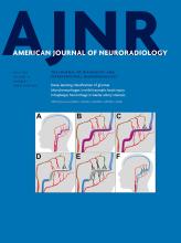
- Fig 1.
Sample orthogonal FLAIR images depicting an FCD lesion (arrows) of a 30-year-old woman. Inserts show the magnification of the lesion on FLAIR contrast and cortical parameter maps of fALFF, regional homogeneity, and wavelet entropy. Increased regional homogeneity, wavelet entropy, and decreased fALFF were observed in the FCD lesion and the immediate perilesional regions in comparison with the regions more distinct from the FCD. Only gray matter is visualized; white matter is masked out.
- Fig 2.
Fractional amplitude of low frequency fluctuations as a function of frequency in the range of 10–250 MHz. Only the lowest sub-band (ie, slow-5; 10–27 MHz, magnified) showed significantly lower amplitudes in the FCD lesion relative to the controls. Error bars represent the standard error of mean.


Tables
No. Age/Sex Seizure Frequencya FCD Location MRI Structural Image Findings 1 30/F High Right insula Transmantle sign, gray-white matter blurring, cortical thickening, T2-FLAIR hyperintensity 2 29/F High Right superior frontal Transmantle sign, gray-white matter blurring, cortical thickening, T2-FLAIR hyperintensity 3 33/M Low Right inferior frontal Transmantle sign, gray-white matter blurring, T2-FLAIR hyperintensity 4 47/F High Right precentral Transmantle sign, gray-white matter blurring, T2-FLAIR hyperintensity 5 21/M Low Left postcentral Gray-white matter blurring, cortical thickening, T2-FLAIR hyperintensity 6 47/M High Left posterior cingulate Transmantle sign, gray-white matter blurring, cortical thickening, T2-FLAIR hyperintensity 7 43/M High Left caudal middle frontal Transmantle sign, gray-white matter blurring, cortical thickening, T2-FLAIR hyperintensity 8 26/M Low Left rostral middle frontal Transmantle sign, gray-white matter blurring, cortical thickening, T2-FLAIR hyperintensity 9 21/F Low Caudal anterior cingulate Transmantle sign, gray-white matter blurring, cortical thickening, T2-FLAIR hyperintensity 10 21/M High Right supramarginal Transmantle sign, gray-white matter blurring, cortical thickening, T2-FLAIR hyperintensity 11 21/M Low Right insular Transmantle sign, gray-white matter blurring, cortical thickening, T2-FLAIR hyperintensity 12 27/M Low Left precentral Transmantle sign, gray-white matter blurring, cortical thickening, T2-FLAIR hyperintensity 13 54/M High Right rostral middle frontal Transmantle sign, gray-white matter blurring, cortical thickening, T2-FLAIR hyperintensity 14 25/M None Left inferior parietal Gray-white matter blurring, cortical thickening, T2-FLAIR hyperintensity ↵a Seizure frequency: low, 1 per month; high, >1 per week.
- Table 3:
The normalized BOLD measures in the FCD lesion, perilesional regions, contralateral homotopic region, and healthy controlsa
Normalized Measure Lesion Perilesional Zones Contralateral Homotopic Control Subjects 1 cm 2 cm 3 cm fALFF 0.922 ± 0.016b 0.941 ± 0.014b 0.947 ± 0.014b 0.981 ± 0.013 0.983 ± 0.012 1.005 ± 0.009 ReHo 1.206 ± 0.066b 0.992 ± 0.032 0.991 ± 0.036 1.021 ± 0.034 1.023 ± 0.032 1.020 ± 0.022 WE 1.018 ± 0.004b 1.013 ± 0.003b 1.009 ± 0.003b 1.003 ± 0.003 1.003 ± 0.002 1.002 ± 0.002 ↵a Data are mean ± standard error. Normalized fALFF was significantly decreased, while WE was significantly higher in FCD lesions and perilesional zones (up to 2 cm) than in the contralateral homotopic cortex and control subjects. ReHo was significantly higher in FCD lesions. BOLD measures were normalized with the entire contralateral cortex.
↵b Significant differences (P < .05) with the contralateral homotopic cortex and the same region in controls, respectively.
- Table 4:
Number of patients with measures outside the 95% confidence interval of the normative (healthy control) valuesa
Normalized Measure Lesion Perilesional Zone Contralateral Homotopic Cortex 1 cm 2 cm 3 cm fALFF 13 (93) 12 (86) 11 (79) 6 (43) 6 (43) ReHo 11 (79) 5 (36) 5 (36) 4 (29) 4 (29) WE 12 (86) 11 (79) 10 (71) 4 (29) 5 (36) ↵a Data are the number of patients (percentage).


Jump to section
Related Articles
Cited By...
- No citing articles found.