
Article Figures & Data
Figures
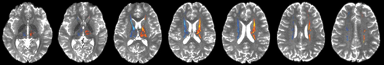
- Fig 1.
Thalamocortical pathways connecting the thalamus with the frontal cortex in a healthy brain. The left thalamocortical pathway is depicted in red/yellow, and the right thalamocortical pathway is depicted in dark/light blue. Axial sections are presented as the tracts progress dorsally from the thalamus to the frontal lobe (left to right).
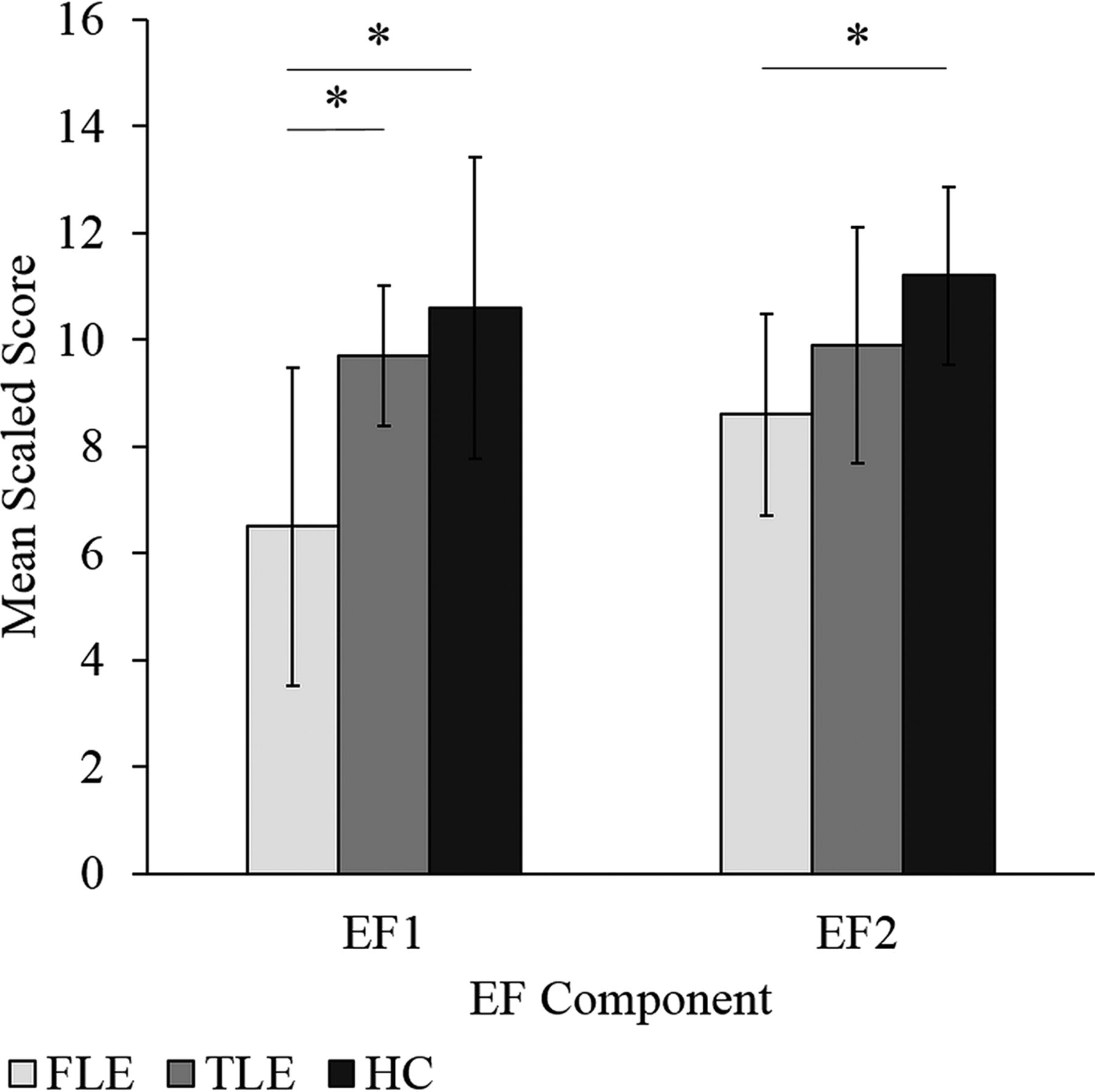
- Fig 2.
Mean composite EF factor scores for the FLE, TLE, and HC groups. Error bars represent SDs. The asterisk indicates a significant group difference (FLE < HC and FLE < TLE for EF1 and FLE < HC for EF2) at the P < .05 level. EF1 represents a mental flexibility/inhibition/switching component, while EF2 represents an attention/cognitive efficiency/problem-solving component.
- Fig 3.
Mean composite EF scores for the left and right FLE, left and right TLE, and HC groups. Error bars represent SDs. The asterisk indicates a significant group difference (EF1: left FLE < HC and right FLE < HC; EF2: left FLE < HC, right FLE < HC, left FLE < left TLE) at the P < .05 level. EF1 represents a mental flexibility/inhibition/switching component, while EF2 represents an attention/cognitive efficiency/problem-solving component.


Tables
FLE (n = 24) TLE (n = 17) HC (n = 25) Sex (male/female) 10:14 8:9 14:11 Age (yr) Mean (SD) 13.10 (3.21) 14.54 (2.65) 13.95 (3.20) Range 6.96–16.96 10.11–19.08 6.97–18.76 Handedness (right/left) 20/4 16/1 22/3 Age at seizure onset (yr) Mean (SD) 7.79 (4.01) 8.75 (3.76) – Range 1.00–14.40 1.30–15.00 – Duration of epilepsy (yr) Mean (SD) 5.20 (2.93) 4.90 (3.62) – Range 1.70–12.00 .33–12.00 – No. of AEDs Mean (SD) 2.00 (.659) 1.94 (.748) – Range 1–3 1–3 – Laterality of epileptogenic focus Left 13 10 – Right 9 4 – Bilateral 2 3 – Region of epileptogenic focus (TLE) Mesial – 3 – Lateral – 12 – Both mesial and lateral – 2 – Region of epileptogenic focus (FLE) Prefrontal 2 – – Inferior frontal 1 – – Frontal-central 9 – – Supplementary motor area 3 – – Unable to localize precisely 9 – – - Table 2:
Factor structure and factor loadings (>.45 and/or highest loading) after varimax rotation of 6 measures of EF in the FLE, TLE, and HC groups (n = 55)
EF Measure EF1 EF2 D-KEFS Verbal Fluency Test: Letter Fluency – .596 D-KEFS Verbal Fluency Test: Category Switching – .450 D-KEFS Color-Word Interference Test: Inhibition .850 – D-KEFS Color-Word Interference Test: Inhibition/Switching .912 – D-KEFS Sorting Test: Confirmed Correct Sorts – .824 CPT3 omission errors – .783 Note:—EF1 indicates executive function component factor 1; EF2, executive function component factor 2.


