Advertisement

Main menu

  • Home
  • Content
    • Current Issue
    • Accepted Manuscripts
    • Article Preview
    • Past Issue Archive
    • AJNR Case Collection
    • Case of the Week Archive
    • Classic Case Archive
    • Case of the Month Archive
  • Special Collections
    • Spinal CSF Leak Articles (Jan 2020-June 2024)
    • 2024 AJNR Journal Awards
    • Most Impactful AJNR Articles
  • Multimedia
    • AJNR Podcast
    • AJNR Scantastics
    • Video Articles
  • For Authors
    • Submit a Manuscript
    • Author Policies
    • Fast publishing of Accepted Manuscripts
    • Graphical Abstract Preparation
    • Manuscript Submission Guidelines
    • Imaging Protocol Submission
    • Submit a Case for the Case Collection
  • About Us
    • About AJNR
    • Editorial Board
  • More
    • Become a Reviewer/Academy of Reviewers
    • Subscribers
    • Permissions
    • Alerts
    • Feedback
    • Advertisers
    • ASNR Home
  • Other Publications
    • ajnr

User menu

  • Alerts
  • Log in

Search

  • Advanced search
American Journal of Neuroradiology

American Journal of Neuroradiology

ASHNR American Society of Functional Neuroradiology ASHNR American Society of Pediatric Neuroradiology ASSR
  • Alerts
  • Log in

Advanced Search

  • Home
  • Content
    • Current Issue
    • Accepted Manuscripts
    • Article Preview
    • Past Issue Archive
    • AJNR Case Collection
    • Case of the Week Archive
    • Classic Case Archive
    • Case of the Month Archive
  • Special Collections
    • Spinal CSF Leak Articles (Jan 2020-June 2024)
    • 2024 AJNR Journal Awards
    • Most Impactful AJNR Articles
  • Multimedia
    • AJNR Podcast
    • AJNR Scantastics
    • Video Articles
  • For Authors
    • Submit a Manuscript
    • Author Policies
    • Fast publishing of Accepted Manuscripts
    • Graphical Abstract Preparation
    • Manuscript Submission Guidelines
    • Imaging Protocol Submission
    • Submit a Case for the Case Collection
  • About Us
    • About AJNR
    • Editorial Board
  • More
    • Become a Reviewer/Academy of Reviewers
    • Subscribers
    • Permissions
    • Alerts
    • Feedback
    • Advertisers
    • ASNR Home
  • Follow AJNR on Twitter
  • Visit AJNR on Facebook
  • Follow AJNR on Instagram
  • Join AJNR on LinkedIn
  • RSS Feeds

Welcome to the new AJNR, Updated Hall of Fame, and more. Read the full announcements.


AJNR is seeking candidates for the position of Associate Section Editor, AJNR Case Collection. Read the full announcement.


Research ArticleSpine

Radiation-Induced Myelitis: Initial and Follow-Up MRI and Clinical Features in Patients at a Single Tertiary Care Institution during 20 Years

M. Khan, P. Ambady, D. Kimbrough, T. Shoemaker, S. Terezakis, J. Blakeley, S.D. Newsome and I. Izbudak
American Journal of Neuroradiology August 2018, 39 (8) 1576-1581; DOI: https://doi.org/10.3174/ajnr.A5671
M. Khan
dDivision of Neuroradiology, Russell H. Morgan Department of Radiology (M.K., I.I.), Johns Hopkins University School of Medicine, Baltimore, Maryland
  • Find this author on Google Scholar
  • Find this author on PubMed
  • Search for this author on this site
  • ORCID record for M. Khan
P. Ambady
aFrom the Brain Cancer Program (P.A., J.B., S.T.)
eNeuro-Oncology Branch (P.A.), National Cancer Institute, Bethesda, Maryland
fBlood Brain Barrier and Neuro-Oncology Program (P.A.), Oregon Health and Science University, Portland, Oregon.
  • Find this author on Google Scholar
  • Find this author on PubMed
  • Search for this author on this site
  • ORCID record for P. Ambady
D. Kimbrough
cDivision of Neuroimmunology and Neuroinfectious Diseases, Department of Neurology (D.K., T.S., S.D.N.)
  • Find this author on Google Scholar
  • Find this author on PubMed
  • Search for this author on this site
  • ORCID record for D. Kimbrough
T. Shoemaker
cDivision of Neuroimmunology and Neuroinfectious Diseases, Department of Neurology (D.K., T.S., S.D.N.)
  • Find this author on Google Scholar
  • Find this author on PubMed
  • Search for this author on this site
  • ORCID record for T. Shoemaker
S. Terezakis
aFrom the Brain Cancer Program (P.A., J.B., S.T.)
bDepartment of Radiation Oncology (S.T.), Sidney Kimmel Comprehensive Cancer Center at Johns Hopkins
  • Find this author on Google Scholar
  • Find this author on PubMed
  • Search for this author on this site
  • ORCID record for S. Terezakis
J. Blakeley
aFrom the Brain Cancer Program (P.A., J.B., S.T.)
  • Find this author on Google Scholar
  • Find this author on PubMed
  • Search for this author on this site
  • ORCID record for J. Blakeley
S.D. Newsome
cDivision of Neuroimmunology and Neuroinfectious Diseases, Department of Neurology (D.K., T.S., S.D.N.)
  • Find this author on Google Scholar
  • Find this author on PubMed
  • Search for this author on this site
  • ORCID record for S.D. Newsome
I. Izbudak
dDivision of Neuroradiology, Russell H. Morgan Department of Radiology (M.K., I.I.), Johns Hopkins University School of Medicine, Baltimore, Maryland
  • Find this author on Google Scholar
  • Find this author on PubMed
  • Search for this author on this site
  • ORCID record for I. Izbudak
  • Article
  • Figures & Data
  • Info & Metrics
  • Responses
  • References
  • PDF
Loading

Article Figures & Data

Figures

  • Tables
  • Fig 1.
    • Download figure
    • Open in new tab
    • Download powerpoint
    Fig 1.

    Study schema describes the method used to identify the patients in this series. It also highlights the rarity of the occurrence of radiation myelitis.
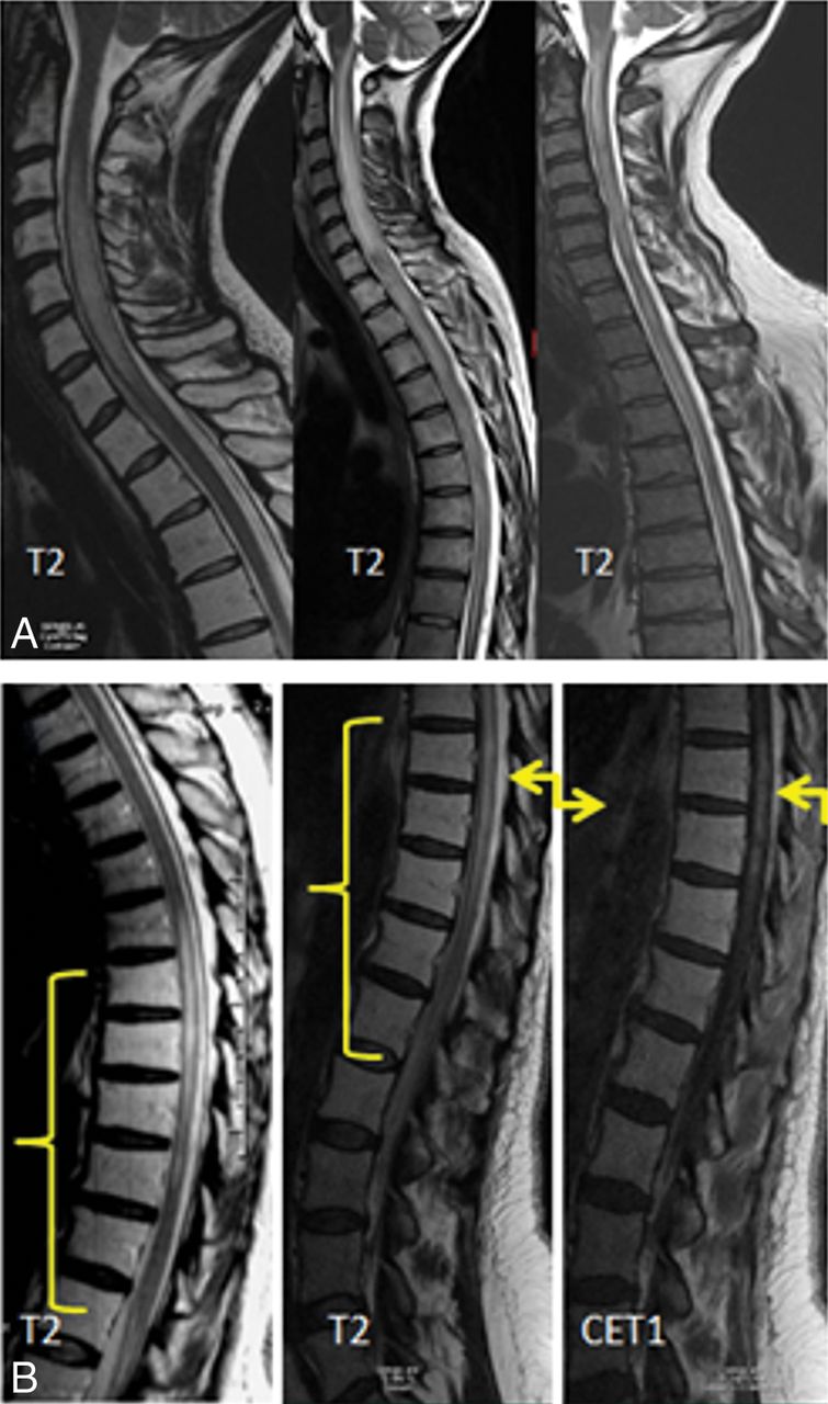
  • Fig 2.
    • Download figure
    • Open in new tab
    • Download powerpoint
    Fig 2.

    Longitudinally extensive myelitis. A, The cervical or thoracic cord or both were involved in all patients in a longitudinally extensive pattern. B, Maximum myelopathic changes (yellow arrows) correspond to the central field of radiation identified by apparent fatty vertebral bone marrow on T1-weighted images (yellow brackets).
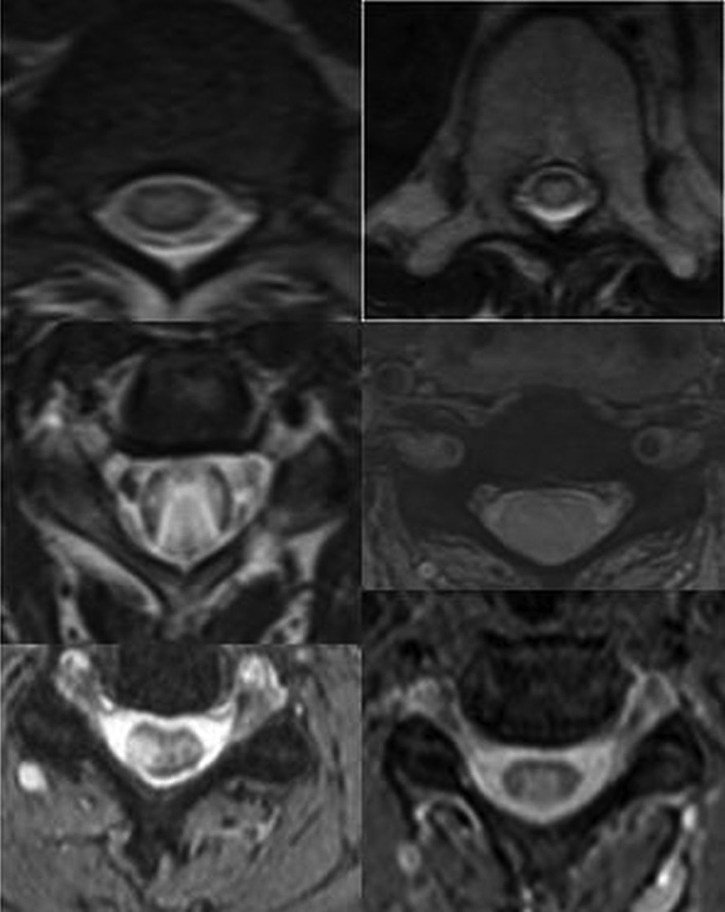
  • Fig 3.
    • Download figure
    • Open in new tab
    • Download powerpoint
    Fig 3.

    Central cord involvement. The central two-thirds of the cord on axial T2-weighted images was involved in 9 patients, and 1 patient presented with a small focal area of central and dorsal cord involvement.

  • Fig 4.
    • Download figure
    • Open in new tab
    • Download powerpoint
    Fig 4.

    Sequential changes on imaging in a patient with severe radiation myelitis. Fatty bone marrow changes present with hyperintense T1 signal involving C7 and the upper thoracic vertebrae on the first image (white arrows). Less extensive longitudinally extensive myelitis at the C7, T1, and T2 levels is noted on the T2-weighted image with hyperintense signal at presentation (yellow arrow). At 3 months, cord expansion and extensive T2/STIR hyperintense signal changes involving the cervical and upper thoracic cord are noted (white arrowhead) with contrast enhancement at the C7, T1, and T2 levels (single large white arrowhead). The patient was paraplegic with urinary and bowel incontinence. She was treated with a high dose of steroids. Follow-up images at 18 months show cord atrophy (blue arrow). She eventually was able to ambulate with a walker after extensive physical therapy. CE indicates contrast enhancement.

Tables

  • Figures
    • View popup
    Table 1:

    Clinical course of myelopathy

    Symptoms at presentationaParesthesia lower limbs: 6/10 (60%)
    Pain (back and legs): 5/10 (50%)
    Lower extremity motor weakness: 7/10 (70%)
    Urinary incontinence: 4/10 (40%)
    Bowel incontinence: 2/10 (20%)
    Treatment receivedSteroids: 9
    IvIg: 1
    Hyperbaric oxygen: 2
    Plasmapheresis: 2
    Bevacizumab (Avastin): 1
    Mean duration of clinical follow-up (mo)22 (range: 4–162)
    Clinical course on follow-up (available in 7 patients)Improved with some sequelae: 5
    Progressive: 2
    Resolved: 0
    • Note:—IvIg indicates intravenous immunoglobulin.

    • ↵a Detailed clinical examination was not available for 1 patient. Hence, only 10 patients were included for evaluation of the clinical course.

    • View popup
    Table 2:

    Demographics of 11 subjects included in the study

    AttributesMedian (Range)
    Sex6 males, 5 females
    Median age (yr)33 (11–60)
    Primary malignancy (No. of patients)1) Recurrent Hodgkin lymphoma: 5
    2) Hodgkin lymphoma: 1
    3) Gastric carcinoma: 1
    4) Acute lymphocytic lymphoma: 1
    5) Brain stem glioma: 1
    6) Medulloblastoma: 1
    7) Recurrent multiple myeloma: 1
    Median time to symptom onset from RT (mo)17 (2–194)
    Median delay in diagnosis from symptom onset (mo)6 (0–14)
    • Note:—RT indicates radiation therapy.

    • View popup
    Table 3:

    Patient demographics and oncology treatment history

    PatientAge (yr)Cancer TypePrior ChemotherapyPrior Chemotherapy RegimenAdjuvant Chemotherapy
    133Recurrent Hodgkin lymphomaYesABVDICE followed by
    cyclophosphamide (Cytoxan) and busulfan
    Autologous bone marrow transplant
    232Recurrent Hodgkin lymphomaYesCHOP/ABVDAra-C, vincristine, cisplatin
    Danazol
    311Brain stem medulloblastomaNo–Vincristine, VP-16, carboplatin
    CCNU, cisplatin
    460Recurrent gastric adenocarcinoma (stage IV)No–5-FU, leucovorin
    542Refractory acute lymphocytic leukemiaYesHyper-CVADIntrathecal cytarabine
    MethotrexateAutologous bone marrow transplant
    6-Mercaptopurine
    641Hodgkin lymphomaNo–No
    729Brain stem gliomaNo–TMZ
    828Recurrent Hodgkin lymphomaYesABVDICE followed by
    cyclophosphamide and busulfan
    Autologous bone marrow transplant
    933Recurrent Hodgkin lymphomaYesABVDICE followed by
    cyclophosphamide and busulfan
    Brentuximab vedotin
    Autologous bone marrow transplant
    1037Recurrent Hodgkin lymphomaYesABVDICE followed by
    cyclophosphamide and busulfan
    Autologous bone marrow transplant
    • Note:—ABVD indicates doxorubicin, bleomycin, vinblastine, dacarbazine; CHOP, cyclophosphamide, doxorubicin hydrochloride, vincristine, prednisone; Hyper-CVAD, cyclophosphamide, vincristine, doxorubicin, and dexamethasone; TMZ, temozolomide; CCNU, lomustine; ICE, ifosfamide, carboplatin, etoposide chemotherapy protocol; Vp-16, etoposide phosphate.

    • View popup
    Table 4:

    Spine MRI findings at initial presentation of myelopathy

    Mean time from symptom onset to first MRI (mo)6 (Range, 0–41)
    Level of longitudinally extensive myelitis (No. of patients) (%)Cervical: 3 (27%)
    Thoracic: 5 (45%)
    Cervical and thoracic: 2 (18%)
    Entire spine: 1 (9%)
    Level of vertebral T1 hyperintensities (No. of patients) (%)Cervical: 2 (18%)
    Cervical and thoracic: 3 (27%)
    Thoracic: 1 (9%)
    Thoracic and lumbar: 1 (9%)
    Entire spine: 2 (18%)
    No changes: 2 (18%)
    Location of axial T2 changes (No. of patients) (%)Central (>2/3): 4 (36%)
    Central and posterior: 1 (9%)
    Entire cord: 5 (45%)
    Indeterminate: 1 (9%)
    Cord expansion (No. of patients) (%)Present: 6 (54%)
    Absent: 3 (27%)
    Indeterminate: 2 (18%)
    Contrast enhancement (No. of patients) (%)Enhancement present: 5 (45%)
    No enhancement: 5 (45%)
    Contrast not given: 1 (9%)
    • View popup
    Table 5:

    Findings on follow-up spine MRI

    Radiographic follow-up availableYes: 8
    No: 3
    Median follow-up (mo)14.5 (4–69)
    Cord atrophy (No. of patients) (%)Present: 2 (25%)
    Absent: 6 (75%)
    T2 findings (No. of patients) (%)Decreased T2 abnormalities: 4 (50%)
    Resolved: 1 (12.5%)
    Hemorrhagic changes: 1 (12.5%)
    Syringohydromyelia: 1 (12.5%)
    Persistent T2 abnormalities: 4 (50%)
PreviousNext
Back to top

In this issue

American Journal of Neuroradiology: 39 (8)
American Journal of Neuroradiology
Vol. 39, Issue 8
1 Aug 2018
  • Table of Contents
  • Index by author
  • Complete Issue (PDF)
Advertisement
Print
Download PDF
Email Article

Thank you for your interest in spreading the word on American Journal of Neuroradiology.

NOTE: We only request your email address so that the person you are recommending the page to knows that you wanted them to see it, and that it is not junk mail. We do not capture any email address.

Enter multiple addresses on separate lines or separate them with commas.
Radiation-Induced Myelitis: Initial and Follow-Up MRI and Clinical Features in Patients at a Single Tertiary Care Institution during 20 Years
(Your Name) has sent you a message from American Journal of Neuroradiology
(Your Name) thought you would like to see the American Journal of Neuroradiology web site.
CAPTCHA
This question is for testing whether or not you are a human visitor and to prevent automated spam submissions.
Cite this article
M. Khan, P. Ambady, D. Kimbrough, T. Shoemaker, S. Terezakis, J. Blakeley, S.D. Newsome, I. Izbudak
Radiation-Induced Myelitis: Initial and Follow-Up MRI and Clinical Features in Patients at a Single Tertiary Care Institution during 20 Years
American Journal of Neuroradiology Aug 2018, 39 (8) 1576-1581; DOI: 10.3174/ajnr.A5671

Citation Manager Formats

  • BibTeX
  • Bookends
  • EasyBib
  • EndNote (tagged)
  • EndNote 8 (xml)
  • Medlars
  • Mendeley
  • Papers
  • RefWorks Tagged
  • Ref Manager
  • RIS
  • Zotero
0 Responses
Respond to this article
Share
Bookmark this article
Radiation-Induced Myelitis: Initial and Follow-Up MRI and Clinical Features in Patients at a Single Tertiary Care Institution during 20 Years
M. Khan, P. Ambady, D. Kimbrough, T. Shoemaker, S. Terezakis, J. Blakeley, S.D. Newsome, I. Izbudak
American Journal of Neuroradiology Aug 2018, 39 (8) 1576-1581; DOI: 10.3174/ajnr.A5671
del.icio.us logo Twitter logo Facebook logo Mendeley logo
  • Tweet Widget
  • Facebook Like
  • Google Plus One
Purchase

Jump to section

  • Article
    • Abstract
    • ABBREVIATION:
    • Discussion
    • Footnotes
    • References
  • Figures & Data
  • Info & Metrics
  • Responses
  • References
  • PDF

Related Articles

  • No related articles found.
  • PubMed
  • Google Scholar

Cited By...

  • Successful treatment of quadriparesis from radiation myelopathy with bevacizumab in a patient with metastatic breast cancer
  • A brief case series of radiation associated myelopathy
  • Crossref (11)
  • Google Scholar

This article has been cited by the following articles in journals that are participating in Crossref Cited-by Linking.

  • Neurological side effects of radiation therapy
    J. Jacob, L. Feuvret, J.-M. Simon, M. Ribeiro, L. Nichelli, C. Jenny, D. Ricard, D. Psimaras, K. Hoang-Xuan, P. Maingon
    Neurological Sciences 2022 43 4
  • Efficacy and toxicity of primary re-irradiation for malignant spinal cord compression based on radiobiological modelling: a phase II clinical trial
    Neil D. Wallace, Mary T. Dunne, Orla McArdle, Cormac Small, Imelda Parker, Aoife M. Shannon, Angela Clayton-Lea, Michael Parker, Conor D. Collins, John G. Armstrong, Charles Gillham, Jerome Coffey, David Fitzpatrick, Osama Salib, Michael Moriarty, Michael R. Stevenson, Alberto Alvarez-Iglesias, Michael McCague, Pierre G. Thirion
    British Journal of Cancer 2023 128 4
  • Manifestations of radiation toxicity in the head, neck, and spine: An image-based review
    Carrie M Carr, John C Benson, David R DeLone, Felix E Diehn, Dong K Kim, Daniel Ma, Alex A Nagelschneider, Ajay A Madhavan, Derek R Johnson
    The Neuroradiology Journal 2022 35 4
  • Successful treatment of quadriparesis from radiation myelopathy with bevacizumab in a patient with metastatic breast cancer
    Nirja Shah, Surabhi Ranjan
    BMJ Case Reports 2022 15 2
  • Management of Metastatic Spinal Disease – A Practical Approach
    Graham Ka Hon Shea, Kenny Yat Hong Kwan
    Global Spine Journal 2024
  • Oncologic Critical Care
    Shalini Moningi, Geoffrey V. Martin, Jay P. Reddy
    2019
  • Oncologic Critical Care
    Shalini Moningi, Geoffrey V. Martin, Jay P. Reddy
    2020
  • Potential Complications of Percutaneous Vertebroplasty Combined with Interstitial Implantation of 125I Seeds for Metastatic Spinal Tumors: A Case Report and Literary Review
    Lunli Xie, Zhenlin Yan, Jun Zhu, Xudong Chen, Changyuan Yang, Xing Su, Keqin Quan, Dan Pu
    Case Reports in Orthopedics 2019 2019
  • Radiopaedia.org
    Mostafa Elfeky, Arlene Campos, Daniel Bell
    2019
  • Radiopaedia.org
    Mostafa Elfeky
    2019

More in this TOC Section

  • Bern Score Validity for SIH
  • MP2RAGE 7T in MS Lesions of the Cervical Spine
  • Deep Learning for STIR Spine MRI Quality
Show more Spine

Similar Articles

Advertisement

Indexed Content

  • Current Issue
  • Accepted Manuscripts
  • Article Preview
  • Past Issues
  • Editorials
  • Editors Choice
  • Fellow Journal Club
  • Letters to the Editor

Cases

  • Case Collection
  • Archive - Case of the Week
  • Archive - Case of the Month
  • Archive - Classic Case

Special Collections

  • Special Collections

Resources

  • News and Updates
  • Turn around Times
  • Submit a Manuscript
  • Author Policies
  • Manuscript Submission Guidelines
  • Evidence-Based Medicine Level Guide
  • Publishing Checklists
  • Graphical Abstract Preparation
  • Imaging Protocol Submission
  • Submit a Case
  • Become a Reviewer/Academy of Reviewers
  • Get Peer Review Credit from Publons

Multimedia

  • AJNR Podcast
  • AJNR SCANtastic
  • Video Articles

About Us

  • About AJNR
  • Editorial Board
  • Not an AJNR Subscriber? Join Now
  • Alerts
  • Feedback
  • Advertise with us
  • Librarian Resources
  • Permissions
  • Terms and Conditions

American Society of Neuroradiology

  • Not an ASNR Member? Join Now

© 2025 by the American Society of Neuroradiology All rights, including for text and data mining, AI training, and similar technologies, are reserved.
Print ISSN: 0195-6108 Online ISSN: 1936-959X

Powered by HighWire