Advertisement

Main menu

  • Home
  • Content
    • Current Issue
    • Accepted Manuscripts
    • Article Preview
    • Past Issue Archive
    • Video Articles
    • AJNR Case Collection
    • Case of the Week Archive
    • Case of the Month Archive
    • Classic Case Archive
  • Special Collections
    • AJNR Awards
    • Low-Field MRI
    • Alzheimer Disease
    • ASNR Foundation Special Collection
    • Photon-Counting CT
    • View All
  • Multimedia
    • AJNR Podcasts
    • AJNR SCANtastic
    • Trainee Corner
    • MRI Safety Corner
    • Imaging Protocols
  • For Authors
    • Submit a Manuscript
    • Submit a Video Article
    • Submit an eLetter to the Editor/Response
    • Manuscript Submission Guidelines
    • Statistical Tips
    • Fast Publishing of Accepted Manuscripts
    • Graphical Abstract Preparation
    • Imaging Protocol Submission
    • Author Policies
  • About Us
    • About AJNR
    • Editorial Board
    • Editorial Board Alumni
  • More
    • Become a Reviewer/Academy of Reviewers
    • Subscribers
    • Permissions
    • Alerts
    • Feedback
    • Advertisers
    • ASNR Home

User menu

  • Alerts
  • Log in

Search

  • Advanced search
American Journal of Neuroradiology

American Journal of Neuroradiology

ASHNR American Society of Functional Neuroradiology ASHNR American Society of Pediatric Neuroradiology ASSR
  • Alerts
  • Log in

Advanced Search

  • Home
  • Content
    • Current Issue
    • Accepted Manuscripts
    • Article Preview
    • Past Issue Archive
    • Video Articles
    • AJNR Case Collection
    • Case of the Week Archive
    • Case of the Month Archive
    • Classic Case Archive
  • Special Collections
    • AJNR Awards
    • Low-Field MRI
    • Alzheimer Disease
    • ASNR Foundation Special Collection
    • Photon-Counting CT
    • View All
  • Multimedia
    • AJNR Podcasts
    • AJNR SCANtastic
    • Trainee Corner
    • MRI Safety Corner
    • Imaging Protocols
  • For Authors
    • Submit a Manuscript
    • Submit a Video Article
    • Submit an eLetter to the Editor/Response
    • Manuscript Submission Guidelines
    • Statistical Tips
    • Fast Publishing of Accepted Manuscripts
    • Graphical Abstract Preparation
    • Imaging Protocol Submission
    • Author Policies
  • About Us
    • About AJNR
    • Editorial Board
    • Editorial Board Alumni
  • More
    • Become a Reviewer/Academy of Reviewers
    • Subscribers
    • Permissions
    • Alerts
    • Feedback
    • Advertisers
    • ASNR Home
  • Follow AJNR on Twitter
  • Visit AJNR on Facebook
  • Follow AJNR on Instagram
  • Join AJNR on LinkedIn
  • RSS Feeds

AJNR Awards, New Junior Editors, and more. Read the latest AJNR updates

Getting new auth cookie, if you see this message a lot, tell someone!
Research ArticleNeurointervention

Toward a Better Understanding of Dural Arteriovenous Fistula Angioarchitecture: Superselective Transvenous Embolization of a Sigmoid Common Arterial Collector

M. Shapiro, E. Raz, M. Litao, T. Becske, H. Riina and P.K. Nelson
American Journal of Neuroradiology September 2018, 39 (9) 1682-1688; DOI: https://doi.org/10.3174/ajnr.A5740
M. Shapiro
aFrom the Departments of Radiology (M.S., E.R., H.R., P.K.N.)
bNeurology (M.S., M.L.)
  • Find this author on Google Scholar
  • Find this author on PubMed
  • Search for this author on this site
  • ORCID record for M. Shapiro
E. Raz
aFrom the Departments of Radiology (M.S., E.R., H.R., P.K.N.)
  • Find this author on Google Scholar
  • Find this author on PubMed
  • Search for this author on this site
  • ORCID record for E. Raz
M. Litao
bNeurology (M.S., M.L.)
  • Find this author on Google Scholar
  • Find this author on PubMed
  • Search for this author on this site
  • ORCID record for M. Litao
T. Becske
dDepartment of Neurology (T.B.), Rochester Regional Health, Rochester, New York.
  • Find this author on Google Scholar
  • Find this author on PubMed
  • Search for this author on this site
  • ORCID record for T. Becske
H. Riina
aFrom the Departments of Radiology (M.S., E.R., H.R., P.K.N.)
cNeurosurgery (H.R., P.K.N.), NYU School of Medicine, New York, New York
  • Find this author on Google Scholar
  • Find this author on PubMed
  • Search for this author on this site
  • ORCID record for H. Riina
P.K. Nelson
aFrom the Departments of Radiology (M.S., E.R., H.R., P.K.N.)
cNeurosurgery (H.R., P.K.N.), NYU School of Medicine, New York, New York
  • Find this author on Google Scholar
  • Find this author on PubMed
  • Search for this author on this site
  • ORCID record for P.K. Nelson
  • Article
  • Figures & Data
  • Info & Metrics
  • Responses
  • References
  • PDF
Loading

Article Figures & Data

Figures

  • Tables
  • Fig 1.
    • Download figure
    • Open in new tab
    • Download powerpoint
    Fig 1.

    Schematic of normal dural arteriovenous architecture (A), with arrows pointing to the arterial arcade in the wall of the transverse/sigmoid sinuses. Schematic of dural fistula angioarchitecture according to a view of multiple arteriovenous shunts (black arrows) draining either separately via multiple discrete veins into the sinus (B) or into a common venous collector/pouch/septation (C) alongside the wall of the sinus proper. D, A low-grade dural fistula according to the common arterial collector view. Multiple arterial feeders converge on a common arterial channel in the sinus wall that empties into the sinus at a single fistulous point (arrowhead). E, Common arterial collector schematic relative to normal anatomy shown in A. The arterial supply converges on the common collector arterial channel within the sinus wall (arrow), with the fistulous point (arrowhead) at the confluence of this channel and the sinus proper.

  • Fig 2.
    • Download figure
    • Open in new tab
    • Download powerpoint
    Fig 2.

    Schematic of typical transvenous sinus sacrifice (A) and incomplete (B) and complete (C) transarterial embolizations, each of which results in an unnecessary closure of arteries or sinus. Targeted occlusion of the shunt (D) at the entrance of the collector channel into the sinus preserves both the sinus and normal arterial structures.
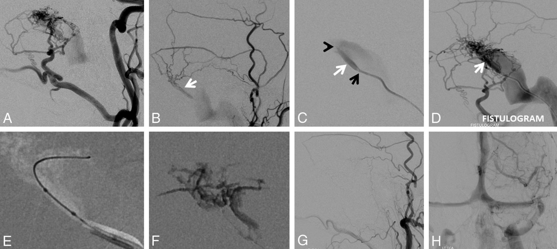
  • Fig 3.
    • Download figure
    • Open in new tab
    • Download powerpoint
    Fig 3.

    A, Left sigmoid sinus type I dural fistula. Posterior meningeal supply (B) proceeds via arteries in the wall of the transverse/sigmoid sinuses (arrows). Subselective external carotid artery injection (C) shows robust occipital and middle meningeal artery supply. D, Inflation of a compliant balloon within the sigmoid sinus reduces transfistulous flow to allow identification of a common collector channel (white arrow) inferior to the sinus proper. E and F, Transvenous microcatheterization of a common collector channel (E, distal catheter tip) and subsequent fistulogram (F) reduce the fistula to a single point where the collector joins the sinus (white arrow). Subselective coiling of this collector (G) results in fistula closure (H and I).

  • Fig 4.
    • Download figure
    • Open in new tab
    • Download powerpoint
    Fig 4.

    A and B, Left type I sigmoid sinus fistula with predominant occipital and middle meningeal supply. C, A 5F VERT (Cook, Bloomington, Indiana) catheter (tip marked by a black arrow) in the sigmoid sinus is wedged into the opening of the common collector (white arrow). Gentle injection of the VERT catheter identifies a second opening of the collector into the sinus shown by the black arrowhead. D, A stronger injection of the VERT (fistulogram) opacifies the entire arterial arcade in a retrograde fashion. E, Scepter C (MicroVention Tustin, California) subselective catheterization of the common collector, with a subsequent n-BCA cast in F. G and H, Postembolization angiography demonstrates occlusion of the fistula with preservation of the left sigmoid sinus.

  • Fig 5.
    • Download figure
    • Open in new tab
    • Download powerpoint
    Fig 5.

    Additional examples of a common collector channel. Case 1 (A–C) of a type IIa fistula shows the collector channel inferior to the sinus (C, white arrow) during Onyx (Covidien, Irvine, California) injection via a Scepter C microcatheter (MicroVention). Case 2 (D–F) illustrates a relatively uncommon presence of a common collector superior to the sinus (white arrows), becoming more obvious after the sinus has been packed with coils. Case 3 (G–I) is another complex-appearing fistula, where the common collector channel (white arrows) is better seen via the left vertebral contributors (H) due to stenosis of the sinus proper. The collector channel is subsequently filled with coils (I, white arrows). In retrospect, cases 2 and 3 might have been cured by superselective embolization rather than sinus sacrifice.

Tables

  • Figures
  • Breakdown by Cognard type, hemorrhage status on presentation, and presence of a common collector channel

    Cognard TypeNo.Hemorrhagic Presentation (No.)Common Channel (No.) (%)
    115014 (93%)
    IIa1205 (42%)
    IIb00NA
    IIab301 (33%)
    III642 (33%)
    • Note:—NA indicates not applicable.

PreviousNext
Back to top

In this issue

American Journal of Neuroradiology: 39 (9)
American Journal of Neuroradiology
Vol. 39, Issue 9
1 Sep 2018
  • Table of Contents
  • Index by author
  • Complete Issue (PDF)
Advertisement
Print
Download PDF
Email Article

Thank you for your interest in spreading the word on American Journal of Neuroradiology.

NOTE: We only request your email address so that the person you are recommending the page to knows that you wanted them to see it, and that it is not junk mail. We do not capture any email address.

Enter multiple addresses on separate lines or separate them with commas.
Toward a Better Understanding of Dural Arteriovenous Fistula Angioarchitecture: Superselective Transvenous Embolization of a Sigmoid Common Arterial Collector
(Your Name) has sent you a message from American Journal of Neuroradiology
(Your Name) thought you would like to see the American Journal of Neuroradiology web site.
CAPTCHA
This question is for testing whether or not you are a human visitor and to prevent automated spam submissions.
Cite this article
M. Shapiro, E. Raz, M. Litao, T. Becske, H. Riina, P.K. Nelson
Toward a Better Understanding of Dural Arteriovenous Fistula Angioarchitecture: Superselective Transvenous Embolization of a Sigmoid Common Arterial Collector
American Journal of Neuroradiology Sep 2018, 39 (9) 1682-1688; DOI: 10.3174/ajnr.A5740

Citation Manager Formats

  • BibTeX
  • Bookends
  • EasyBib
  • EndNote (tagged)
  • EndNote 8 (xml)
  • Medlars
  • Mendeley
  • Papers
  • RefWorks Tagged
  • Ref Manager
  • RIS
  • Zotero
0 Responses
Respond to this article
Share
Bookmark this article
Toward a Better Understanding of Dural Arteriovenous Fistula Angioarchitecture: Superselective Transvenous Embolization of a Sigmoid Common Arterial Collector
M. Shapiro, E. Raz, M. Litao, T. Becske, H. Riina, P.K. Nelson
American Journal of Neuroradiology Sep 2018, 39 (9) 1682-1688; DOI: 10.3174/ajnr.A5740
del.icio.us logo Twitter logo Facebook logo Mendeley logo
  • Tweet Widget
  • Facebook Like
  • Google Plus One
Purchase

Jump to section

  • Article
    • Abstract
    • ABBREVIATION:
    • Materials and Methods
    • Results
    • Discussion
    • Conclusions
    • Footnotes
    • References
  • Figures & Data
  • Info & Metrics
  • Responses
  • References
  • PDF

Related Articles

  • No related articles found.
  • PubMed
  • Google Scholar

Cited By...

  • Principles, techniques and applications of high resolution cone beam CT angiography in the neuroangio suite
  • Cerebral venous anatomy: implications for the neurointerventionalist
  • Transarterial embolization of dural arteriovenous fistulas of the lateral sinuses with stent-assisted sinus protection
  • Cerebral venous anatomy: implications for the neurointerventionalist
  • Principles, techniques and applications of high resolution cone beam CT angiography in the neuroangio suite
  • Endovascular Management of Intracranial Dural AVFs: Transvenous Approach
  • Endovascular Management of Intracranial Dural Arteriovenous Fistulas: Transarterial Approach
  • Dural venous system: angiographic technique and correlation with ex vivo investigations
  • Crossref
  • Google Scholar

This article has not yet been cited by articles in journals that are participating in Crossref Cited-by Linking.

More in this TOC Section

  • A Retrospective Study in Tentorial DAVFs
  • Proximal Protection Devices for Carotid Stenting
  • Guided vs Conventional Angiography Systems
Show more NEUROINTERVENTION

Similar Articles

Advertisement

Indexed Content

  • Current Issue
  • Accepted Manuscripts
  • Article Preview
  • Past Issues
  • Editorials
  • Editor's Choice
  • Fellows' Journal Club
  • Letters to the Editor
  • Video Articles

Cases

  • Case Collection
  • Archive - Case of the Week
  • Archive - Case of the Month
  • Archive - Classic Case

More from AJNR

  • Trainee Corner
  • Imaging Protocols
  • MRI Safety Corner
  • Book Reviews

Multimedia

  • AJNR Podcasts
  • AJNR Scantastics

Resources

  • Turnaround Time
  • Submit a Manuscript
  • Submit a Video Article
  • Submit an eLetter to the Editor/Response
  • Manuscript Submission Guidelines
  • Statistical Tips
  • Fast Publishing of Accepted Manuscripts
  • Graphical Abstract Preparation
  • Imaging Protocol Submission
  • Evidence-Based Medicine Level Guide
  • Publishing Checklists
  • Author Policies
  • Become a Reviewer/Academy of Reviewers
  • News and Updates

About Us

  • About AJNR
  • Editorial Board
  • Editorial Board Alumni
  • Alerts
  • Permissions
  • Not an AJNR Subscriber? Join Now
  • Advertise with Us
  • Librarian Resources
  • Feedback
  • Terms and Conditions
  • AJNR Editorial Board Alumni

American Society of Neuroradiology

  • Not an ASNR Member? Join Now

© 2025 by the American Society of Neuroradiology All rights, including for text and data mining, AI training, and similar technologies, are reserved.
Print ISSN: 0195-6108 Online ISSN: 1936-959X

Powered by HighWire