Advertisement

Main menu

  • Home
  • Content
    • Current Issue
    • Accepted Manuscripts
    • Article Preview
    • Past Issue Archive
    • AJNR Case Collection
    • Case of the Week Archive
    • Classic Case Archive
    • Case of the Month Archive
  • Special Collections
    • Spinal CSF Leak Articles (Jan 2020-June 2024)
    • 2024 AJNR Journal Awards
    • Most Impactful AJNR Articles
  • Multimedia
    • AJNR Podcast
    • AJNR Scantastics
    • Video Articles
  • For Authors
    • Submit a Manuscript
    • Author Policies
    • Fast publishing of Accepted Manuscripts
    • Graphical Abstract Preparation
    • Manuscript Submission Guidelines
    • Imaging Protocol Submission
    • Submit a Case for the Case Collection
  • About Us
    • About AJNR
    • Editorial Board
  • More
    • Become a Reviewer/Academy of Reviewers
    • Subscribers
    • Permissions
    • Alerts
    • Feedback
    • Advertisers
    • ASNR Home
  • Other Publications
    • ajnr

User menu

  • Alerts
  • Log in

Search

  • Advanced search
American Journal of Neuroradiology

American Journal of Neuroradiology

ASHNR American Society of Functional Neuroradiology ASHNR American Society of Pediatric Neuroradiology ASSR
  • Alerts
  • Log in

Advanced Search

  • Home
  • Content
    • Current Issue
    • Accepted Manuscripts
    • Article Preview
    • Past Issue Archive
    • AJNR Case Collection
    • Case of the Week Archive
    • Classic Case Archive
    • Case of the Month Archive
  • Special Collections
    • Spinal CSF Leak Articles (Jan 2020-June 2024)
    • 2024 AJNR Journal Awards
    • Most Impactful AJNR Articles
  • Multimedia
    • AJNR Podcast
    • AJNR Scantastics
    • Video Articles
  • For Authors
    • Submit a Manuscript
    • Author Policies
    • Fast publishing of Accepted Manuscripts
    • Graphical Abstract Preparation
    • Manuscript Submission Guidelines
    • Imaging Protocol Submission
    • Submit a Case for the Case Collection
  • About Us
    • About AJNR
    • Editorial Board
  • More
    • Become a Reviewer/Academy of Reviewers
    • Subscribers
    • Permissions
    • Alerts
    • Feedback
    • Advertisers
    • ASNR Home
  • Follow AJNR on Twitter
  • Visit AJNR on Facebook
  • Follow AJNR on Instagram
  • Join AJNR on LinkedIn
  • RSS Feeds

Welcome to the new AJNR, Updated Hall of Fame, and more. Read the full announcements.


AJNR is seeking candidates for the position of Associate Section Editor, AJNR Case Collection. Read the full announcement.


Research ArticleAdult Brain
Open Access

Automated ASPECTS on Noncontrast CT Scans in Patients with Acute Ischemic Stroke Using Machine Learning

H. Kuang, M. Najm, D. Chakraborty, N. Maraj, S.I. Sohn, M. Goyal, M.D. Hill, A.M. Demchuk, B.K. Menon and W. Qiu
American Journal of Neuroradiology January 2019, 40 (1) 33-38; DOI: https://doi.org/10.3174/ajnr.A5889
H. Kuang
aFrom the Calgary Stroke Program (H.K., W.Q., M.N., D.C., N.M., M.G., M.D.H., A.M.D., B.K.M.)
  • Find this author on Google Scholar
  • Find this author on PubMed
  • Search for this author on this site
  • ORCID record for H. Kuang
M. Najm
aFrom the Calgary Stroke Program (H.K., W.Q., M.N., D.C., N.M., M.G., M.D.H., A.M.D., B.K.M.)
  • Find this author on Google Scholar
  • Find this author on PubMed
  • Search for this author on this site
  • ORCID record for M. Najm
D. Chakraborty
aFrom the Calgary Stroke Program (H.K., W.Q., M.N., D.C., N.M., M.G., M.D.H., A.M.D., B.K.M.)
  • Find this author on Google Scholar
  • Find this author on PubMed
  • Search for this author on this site
  • ORCID record for D. Chakraborty
N. Maraj
aFrom the Calgary Stroke Program (H.K., W.Q., M.N., D.C., N.M., M.G., M.D.H., A.M.D., B.K.M.)
  • Find this author on Google Scholar
  • Find this author on PubMed
  • Search for this author on this site
  • ORCID record for N. Maraj
S.I. Sohn
eDepartment of Neurology (S.I.S.), Keimyung University, Daegu, South Korea.
  • Find this author on Google Scholar
  • Find this author on PubMed
  • Search for this author on this site
  • ORCID record for S.I. Sohn
M. Goyal
aFrom the Calgary Stroke Program (H.K., W.Q., M.N., D.C., N.M., M.G., M.D.H., A.M.D., B.K.M.)
bDepartment of Clinical Neurosciences, Department of Radiology (M.D.H., A.M.D., M.G., B.K.M.)
dHotchkiss Brain Institute, Calgary, Alberta, Canada (M.D.H., A.M.D., M.G., B.K.M.)
  • Find this author on Google Scholar
  • Find this author on PubMed
  • Search for this author on this site
  • ORCID record for M. Goyal
M.D. Hill
aFrom the Calgary Stroke Program (H.K., W.Q., M.N., D.C., N.M., M.G., M.D.H., A.M.D., B.K.M.)
bDepartment of Clinical Neurosciences, Department of Radiology (M.D.H., A.M.D., M.G., B.K.M.)
cDepartment of Community Health Sciences (M.D.H., B.K.M.), University of Calgary, Calgary, Alberta, Canada
dHotchkiss Brain Institute, Calgary, Alberta, Canada (M.D.H., A.M.D., M.G., B.K.M.)
  • Find this author on Google Scholar
  • Find this author on PubMed
  • Search for this author on this site
  • ORCID record for M.D. Hill
A.M. Demchuk
aFrom the Calgary Stroke Program (H.K., W.Q., M.N., D.C., N.M., M.G., M.D.H., A.M.D., B.K.M.)
bDepartment of Clinical Neurosciences, Department of Radiology (M.D.H., A.M.D., M.G., B.K.M.)
dHotchkiss Brain Institute, Calgary, Alberta, Canada (M.D.H., A.M.D., M.G., B.K.M.)
  • Find this author on Google Scholar
  • Find this author on PubMed
  • Search for this author on this site
  • ORCID record for A.M. Demchuk
B.K. Menon
aFrom the Calgary Stroke Program (H.K., W.Q., M.N., D.C., N.M., M.G., M.D.H., A.M.D., B.K.M.)
bDepartment of Clinical Neurosciences, Department of Radiology (M.D.H., A.M.D., M.G., B.K.M.)
cDepartment of Community Health Sciences (M.D.H., B.K.M.), University of Calgary, Calgary, Alberta, Canada
dHotchkiss Brain Institute, Calgary, Alberta, Canada (M.D.H., A.M.D., M.G., B.K.M.)
  • Find this author on Google Scholar
  • Find this author on PubMed
  • Search for this author on this site
  • ORCID record for B.K. Menon
W. Qiu
aFrom the Calgary Stroke Program (H.K., W.Q., M.N., D.C., N.M., M.G., M.D.H., A.M.D., B.K.M.)
  • Find this author on Google Scholar
  • Find this author on PubMed
  • Search for this author on this site
  • ORCID record for W. Qiu
  • Article
  • Figures & Data
  • Supplemental
  • Info & Metrics
  • Responses
  • References
  • PDF
Loading

Article Figures & Data

Figures

  • Tables
  • Fig 1.
    • Download figure
    • Open in new tab
    • Download powerpoint
    Fig 1.

    Examples of each ASPECTS region. L indicates lentiform; I, insula; C, caudate; IC, internal capsule; M, MCA.

  • Fig 2.
    • Download figure
    • Open in new tab
    • Download powerpoint
    Fig 2.

    A flowchart of the training and testing processes used in the study for each ASPECTS region.
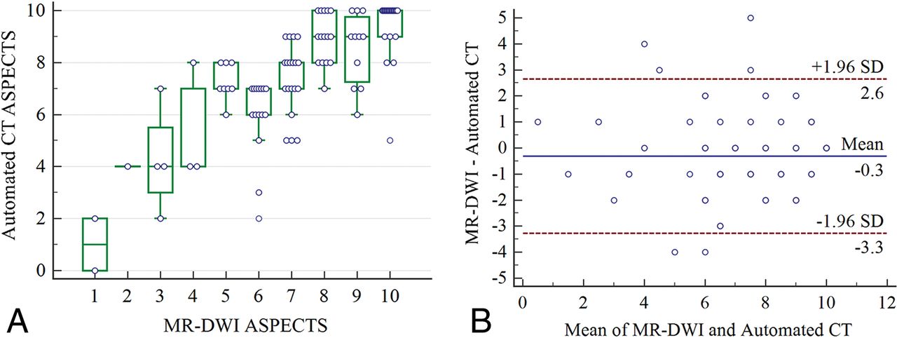
  • Fig 3.
    • Download figure
    • Open in new tab
    • Download powerpoint
    Fig 3.

    A, Boxplot with a scatterplot showing the distribution of the automated CT ASPECTS at each individual ASPECTS on DWI. B, A Bland-Altman plot illustrating agreement between a total automated ASPECTS score and ASPECTS scores on DWI. Random jitter has been added to illustrate the number of measurements at each ASPECTS point. The horizontal black line represents the mean difference in the ASPECTS score between the 2 methods, while the dotted lines represent a 1.96 SD around the difference.
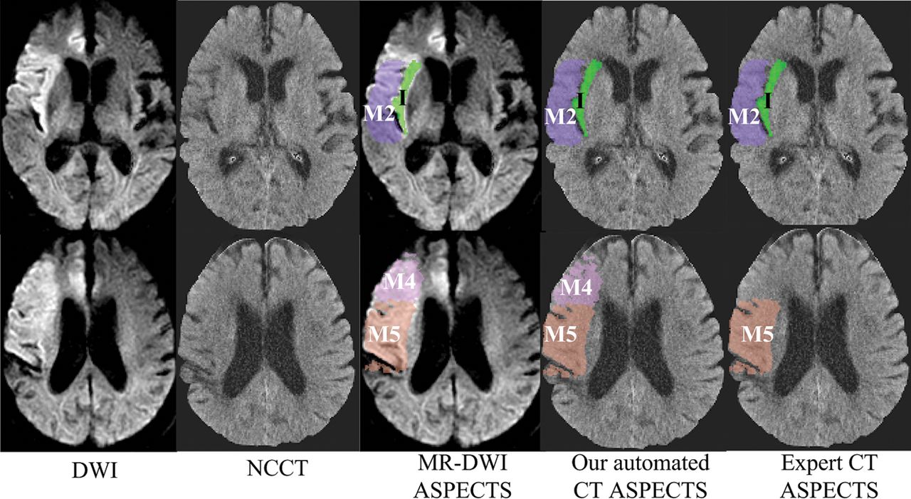
  • Fig 4.
    • Download figure
    • Open in new tab
    • Download powerpoint
    Fig 4.

    Examples of DWI ASPECTS, the automated CT ASPECTS derived in this study, and expert-read CT ASPECTS. ASPECTS regions with ischemic changes are shown in color.

Tables

  • Figures
    • View popup
    Table 1:

    κ, accuracy, F1 measure, sensitivity, specificity, and AUC on each ASPECTS region

    Regionκ (95% CI)Accuracy (%) (95% CI)F1 MeasureSensitivity (%) (95% CI)Specificity (%) (95% CI)AUC (95% CI)
    M10.59 (0.38–0.81)90 (90/100) (84.1–95.9)0.6447.4 (9/19) (24.4–71.1)100 (81/81) (95.5–100)0.74 (0.64–0.82)
    M20.52 (0.35–0.68)76 (76/100) (67.6–84.4)0.7376.2 (32/42) (60.5–87.9)75.9 (44/58) (62.8–86.1)0.76 (0.67–0.84)
    M30.47 (0.21–0.73)88 (88/100) (81.6–94.4)0.5450 (7/14) (23–77)94.2 (81/86) (87–98.1)0.72 (0.62–0.81)
    M40.36 (0.13–0.63)85 (85/100) (78–92)0.3536.4 (4/11) (10.9–69.2)91.1 (81/89) (83.1–96)0.64 (0.54–0.73)
    M50.54 (0.37–0.7)77 (77/100) (68.8–85.3)0.7468.1 (32/47) (52.9–80.9)84.9 (45/53) (72.4–93.3)0.77 (0.67–0.84)
    M60.39 (0.14–0.64)86 (86/100) (79.2–92.8)0.4635.3 (6/17) (14.2–61.7)96.4 (80/83) (89.8–99.2)0.66 (0.56–0.75)
    Lentiform0.64 (0.47–0.81)85 (85/100) (78–92)0.7571.0 (22/31) (52–85.8)91.3 (63/69) (82–96.7)0.81 (0.72–0.88)
    Insula0.62 (0.46–0.77)81 (81/100) (73.3–88.7)0.8385.5 (47/55) (73.3–93.5)75.6 (34/45) (60.5–87.1)0.81 (0.71–0.88)
    Caudate0.63 (0.42–0.84)90 (90/100) (84.1–95.9)0.6957.9 (11/19) (33.5–79.7)97.5 (79/81) (91.4–99.7)0.78 (0.68–0.85)
    Internal capsule0.59 (0.35–0.83)91 (91/100) (85.4–96.6)0.6457.1 (8/14) (28.9–82.3)96.5 (83/86) (90.1–99.3)0.77 (0.67–0.85)
    All regions0.60 (0.54–0.66)84.9 (849/1000) (82.7–87.1)0.7066.2 (178/269) (60.2–71.8)91.8 (671/731) (89.6–93.7)0.79 (0.75–0.83)
    • View popup
    Table 2:

    Agreement on ASPECTS interpretation at a regional level and for dichotomized ASPECTS (>4 vs. ≤4) between the automated ASPECTS method and expert-read DWI ASPECTS using different DWI ASPECTS region-involvement thresholds

    DWI ASPECTS Region-Involvement Thresholdsκ (95% CI)Accuracy (%) (95% CI)F1 MeasureSensitivity (%) (95% CI)Specificity (%) (95% CI)AUC (95% CI)
    20%
        All regions0.6 (0.54–0.66)84.9 (849/1000) (82.7–87.1)0.7066.2 (178/269) (60.2–71.8)91.8 (671/731) (89.6–93.7)0.79 (0.75–0.83)
        >4 and ≤40.78 (0.57–0.99)96 (96/100) (92.2–99.8)0.9897.8 (88/90) (92.2–99.7)80 (8/10) (34.8–93.3)0.89 (0.81–0.94)
    50%
        All regions0.64 (0.57–0.70)88.8 (888/1000) (86.9–90.8)0.7168.2 (133/195) (61.2–74.7)93.8 (755/805) (91.9–95.4)0.81 (0.77–0.85)
        >4 and ≤41 (1–1)100 (100/100) (100–100)1100 (94/94) (96.2–100)100 (6/6) (54.1–100)1 (0.96–1)
    0%
        All regions0.56 (0.51–0.61)79.5 (795/1000) (77–82)0.7269.7 (264/379) (64.8–74.2)85.5 (531/621) (82.5–88.2)0.78 (0.76–0.79)
        >4 and ≤40.46 (0.26–0.66)81 (81/100) (73.3–88.7)0.8893.2 (68/73) (84.7–97.7)48.2 (13/27) (28.7–68.1)0.71 (0.61–0.79)
PreviousNext
Back to top

In this issue

American Journal of Neuroradiology: 40 (1)
American Journal of Neuroradiology
Vol. 40, Issue 1
1 Jan 2019
  • Table of Contents
  • Index by author
  • Complete Issue (PDF)
Advertisement
Print
Download PDF
Email Article

Thank you for your interest in spreading the word on American Journal of Neuroradiology.

NOTE: We only request your email address so that the person you are recommending the page to knows that you wanted them to see it, and that it is not junk mail. We do not capture any email address.

Enter multiple addresses on separate lines or separate them with commas.
Automated ASPECTS on Noncontrast CT Scans in Patients with Acute Ischemic Stroke Using Machine Learning
(Your Name) has sent you a message from American Journal of Neuroradiology
(Your Name) thought you would like to see the American Journal of Neuroradiology web site.
CAPTCHA
This question is for testing whether or not you are a human visitor and to prevent automated spam submissions.
Cite this article
H. Kuang, M. Najm, D. Chakraborty, N. Maraj, S.I. Sohn, M. Goyal, M.D. Hill, A.M. Demchuk, B.K. Menon, W. Qiu
Automated ASPECTS on Noncontrast CT Scans in Patients with Acute Ischemic Stroke Using Machine Learning
American Journal of Neuroradiology Jan 2019, 40 (1) 33-38; DOI: 10.3174/ajnr.A5889

Citation Manager Formats

  • BibTeX
  • Bookends
  • EasyBib
  • EndNote (tagged)
  • EndNote 8 (xml)
  • Medlars
  • Mendeley
  • Papers
  • RefWorks Tagged
  • Ref Manager
  • RIS
  • Zotero
0 Responses
Respond to this article
Share
Bookmark this article
Automated ASPECTS on Noncontrast CT Scans in Patients with Acute Ischemic Stroke Using Machine Learning
H. Kuang, M. Najm, D. Chakraborty, N. Maraj, S.I. Sohn, M. Goyal, M.D. Hill, A.M. Demchuk, B.K. Menon, W. Qiu
American Journal of Neuroradiology Jan 2019, 40 (1) 33-38; DOI: 10.3174/ajnr.A5889
del.icio.us logo Twitter logo Facebook logo Mendeley logo
  • Tweet Widget
  • Facebook Like
  • Google Plus One
Purchase

Jump to section

  • Article
    • Abstract
    • ABBREVIATIONS:
    • Materials and Methods
    • Results
    • Discussion
    • Conclusions
    • Footnotes
    • References
  • Figures & Data
  • Supplemental
  • Info & Metrics
  • Responses
  • References
  • PDF

Related Articles

  • PubMed
  • Google Scholar

Cited By...

  • Clinical evaluation of a deep-learning model for automatic scoring of the Alberta stroke program early CT score on non-contrast CT
  • Recent developments in pre-hospital and in-hospital triage for endovascular stroke treatment
  • Artificial intelligence-driven ASPECTS for the detection of early stroke changes in non-contrast CT: a systematic review and meta-analysis
  • Clinical evaluation of a deep-learning model for automatic scoring of the Alberta stroke program early CT score on non-contrast CT
  • Clinical evaluation of a deep-learning model for automatic scoring of the Alberta stroke program early CT score on non-contrast CT
  • Benefit and risk of intravenous alteplase in patients with acute large vessel occlusion stroke and low ASPECTS
  • Artificial Intelligence and Acute Stroke Imaging
  • Management of Acute Ischemic Stroke Due to Large-Vessel Occlusion: JACC Focus Seminar
  • Artificial intelligence to diagnose ischemic stroke and identify large vessel occlusions: a systematic review
  • Automated ASPECTS in Acute Ischemic Stroke: A Comparative Analysis with CT Perfusion
  • Crossref (98)
  • Google Scholar

This article has been cited by the following articles in journals that are participating in Crossref Cited-by Linking.

  • Artificial intelligence to diagnose ischemic stroke and identify large vessel occlusions: a systematic review
    Nick M Murray, Mathias Unberath, Gregory D Hager, Ferdinand K Hui
    Journal of NeuroInterventional Surgery 2020 12 2
  • Artificial Intelligence and Acute Stroke Imaging
    J.E. Soun, D.S. Chow, M. Nagamine, R.S. Takhtawala, C.G. Filippi, W. Yu, P.D. Chang
    American Journal of Neuroradiology 2021 42 1
  • Machine Learning for Detecting Early Infarction in Acute Stroke with Non–Contrast-enhanced CT
    Wu Qiu, Hulin Kuang, Ericka Teleg, Johanna M. Ospel, Sung Il Sohn, Mohammed Almekhlafi, Mayank Goyal, Michael D. Hill, Andrew M. Demchuk, Bijoy K. Menon
    Radiology 2020 294 3
  • CT for Treatment Selection in Acute Ischemic Stroke: A Code Stroke Primer
    Christopher A. Potter, Achala S. Vagal, Mayank Goyal, Diego B. Nunez, Thabele M. Leslie-Mazwi, Michael H. Lev
    RadioGraphics 2019 39 6
  • Management of Acute Ischemic Stroke Due to Large-Vessel Occlusion
    Johanna M. Ospel, Jessalyn K. Holodinsky, Mayank Goyal
    Journal of the American College of Cardiology 2020 75 15
  • Artificial intelligence in stroke imaging: Current and future perspectives
    Vivek S. Yedavalli, Elizabeth Tong, Dann Martin, Kristen W. Yeom, Nils D. Forkert
    Clinical Imaging 2021 69
  • Computer-aided imaging analysis in acute ischemic stroke – background and clinical applications
    Yahia Mokli, Johannes Pfaff, Daniel Pinto dos Santos, Christian Herweh, Simon Nagel
    Neurological Research and Practice 2019 1 1
  • EIS-Net: Segmenting early infarct and scoring ASPECTS simultaneously on non-contrast CT of patients with acute ischemic stroke
    Hulin Kuang, Bijoy K. Menon, Sung IL Sohn, Wu Qiu
    Medical Image Analysis 2021 70
  • Artificial intelligence in emergency radiology: A review of applications and possibilities
    Benjamin D. Katzman, Christian B. van der Pol, Philippe Soyer, Michael N. Patlas
    Diagnostic and Interventional Imaging 2023 104 1
  • Automated ASPECT scoring in acute ischemic stroke: comparison of three software tools
    Philip Hoelter, Iris Muehlen, Philipp Goelitz, Vanessa Beuscher, Stefan Schwab, Arnd Doerfler
    Neuroradiology 2020 62 10

More in this TOC Section

Adult Brain

  • Diagnostic Neuroradiology of Monoclonal Antibodies
  • Cerebral ADC Changes in Fabry Disease
  • ML for Glioma Molecular Subtype Prediction
Show more Adult Brain

Functional

  • Glutaric Aciduria Type 1: DK vs. Conventional MRI
  • Kurtosis and Epileptogenic Tubers: A Pilot Study
  • Predicting Outcomes in Tuberous Sclerosis Epilepsy
Show more Functional

Similar Articles

Advertisement

Indexed Content

  • Current Issue
  • Accepted Manuscripts
  • Article Preview
  • Past Issues
  • Editorials
  • Editors Choice
  • Fellow Journal Club
  • Letters to the Editor

Cases

  • Case Collection
  • Archive - Case of the Week
  • Archive - Case of the Month
  • Archive - Classic Case

Special Collections

  • Special Collections

Resources

  • News and Updates
  • Turn around Times
  • Submit a Manuscript
  • Author Policies
  • Manuscript Submission Guidelines
  • Evidence-Based Medicine Level Guide
  • Publishing Checklists
  • Graphical Abstract Preparation
  • Imaging Protocol Submission
  • Submit a Case
  • Become a Reviewer/Academy of Reviewers
  • Get Peer Review Credit from Publons

Multimedia

  • AJNR Podcast
  • AJNR SCANtastic
  • Video Articles

About Us

  • About AJNR
  • Editorial Board
  • Not an AJNR Subscriber? Join Now
  • Alerts
  • Feedback
  • Advertise with us
  • Librarian Resources
  • Permissions
  • Terms and Conditions

American Society of Neuroradiology

  • Not an ASNR Member? Join Now

© 2025 by the American Society of Neuroradiology All rights, including for text and data mining, AI training, and similar technologies, are reserved.
Print ISSN: 0195-6108 Online ISSN: 1936-959X

Powered by HighWire