
Article Figures & Data
Figures
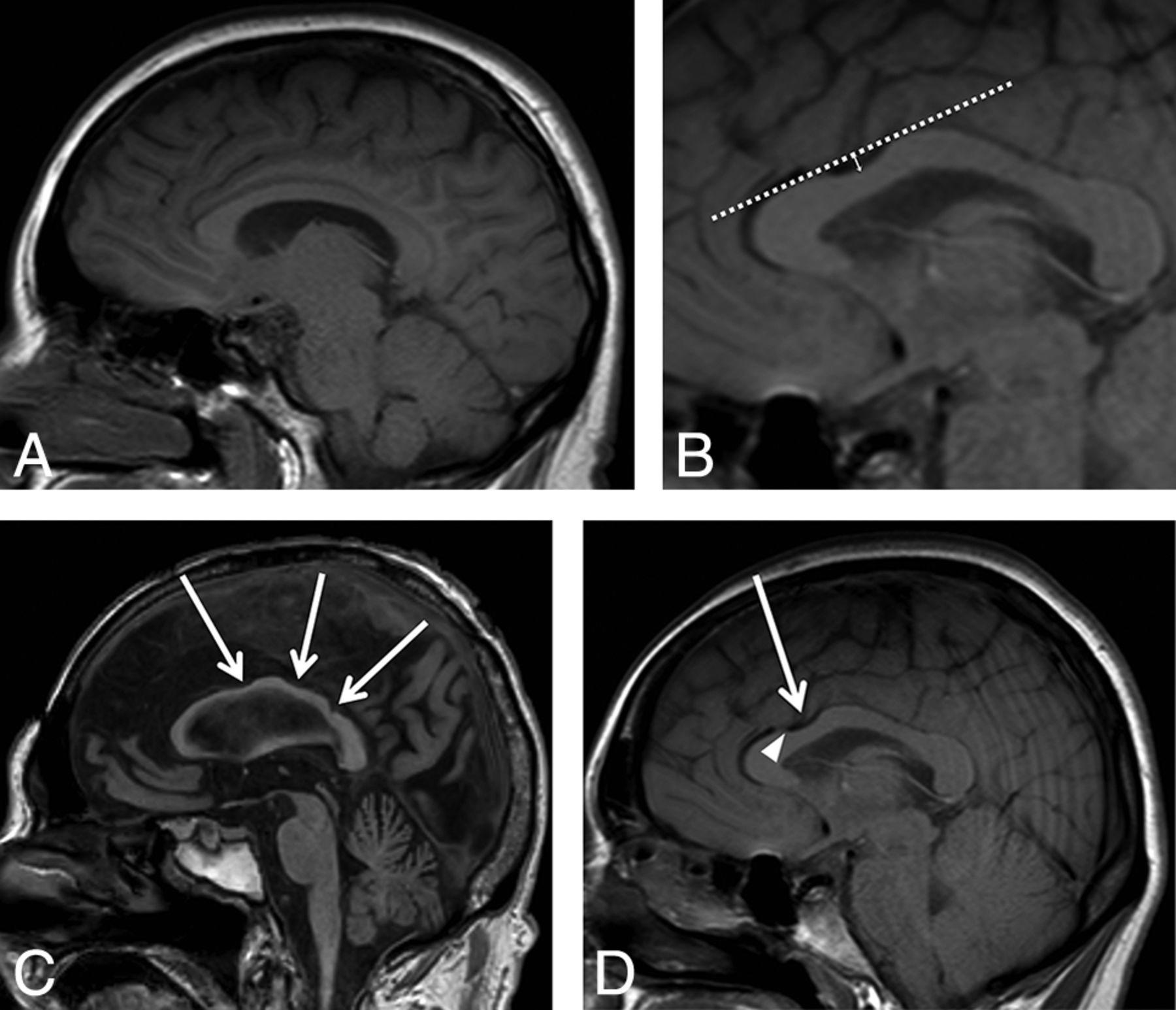
- Fig 1.
A, Midline sagittal T1-weighted image shows an example of a normal corpus callosum without evidence of anterior or posterior notching. B, A “notch” was defined as a depression in the dorsal surface whose depth was at least 1 mm from a tangential (dashed) line to the surface of the CC. The solid arrow line shows a 3-mm-deep anterior notch. C, Three areas of notching indicated by the white arrows correspond to an undulating configuration. D, Midline sagittal T1-weighted image shows that a 3-mm anterior notch (arrow) is present. The flow void from the pericallosal artery is visualized extending into the notch (arrowhead).
- Fig 2.
Scatterplot of prevalence versus age in 5-year increments. The posterior notch prevalence (red squares and red line) decreases with time, R2 = 0.29. The anterior notch prevalence (blue diamonds and blue line) increases with time, R2 = 0.60. The undulating pattern prevalence (green triangles and green line) also increases with time, R2 = 0.28.
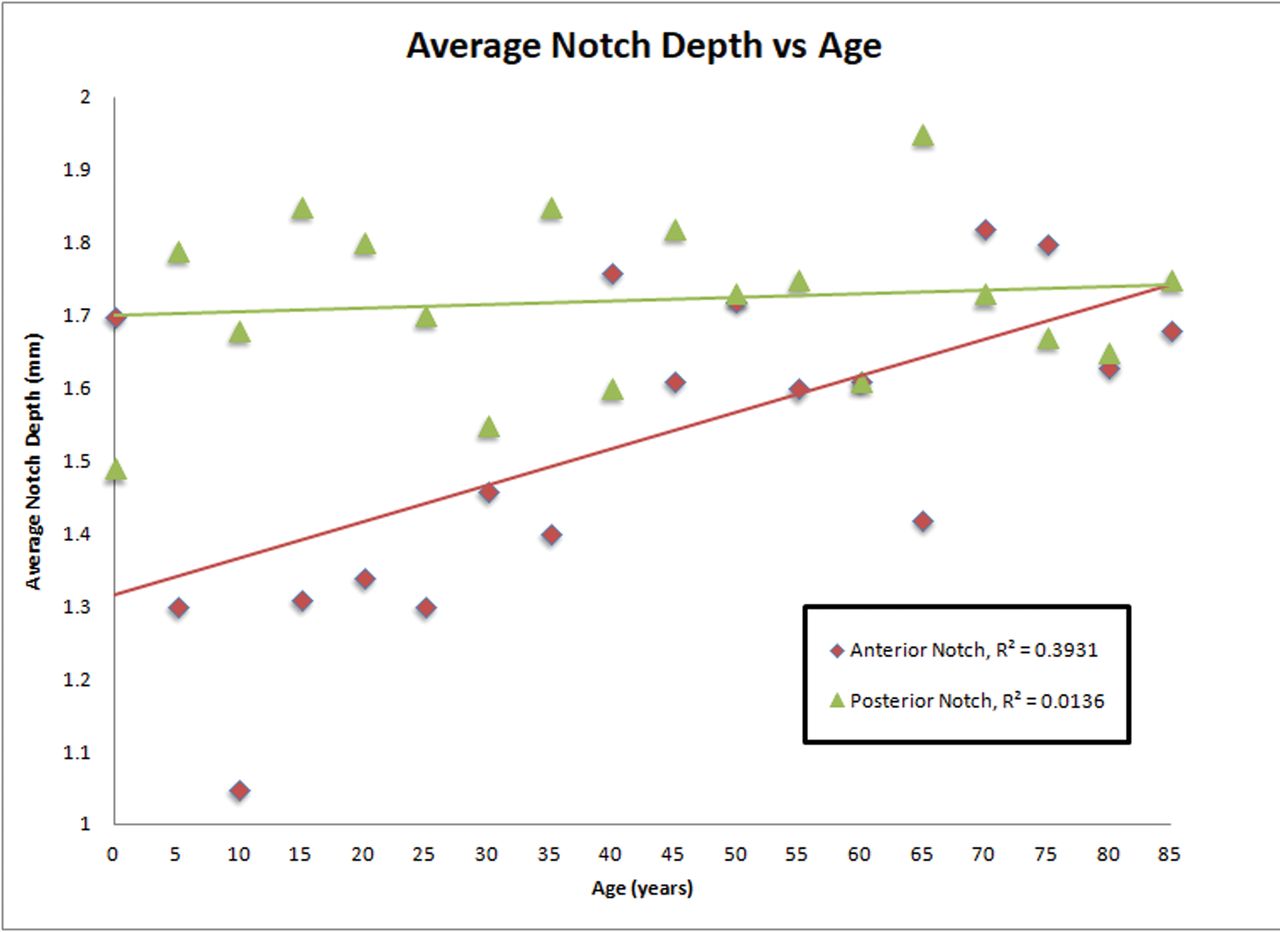
- Fig 3.
Scatterplot of notch depth (millimeters) versus age. No significant difference is seen in the depth of the posterior notch with increasing age (R2 = 0.01). There is a significant increase in the depth of the anterior notch with increasing age (R2 = 0.39).


Tables
Retrospective review summary of brain MR imaging studies, notch count, and position
Review Summary Brain MR imaging studies reviewed (No.) 1639 Brain MR imaging studies, with notches (No.) 823 Notches identified in 823 MR imaging studies (No.) 1102 Female (No.) (%) 919; 56 Notch identified 460; 50 Male (No.) (%) 720; 44 Notch identified 363; 50 Anterior position (No.) (%) 344; 31 Posterior position (No.) (%) 660; 60 Undulating, >2 notches (No.) (%) 98; 9 Pericallosal artery notch association (No.) (%) 490; 60


