
Article Figures & Data
Figures
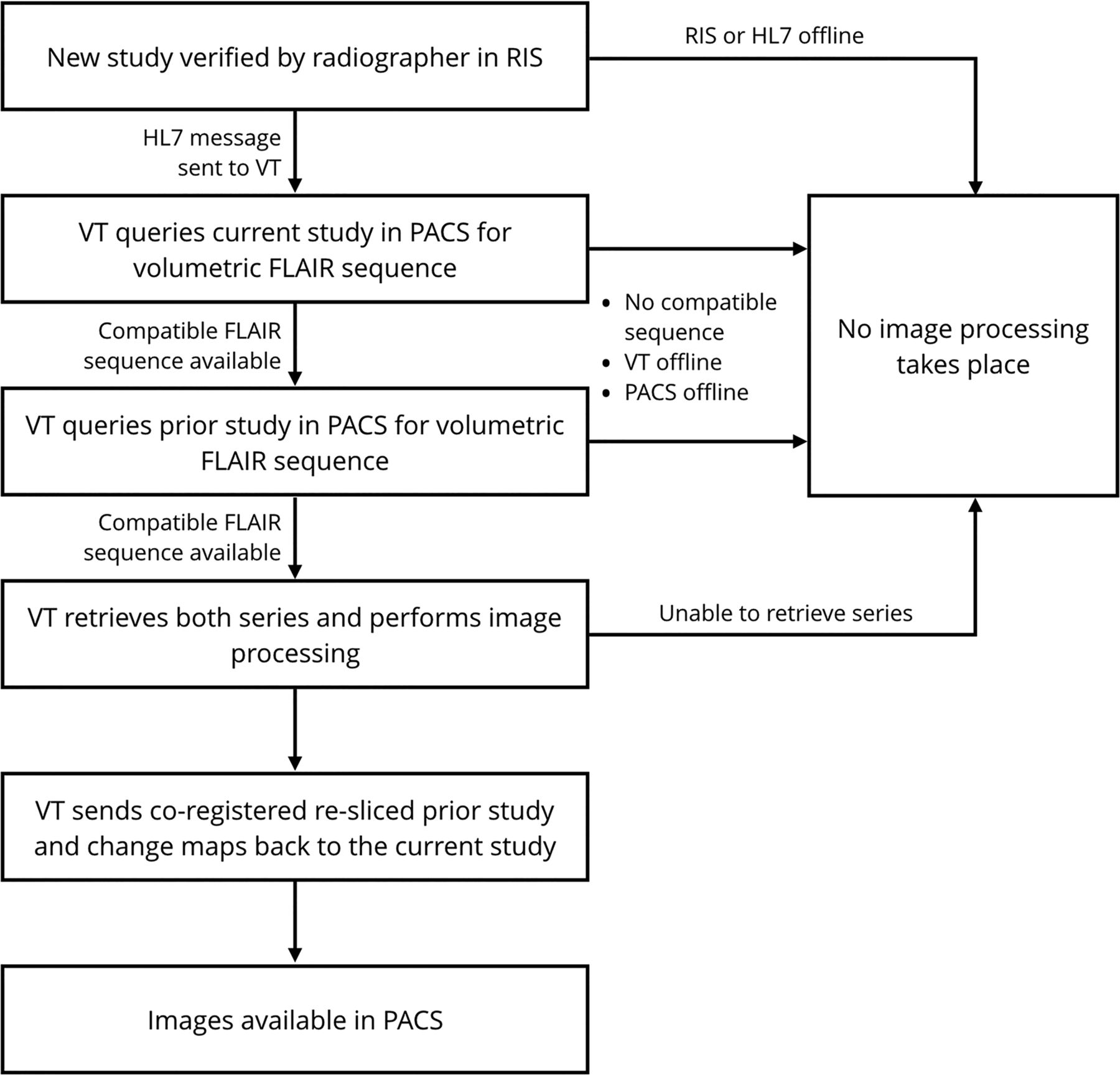
- Fig 1.
Software integration into PACS workflow. This flow diagram outlines how the new MR imaging studies for patients with MS are processed by the VisTarsier software in a virtual machine once they are signed off in the radiology information system (RIS) by the radiographer. Successful processing requires all systems to be operational and compatible sequences to be available.
- Fig 2.
The proportion of scans showing MS progression within each year. This scatterplot highlights the number of scans and the proportion in which new and enlarging lesions were detected for each study group during each year. The position on the vertical axis corresponds to the proportion of scans showing progression. The position along the horizontal axis corresponds to the study year. The lighter shade corresponds to scans generated with the software.


Tables
Proportion CSSC Software-Assisted P Value Scans belonging to female patients 52/65 (80.00%) 599/841 (71.22%) .23 Primary reporting doctors with fellowship certification 47/65 (72.31%) 586/841 (69.68%) .36 Age at scan Mean, 44 yr, 139 days SD, 11 yr, 223 days Mean, 43 yr, 15 days SD,11 yr, 256 days .72 Time since diagnosis Mean, 10 yr, 47 days SD, 6 yr, 230 days Mean, 9 yr, 58 days SD, 6 yr, 234 days .61 EDSS Median, 2.0 Quartiles, 25% = 1.9, 75% = 3.1 Median, 2.0 Quartiles, 25% = 1.0, 75% = 3.5 .45 ↵a This table summarizes the demographic and clinical details for all eligible patients who underwent an MR imaging brain scan at the Royal Melbourne Hospital from July 1, 2015, until June 30, 2017. χ2 and t test statistics were performed to confirm group similarities.31
Medication CSSC (No.) Software-Assisted (No.) Fingolimod 35.85% (19) 37.30% (282) Natalizumab 32.08% (17) 27.12% (205) Dimethyl fumarate 3.77% (2) 7.80% (59) Alemtuzumab 1.89% (1) 3.44% (26) Glatiramer acetate 5.66% (3) 2.78% (21) Interferon β 5.66% (3) 3.57% (27) Otherb 9.43% (5) 6.61% (50) No active treatment 5.66% (3) 11.38% (86) Total 100% (53) 100% (756) Proportion on higher efficacy therapies (fingolimod/natalizumab/alemtuzumab) 69.87% 67.84%


