Advertisement

Main menu

  • Home
  • Content
    • Current Issue
    • Accepted Manuscripts
    • Article Preview
    • Past Issue Archive
    • Video Articles
    • AJNR Case Collection
    • Case of the Week Archive
    • Case of the Month Archive
    • Classic Case Archive
  • Special Collections
    • AJNR Awards
    • Low-Field MRI
    • Alzheimer Disease
    • ASNR Foundation Special Collection
    • Photon-Counting CT
    • View All
  • Multimedia
    • AJNR Podcasts
    • AJNR SCANtastic
    • Trainee Corner
    • MRI Safety Corner
    • Imaging Protocols
  • For Authors
    • Submit a Manuscript
    • Submit a Video Article
    • Submit an eLetter to the Editor/Response
    • Manuscript Submission Guidelines
    • Statistical Tips
    • Fast Publishing of Accepted Manuscripts
    • Graphical Abstract Preparation
    • Imaging Protocol Submission
    • Author Policies
  • About Us
    • About AJNR
    • Editorial Board
    • Editorial Board Alumni
  • More
    • Become a Reviewer/Academy of Reviewers
    • Subscribers
    • Permissions
    • Alerts
    • Feedback
    • Advertisers
    • ASNR Home

User menu

  • Alerts
  • Log in

Search

  • Advanced search
American Journal of Neuroradiology

American Journal of Neuroradiology

ASHNR American Society of Functional Neuroradiology ASHNR American Society of Pediatric Neuroradiology ASSR
  • Alerts
  • Log in

Advanced Search

  • Home
  • Content
    • Current Issue
    • Accepted Manuscripts
    • Article Preview
    • Past Issue Archive
    • Video Articles
    • AJNR Case Collection
    • Case of the Week Archive
    • Case of the Month Archive
    • Classic Case Archive
  • Special Collections
    • AJNR Awards
    • Low-Field MRI
    • Alzheimer Disease
    • ASNR Foundation Special Collection
    • Photon-Counting CT
    • View All
  • Multimedia
    • AJNR Podcasts
    • AJNR SCANtastic
    • Trainee Corner
    • MRI Safety Corner
    • Imaging Protocols
  • For Authors
    • Submit a Manuscript
    • Submit a Video Article
    • Submit an eLetter to the Editor/Response
    • Manuscript Submission Guidelines
    • Statistical Tips
    • Fast Publishing of Accepted Manuscripts
    • Graphical Abstract Preparation
    • Imaging Protocol Submission
    • Author Policies
  • About Us
    • About AJNR
    • Editorial Board
    • Editorial Board Alumni
  • More
    • Become a Reviewer/Academy of Reviewers
    • Subscribers
    • Permissions
    • Alerts
    • Feedback
    • Advertisers
    • ASNR Home
  • Follow AJNR on Twitter
  • Visit AJNR on Facebook
  • Follow AJNR on Instagram
  • Join AJNR on LinkedIn
  • RSS Feeds

AJNR Awards, New Junior Editors, and more. Read the latest AJNR updates

Research ArticleAdult Brain
Open Access

Automated ASPECTS in Acute Ischemic Stroke: A Comparative Analysis with CT Perfusion

V.K. Sundaram, J. Goldstein, D. Wheelwright, A. Aggarwal, P.S. Pawha, A. Doshi, J.T. Fifi, R. De Leacy, J. Mocco, J. Puig and K. Nael
American Journal of Neuroradiology December 2019, 40 (12) 2033-2038; DOI: https://doi.org/10.3174/ajnr.A6303
V.K. Sundaram
aFrom the Department of Radiology (V.K.S., J.G., A.A., P.P., A.D., K.N.)
  • Find this author on Google Scholar
  • Find this author on PubMed
  • Search for this author on this site
  • ORCID record for V.K. Sundaram
J. Goldstein
aFrom the Department of Radiology (V.K.S., J.G., A.A., P.P., A.D., K.N.)
  • Find this author on Google Scholar
  • Find this author on PubMed
  • Search for this author on this site
  • ORCID record for J. Goldstein
D. Wheelwright
bNeuroimaging Advanced and Exploratory Lab, Department of Neurology (D.W., J.T.F., R.D.L.)
  • Find this author on Google Scholar
  • Find this author on PubMed
  • Search for this author on this site
  • ORCID record for D. Wheelwright
A. Aggarwal
aFrom the Department of Radiology (V.K.S., J.G., A.A., P.P., A.D., K.N.)
  • Find this author on Google Scholar
  • Find this author on PubMed
  • Search for this author on this site
  • ORCID record for A. Aggarwal
P.S. Pawha
aFrom the Department of Radiology (V.K.S., J.G., A.A., P.P., A.D., K.N.)
  • Find this author on Google Scholar
  • Find this author on PubMed
  • Search for this author on this site
  • ORCID record for P.S. Pawha
A. Doshi
aFrom the Department of Radiology (V.K.S., J.G., A.A., P.P., A.D., K.N.)
  • Find this author on Google Scholar
  • Find this author on PubMed
  • Search for this author on this site
  • ORCID record for A. Doshi
J.T. Fifi
bNeuroimaging Advanced and Exploratory Lab, Department of Neurology (D.W., J.T.F., R.D.L.)
cDepartment of Neurosurgery (J.T.F., R.D.L., J.M.), Icahn School of Medicine at Mount Sinai, New York, New York
  • Find this author on Google Scholar
  • Find this author on PubMed
  • Search for this author on this site
  • ORCID record for J.T. Fifi
R. De Leacy
bNeuroimaging Advanced and Exploratory Lab, Department of Neurology (D.W., J.T.F., R.D.L.)
cDepartment of Neurosurgery (J.T.F., R.D.L., J.M.), Icahn School of Medicine at Mount Sinai, New York, New York
  • Find this author on Google Scholar
  • Find this author on PubMed
  • Search for this author on this site
  • ORCID record for R. De Leacy
J. Mocco
cDepartment of Neurosurgery (J.T.F., R.D.L., J.M.), Icahn School of Medicine at Mount Sinai, New York, New York
  • Find this author on Google Scholar
  • Find this author on PubMed
  • Search for this author on this site
  • ORCID record for J. Mocco
J. Puig
dDepartment of Radiology (J.P.). University of Manitoba, Winnipeg, Manitoba, Canada.
  • Find this author on Google Scholar
  • Find this author on PubMed
  • Search for this author on this site
  • ORCID record for J. Puig
K. Nael
aFrom the Department of Radiology (V.K.S., J.G., A.A., P.P., A.D., K.N.)
  • Find this author on Google Scholar
  • Find this author on PubMed
  • Search for this author on this site
  • ORCID record for K. Nael
  • Article
  • Figures & Data
  • Info & Metrics
  • Responses
  • References
  • PDF
Loading

Article Figures & Data

Figures

  • Tables
  • Fig 1.
    • Download figure
    • Open in new tab
    • Download powerpoint
    Fig 1.

    Comparative illustration of the distribution of ASPECTS from consensus reads of 2 neuroradiologists, software-based automated ASPECTS, and CTP-CBV for all patients in our study population (each marker represents a patient’s score). Automated ASPECTS showed excellent agreement (κ = 0.84; 95% CI, 0.62–1.0) with both consensus and CTP-CBV ASPECTS. The intraclass correlation coefficient was 0.84 (95% CI, 0.76–0.90) across all 3 groups.

  • Fig 2.
    • Download figure
    • Open in new tab
    • Download powerpoint
    Fig 2.

    An 82-year-old woman who presented with right MCA M1 occlusion and an NIHSS score of 18. She underwent successful mechanical thrombectomy (TICI 3) with a CT-to-recanalization time of 50 minutes. Axial NCCT (A), automated ASPECTS (B), axial CTP-CBV (C), and axial NCCT 48 hours after endovascular treatment (D) are shown. For the 2 human readers, one scored 6 and the other, 7 (consensus ASPECTS, 6). B, Automated software assigned an ASPECTS of 6. CTP-CBV ASPECTS was 7. There is good topographic correlation with the final infarction volume.
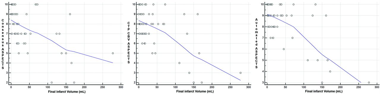
  • Fig 3.
    • Download figure
    • Open in new tab
    • Download powerpoint
    Fig 3.

    Scatterplots for correlation between ASPECTS and final infarction volume for all 3 ASPECTS groups showing significant (P < .001) negative correlation (r = −0.52 for the consensus reads, −0.58 for CTP-CBV, and −0.66 for automated ASPECTS).

Tables

  • Figures
    • View popup
    Table 1:

    Comparative analysis among ASPECTS scores categorized on the basis of time of symptom onseta

    Onset Symptoms ≤6 hrs (24)Onset Symptoms >6 hrs (34)P Value
    Consensus ASPECTS (2 neuroradiologists)8 (6–9)7 (7–9).46
    Automated ASPECTS9 (7–10)9 (8–9).64
    CTP-CBV ASPECTS8 (6–9)8 (7–9).73
    • ↵a Data are presented in median (IQR).

    • View popup
    Table 2:

    Clinical and imaging data in patients with good-versus-poor functional outcome using mRS >2 as a cutoff

    VariableOverall (n = 50)Good Outcome (n = 22)Poor Outcome (n = 28)P Value
    Age (mean) (SD) (yr)70.0 (13.0)65.1 (12.8)73.8 (12.1).02
    Sex (M/F)23:2713:910:18.10
    Baseline NIHSS (median) (IQR)15 (11–21)11 (8–14)20 (14–22).001
    Time from symptom onset (mean) (SD) (hr)8.4 (5.6)8.8 (6.7)8.1 (4.6).62
    Consensus ASPECTS ≥ 6 (No.) (%)4321 (95%)22 (78%).10
    Automated ASPECTS ≥ 6 (No.) (%)4521 (95%)24 (85%).26
    CTP-CBV ASPECTS ≥ 6 (No.) (%)4321 (95%)22 (78%).10
    Final infarction volume (mean) (SD) (mL)4924.4 (29.0)57.5 (64.7).03
PreviousNext
Back to top

In this issue

American Journal of Neuroradiology: 40 (12)
American Journal of Neuroradiology
Vol. 40, Issue 12
1 Dec 2019
  • Table of Contents
  • Index by author
  • Complete Issue (PDF)
Advertisement
Print
Download PDF
Email Article

Thank you for your interest in spreading the word on American Journal of Neuroradiology.

NOTE: We only request your email address so that the person you are recommending the page to knows that you wanted them to see it, and that it is not junk mail. We do not capture any email address.

Enter multiple addresses on separate lines or separate them with commas.
Automated ASPECTS in Acute Ischemic Stroke: A Comparative Analysis with CT Perfusion
(Your Name) has sent you a message from American Journal of Neuroradiology
(Your Name) thought you would like to see the American Journal of Neuroradiology web site.
CAPTCHA
This question is for testing whether or not you are a human visitor and to prevent automated spam submissions.
Cite this article
V.K. Sundaram, J. Goldstein, D. Wheelwright, A. Aggarwal, P.S. Pawha, A. Doshi, J.T. Fifi, R. De Leacy, J. Mocco, J. Puig, K. Nael
Automated ASPECTS in Acute Ischemic Stroke: A Comparative Analysis with CT Perfusion
American Journal of Neuroradiology Dec 2019, 40 (12) 2033-2038; DOI: 10.3174/ajnr.A6303

Citation Manager Formats

  • BibTeX
  • Bookends
  • EasyBib
  • EndNote (tagged)
  • EndNote 8 (xml)
  • Medlars
  • Mendeley
  • Papers
  • RefWorks Tagged
  • Ref Manager
  • RIS
  • Zotero
0 Responses
Respond to this article
Share
Bookmark this article
Automated ASPECTS in Acute Ischemic Stroke: A Comparative Analysis with CT Perfusion
V.K. Sundaram, J. Goldstein, D. Wheelwright, A. Aggarwal, P.S. Pawha, A. Doshi, J.T. Fifi, R. De Leacy, J. Mocco, J. Puig, K. Nael
American Journal of Neuroradiology Dec 2019, 40 (12) 2033-2038; DOI: 10.3174/ajnr.A6303
del.icio.us logo Twitter logo Facebook logo Mendeley logo
  • Tweet Widget
  • Facebook Like
  • Google Plus One
Purchase

Jump to section

  • Article
    • Abstract
    • ABBREVIATIONS:
    • MATERIALS AND METHODS
    • RESULTS
    • DISCUSSION
    • CONCLUSIONS
    • Footnotes
    • References
  • Figures & Data
  • Info & Metrics
  • Responses
  • References
  • PDF

Related Articles

  • No related articles found.
  • PubMed
  • Google Scholar

Cited By...

  • Automated assessment of ischemic core on non-contrast computed tomography: a multicenter comparative analysis with CT perfusion
  • Real-world evaluation of Brainomix e-Stroke software
  • MR microscopy to assess clot composition following mechanical thrombectomy predicts recanalization and clinical outcome
  • Alberta Stroke Program Early CT Score and collateral status predict target mismatch in large vessel occlusion with delayed time windows
  • Performance of Automated ASPECTS Software and Value as a Computer-Aided Detection Tool
  • Benefit and risk of intravenous alteplase in patients with acute large vessel occlusion stroke and low ASPECTS
  • Emerging Artificial Intelligence Imaging Applications for Stroke Interventions
  • Innovative use of artificial intelligence and digital communication in acute stroke pathway in response to COVID-19
  • ASPECTS Distorts Infarct Volume Measurement
  • Crossref
  • Google Scholar

This article has not yet been cited by articles in journals that are participating in Crossref Cited-by Linking.

More in this TOC Section

  • Diagnostic Neuroradiology of Monoclonal Antibodies
  • Clinical Outcomes After Chiari I Decompression
  • Segmentation of Brain Metastases with BLAST
Show more Adult Brain

Similar Articles

Advertisement

Indexed Content

  • Current Issue
  • Accepted Manuscripts
  • Article Preview
  • Past Issues
  • Editorials
  • Editor's Choice
  • Fellows' Journal Club
  • Letters to the Editor
  • Video Articles

Cases

  • Case Collection
  • Archive - Case of the Week
  • Archive - Case of the Month
  • Archive - Classic Case

More from AJNR

  • Trainee Corner
  • Imaging Protocols
  • MRI Safety Corner
  • Book Reviews

Multimedia

  • AJNR Podcasts
  • AJNR Scantastics

Resources

  • Turnaround Time
  • Submit a Manuscript
  • Submit a Video Article
  • Submit an eLetter to the Editor/Response
  • Manuscript Submission Guidelines
  • Statistical Tips
  • Fast Publishing of Accepted Manuscripts
  • Graphical Abstract Preparation
  • Imaging Protocol Submission
  • Evidence-Based Medicine Level Guide
  • Publishing Checklists
  • Author Policies
  • Become a Reviewer/Academy of Reviewers
  • News and Updates

About Us

  • About AJNR
  • Editorial Board
  • Editorial Board Alumni
  • Alerts
  • Permissions
  • Not an AJNR Subscriber? Join Now
  • Advertise with Us
  • Librarian Resources
  • Feedback
  • Terms and Conditions
  • AJNR Editorial Board Alumni

American Society of Neuroradiology

  • Not an ASNR Member? Join Now

© 2025 by the American Society of Neuroradiology All rights, including for text and data mining, AI training, and similar technologies, are reserved.
Print ISSN: 0195-6108 Online ISSN: 1936-959X

Powered by HighWire