
Article Figures & Data
Figures
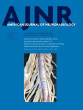
- Fig 1.
Boxplot showing the growth rate per aneurysm location. ACA indicates anterior cerebral artery; PcomA, posterior communicating artery; AcomA, anterior communicating artery; BA, basilar artery; SCA, superior cerebellar artery; VA, vertebral artery.
- Fig 2.
Dumbbell plot depicting the ratio between the median value of growing and stable aneurysms at baseline (blue) and last imaging (red). Characteristics are sorted according to baseline size. ICI indicates inflow concentration index; SR, mean aneurysm shear rate; VE, mean aneurysm velocity; SCI, shear concentration index; AR, aspect ratio; BF, bottleneck factor; BL, bulge location; IPR, isoperimetric ratio; UI, undulation index; NSI, nonsphericity index; Avol, aneurysm Volume; Nsize, Neck Size; HWR, height-to-width ratio; Asize, Aneurysm Size; podent, proper orthogonal decomposition entropy; max, maximum; Vdiam, Vessel diameter; corlean, core line length.
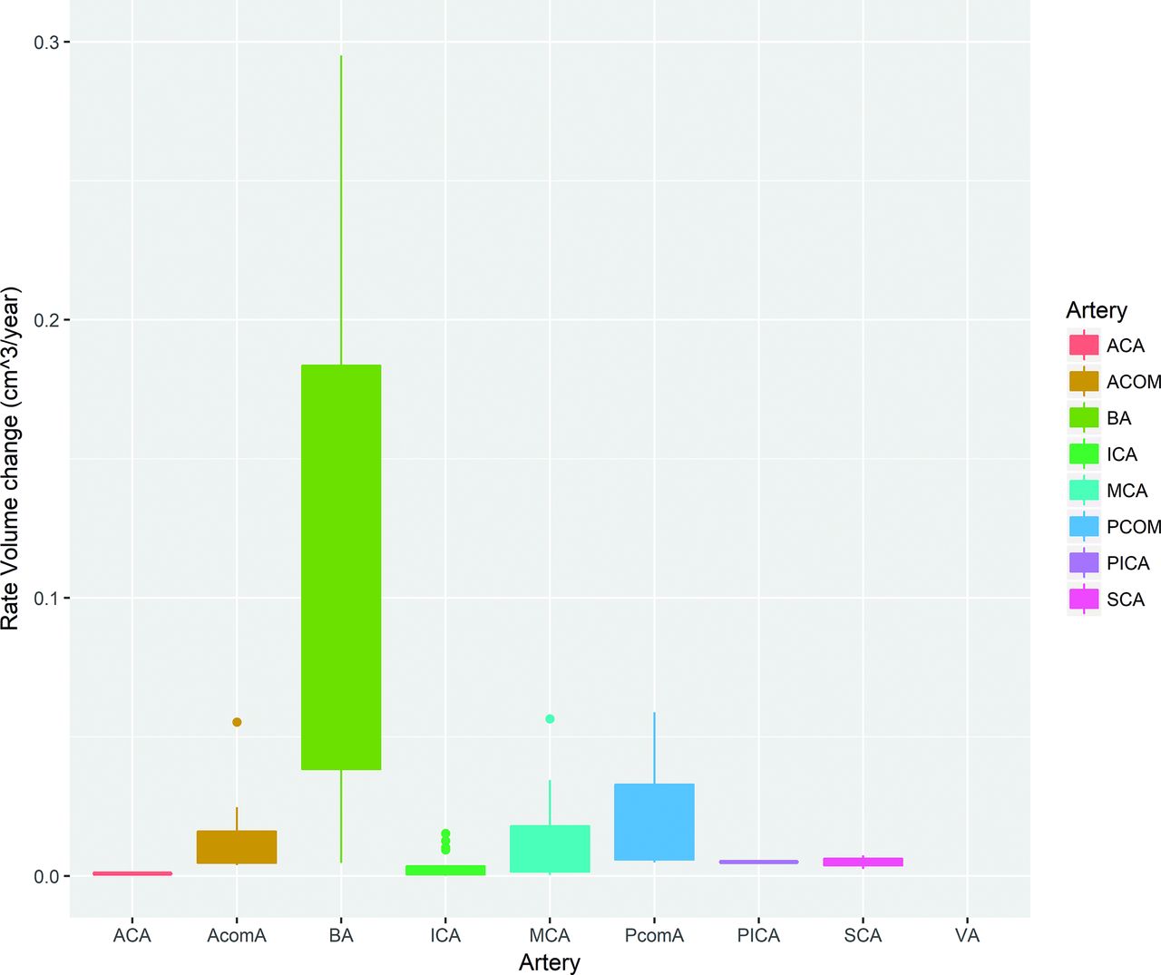
- Fig 3.
Boxplot comparing core-line length (left) and low shear area (right) in the different location categories according to the ELAPSS (upper row) and PHASES (lower row) scores. ACA indicates anterior cerebral artery; PcomA, posterior communicating artery; AcomA, anterior communicating artery.


Tables
Name Description Unit Morphologic characteristics Aneurysm volume Volume of the aneurysm cm3 Aneurysm size Maximum Euclidean distance of the aneurysm surface cm Neck size Maximum Euclidean distance of the neck surface cm Parent vessel diameter Diameter of the vessel nearest to the aneurysm neck cm AR Aspect ratio; aneurysm height/neck size Ratio HWR Height-to-width ratio; aneurysm height/aneurysm width Ratio BF Bottleneck factor; aneurysm width/neck size Ratio BL Bulge location; distance of plane with largest diameter from neck/aneurysm height Ratio SizeR Size ratio Ratio VOR Volume to ostium ratio; aneurysm volume/neck area Ratio IPR Isoperimetric ratio; aneurysm area/aneurysm volume2/3 Ratio UI Undulation index Index EI Ellipticity index Index NSI Nonsphericity index Index GAA Area weighted average of the Gaussian curvature cm–2 Hemodynamic characteristics ICI Inflow concentration index Index SR Shear rate 1/s VE Mean velocity cm/s VO Mean vorticity 1/s WSS Wall shear stress (time averaged mean and maximum) dyne/cm2 LSA Low shear stress area % SCI Shear concentration index Index OSI Oscilatory shear index (time averaged mean and maximum) Index CLL Time-averaged vortex core-line length cm PODE Proper orthogonal decomposition entropy Stable Aneurysms Growing Aneurysms: Baseline Grown Aneurysms: Follow-Up Imaging No. 81 56 56 Sex b Male 11 (14%) 7 (12%) Female 53 (65%) 48 (86%) Unknown 17 (21%) 1 (2%) Artery (%) ACA 5 (6%) 1 (2%) AcomA 8 (10%) 8 (14%) ICA 37 (46%) 18 (32%) MCA 16 (20%) 19 (34%) PcomA 12 (15%) 3 (5%) Posterior circulation 3 (3%) 7 (13%) Previous SAH = yes (%) 2 (2%) 14 (25%)a Configuration = lateral (%) 33 (41%) 18 (32%) Age (mean) (SD) (yr) 59 (18) 55 (14) 59 (13) PHASES (mean) (SD) 3 (2) 4 (3)c 5 (3)a Without hypertension and population 3 (3) 4 (3) 5 (3)c ELAPSS (mean) (SD) 14 (6) 15 (7) 17 (6)b Without population 14 (6) 15 (7) 17 (6)b Hemodynamic characteristics ICI (median) (IQR) 0.4 (0.2–1.0) 0.5 (0.3–1.2) 0.6 (0.3–1.2) SR (median) (IQR) (1/s) 207.3 (124.0–317.5) 190.1 (67.8–273.7) 126.3 (65.1–177.0)a VE (median) (IQR) (cm/s) 9.4 (5.9–13.6) 8.0 (4.6–11.4) 6.6 (3.8–10.2)b VO (median) (IQR) (1/s) 270.5 (165.5–416.2) 256.9 (93.9–384.5) 177.5 (88.3–255.0)a Max WSS (median) (IQR) (dyne/cm2) 175.2 (126.8–239.2) 149.5 (107.6–194.5)c 146.8 (94.7–186.1)b Mean WSS (median) (IQR) (dyne/cm2) 17.5 (11.2–29.9) 16.5 (6.5–24.2) 11.5 (5.4–18.8)a LSA (median) (IQR) (%) 54 (29–74) 53 (31–77) 68 (50–89)c SCI (median) (IQR) 3.2 (1.9–4.6) 2.7 (1.0–4.2) 3.2 (0.2–5.7) Max OSI (median) (IQR) 0.3 (0.1–0.4) 0.2 (0.2–0.4) 0.3 (0.2–0.4) Mean OSI (median) (IQR) 0.01 (0.01–0.01) 0.01 (0.0–0.01) 0.01 (0.01–0.02) CLL (median) (IQR) 0.7 (0.2–1.4) 0.6 (0.4–1.5) 1.1 (0.5–2.0)c PODE (median) (IQR) 0.1 (0.1–0.2) 0.1 (0.1–0.2) 0.2 (0.1–0.3) Note:–ACA indicates anterior cerebral artery; AcomA, anterior communicating artery; PcomA, posterior communicating artery; Q, flow rate; ICI, inflow concentration index; SR, mean aneurysm shear rate; SCI, shear concentration index; max, maximum. P-value compared to stable aneurysms.
↵a P < .001.
↵b P < .02.
↵c P < .05.
Stable Aneurysms Growing Aneurysms: Baseline Grown Aneurysms: Follow-Up Imaging No. 81 56 56 Aneurysm volume (median) (IQR) (cm) 0.02 (0.01–0.11) 0.03 (0.01–0.12) 0.05 (0.02–0.19)b Aneurysm size (median) (IQR) (cm) 0.53 (0.36–0.76) 0.53 (0.41–0.81) 0.64 (0.46–0.97)b Neck size (median) (IQR) (cm) 0.42 (0.30–0.55) 0.43 (0.33–0.62) 0.48 (0.38–0.69)b Parent vessel diameter (median) (IQR) (cm) 0.37 (0.29–0.45) 0.33 (0.28–0.38)b 0.33 (0.28–0.38)b AR (median) (IQR) 0.73 (0.53–1.10) 0.76 (0.56–0.95) 0.88 (0.71–1.08)c HWR (median) (IQR) 0.82 (0.64–0.97) 0.84 (0.70–0.95) 0.90 (0.80–1.08)b BF (median) (IQR) 1.00 (0.87–1.25) 1.03 (0.89–1.25) 1.15 (0.97–1.32)c BL (median) (IQR) 0.30 (0.13–0.44) 0.32 (0.16–0.45) 0.39 (0.26–0.48)b SizeR (median) (IQR) 0.37 (0.29–0.45) 1.82 (1.12–2.49) 2.14 (1.44–3.01)a VOR (median) (IQR) 1.38 (1.07–2.04) 0.25 (0.14–0.59) 0.35 (0.20–0.89)b IPR (median) (IQR) 0.22 (0.10–0.67) 4.71 (4.60–4.85) 4.71 (4.61–4.93) UI (median) (IQR) 4.74 (4.61–4.90) 0.23 (0.14–0.36) 0.19 (0.13–0.27) EI (median) (IQR) 0.21 (0.14–0.35) 0.26 (0.25–0.28) 0.26 (0.24–0.27) NSI (median) (IQR) 0.26 (0.25–0.29) 0.19 (0.17–0.21) 0.19 (0.17–0.22) GAA (median) (IQR) (cm–2) 10.4 (5.0–21.2) 8.7 (4.4–15.9) 6.0 (3.2–10.7)b


