
Article Figures & Data
Figures
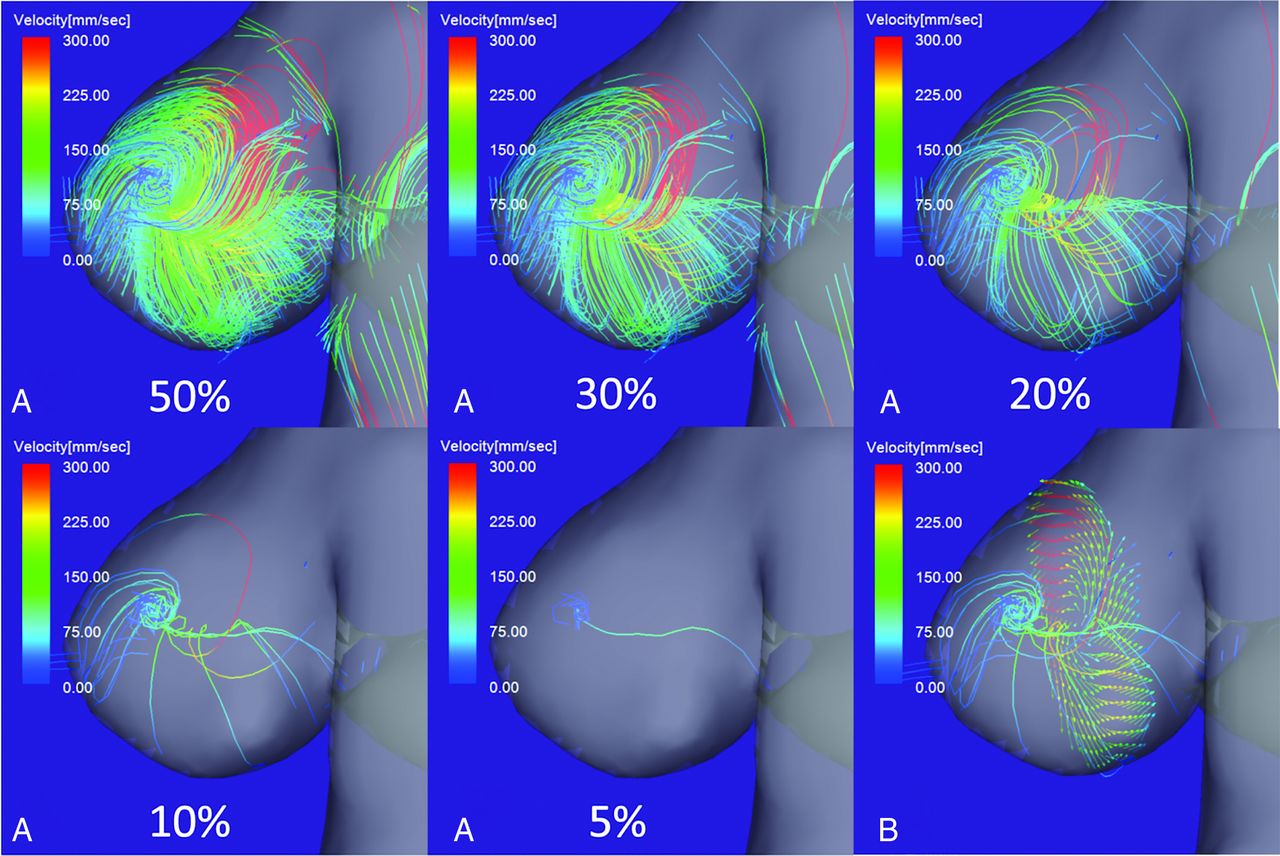
- Fig 1.
4D flow MR images of flow streamlines with velocities below the threshold determined by the percentage value of the maximum inflow velocity. A, When the threshold was decreased from 30% to 10% of the maximum inflow velocity, a single vortex core was visualized as a thin, streamline bundle. At a threshold of 5%, the vortex core was a single line. B, A thin, streamline bundle passed through the center of vortical flow vectors on a cutting plane of the aneurysm dome.
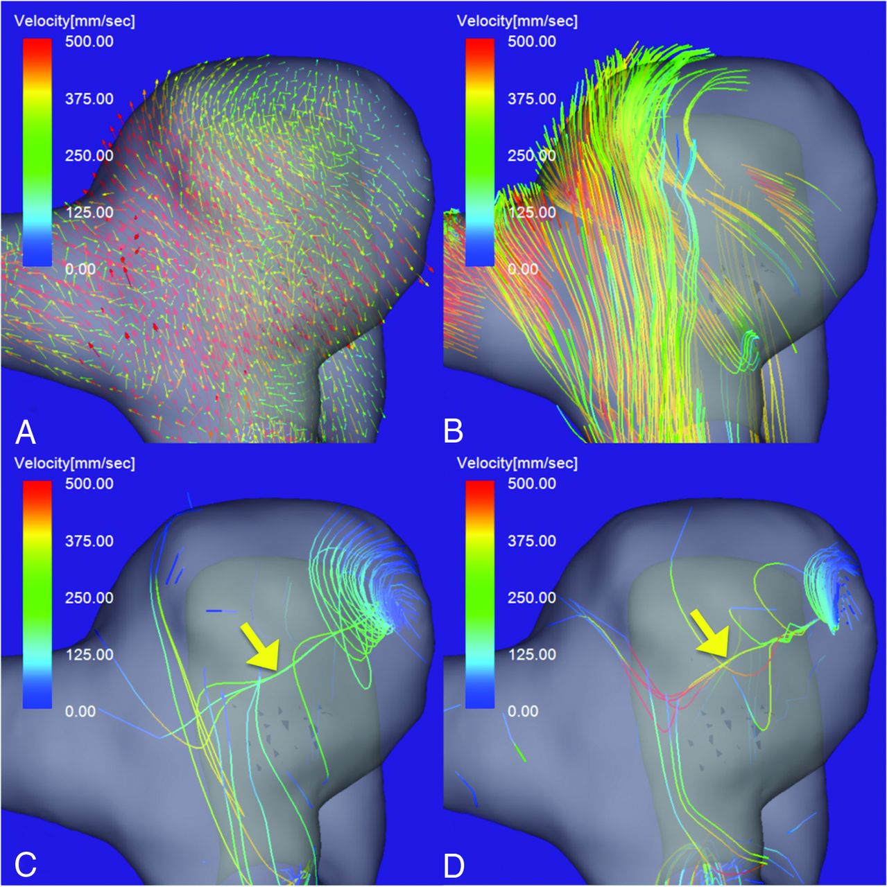
- Fig 2.
4D flow MR images of an unruptured aneurysm on the paraclinoid segment of the right ICA. A, Flow vector map. B, The inflow jet is visualized as a layer of streamlines with high velocities. A single stable vortex core (yellow arrow) is visualized in the diastolic (C) and systolic (D) phases of the cardiac cycle. The vortex core is visualized as a bundle of streamlines with velocities below 7% (C) and 10% (D) of the maximum inflow velocity. The aneurysm flow pattern is simple and stable.
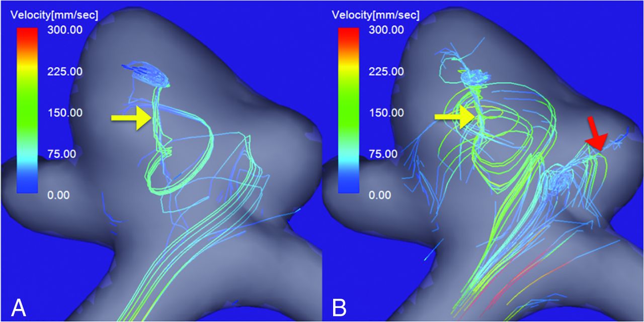
- Fig 3.
An unruptured right MCA bifurcation aneurysm with a daughter sac. Vortex cores in the diastolic (A) and systolic (B) phases of the cardiac cycle. A single vortex core is visualized in the diastolic phase (A, yellow arrow), and another, in the systolic phase (B, red arrow). The vortex cores are visualized as bundles of streamlines with velocities below 4% and 7% of the maximum inflow velocity in the diastolic and systolic phases, respectively. The flow pattern was recorded as complex.
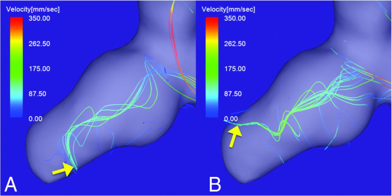
- Fig 4.
A ruptured right ICA aneurysm with a daughter sac. The vortex core in the diastolic (A) and systolic (B) phases of the cardiac cycle. The vortex core in both phases is visualized as a bundle of streamlines with velocities below 4% of the maximum inflow velocity. The direction of the tip of the vortex core markedly changes during the cardiac cycle (yellow arrows). The flow pattern was recorded as unstable.
- Fig 5.
A large ruptured aneurysm on the tip of the basilar artery. A, Flow vector map. Streamlines with velocities below 7% of the maximum inflow velocity in the diastolic (B) and systolic (C) phases. Although multiple small vortices beneath the aneurysm surface and irregular streamlines are visualized, no vortex cores are visualized through the cardiac cycle.


Tables
Aneurysm Complexity Simple/Complex Stability Stable/Unstable Smooth wall (n = 25) 21 (84.0%)/4 (16.0%) 22 (88.0%)/3 (12.0%) Irregular wall (n = 15) 6 (40.0%)/9 (60.0%) 10 (66.7%)/5 (33.3%) P value .006 (S) .126 (NS) Note:—NS indicates not significant; S, significant on the Fisher exact test.
↵a Simple and complex flow patterns were defined as exhibiting a single vortex core or multiple or nonvisualized vortex cores through the cardiac cycle, respectively. Stable and unstable flow patterns were defined as exhibiting persistent vortex cores or moving or nonvisualized vortex cores, respectively. The designation of “irregular wall” was recorded when the wall had protruding blebs or daughter sacs. Differences of P < .05 were considered significant.


