Advertisement

Main menu

  • Home
  • Content
    • Current Issue
    • Accepted Manuscripts
    • Article Preview
    • Past Issue Archive
    • Video Articles
    • AJNR Case Collection
    • Case of the Week Archive
    • Case of the Month Archive
    • Classic Case Archive
  • Special Collections
    • AJNR Awards
    • Low-Field MRI
    • Alzheimer Disease
    • ASNR Foundation Special Collection
    • Photon-Counting CT
    • View All
  • Multimedia
    • AJNR Podcasts
    • AJNR SCANtastic
    • Trainee Corner
    • MRI Safety Corner
    • Imaging Protocols
  • For Authors
    • Submit a Manuscript
    • Submit a Video Article
    • Submit an eLetter to the Editor/Response
    • Manuscript Submission Guidelines
    • Statistical Tips
    • Fast Publishing of Accepted Manuscripts
    • Graphical Abstract Preparation
    • Imaging Protocol Submission
    • Author Policies
  • About Us
    • About AJNR
    • Editorial Board
    • Editorial Board Alumni
  • More
    • Become a Reviewer/Academy of Reviewers
    • Subscribers
    • Permissions
    • Alerts
    • Feedback
    • Advertisers
    • ASNR Home

User menu

  • Alerts
  • Log in

Search

  • Advanced search
American Journal of Neuroradiology

American Journal of Neuroradiology

ASHNR American Society of Functional Neuroradiology ASHNR American Society of Pediatric Neuroradiology ASSR
  • Alerts
  • Log in

Advanced Search

  • Home
  • Content
    • Current Issue
    • Accepted Manuscripts
    • Article Preview
    • Past Issue Archive
    • Video Articles
    • AJNR Case Collection
    • Case of the Week Archive
    • Case of the Month Archive
    • Classic Case Archive
  • Special Collections
    • AJNR Awards
    • Low-Field MRI
    • Alzheimer Disease
    • ASNR Foundation Special Collection
    • Photon-Counting CT
    • View All
  • Multimedia
    • AJNR Podcasts
    • AJNR SCANtastic
    • Trainee Corner
    • MRI Safety Corner
    • Imaging Protocols
  • For Authors
    • Submit a Manuscript
    • Submit a Video Article
    • Submit an eLetter to the Editor/Response
    • Manuscript Submission Guidelines
    • Statistical Tips
    • Fast Publishing of Accepted Manuscripts
    • Graphical Abstract Preparation
    • Imaging Protocol Submission
    • Author Policies
  • About Us
    • About AJNR
    • Editorial Board
    • Editorial Board Alumni
  • More
    • Become a Reviewer/Academy of Reviewers
    • Subscribers
    • Permissions
    • Alerts
    • Feedback
    • Advertisers
    • ASNR Home
  • Follow AJNR on Twitter
  • Visit AJNR on Facebook
  • Follow AJNR on Instagram
  • Join AJNR on LinkedIn
  • RSS Feeds

AJNR Awards, New Junior Editors, and more. Read the latest AJNR updates

Research ArticleNeurointervention

The Influence of Angioarchitectural Features on the Success of Endovascular Embolization of Cranial Dural Arteriovenous Fistulas with Onyx

D.F. Vollherbst, C. Herweh, S. Schönenberger, F. Seker, S. Nagel, P.A. Ringleb, M. Bendszus and M.A. Möhlenbruch
American Journal of Neuroradiology December 2019, 40 (12) 2130-2136; DOI: https://doi.org/10.3174/ajnr.A6326
D.F. Vollherbst
aFrom the Departments of Neuroradiology (D.F.V., C.H., F.S., M.B., M.A.M.)
  • Find this author on Google Scholar
  • Find this author on PubMed
  • Search for this author on this site
  • ORCID record for D.F. Vollherbst
C. Herweh
aFrom the Departments of Neuroradiology (D.F.V., C.H., F.S., M.B., M.A.M.)
  • Find this author on Google Scholar
  • Find this author on PubMed
  • Search for this author on this site
  • ORCID record for C. Herweh
S. Schönenberger
bNeurology (S.S., S.N., P.A.R.), Heidelberg University Hospital, Heidelberg, Germany.
  • Find this author on Google Scholar
  • Find this author on PubMed
  • Search for this author on this site
  • ORCID record for S. Schönenberger
F. Seker
aFrom the Departments of Neuroradiology (D.F.V., C.H., F.S., M.B., M.A.M.)
  • Find this author on Google Scholar
  • Find this author on PubMed
  • Search for this author on this site
  • ORCID record for F. Seker
S. Nagel
bNeurology (S.S., S.N., P.A.R.), Heidelberg University Hospital, Heidelberg, Germany.
  • Find this author on Google Scholar
  • Find this author on PubMed
  • Search for this author on this site
  • ORCID record for S. Nagel
P.A. Ringleb
bNeurology (S.S., S.N., P.A.R.), Heidelberg University Hospital, Heidelberg, Germany.
  • Find this author on Google Scholar
  • Find this author on PubMed
  • Search for this author on this site
  • ORCID record for P.A. Ringleb
M. Bendszus
aFrom the Departments of Neuroradiology (D.F.V., C.H., F.S., M.B., M.A.M.)
  • Find this author on Google Scholar
  • Find this author on PubMed
  • Search for this author on this site
  • ORCID record for M. Bendszus
M.A. Möhlenbruch
aFrom the Departments of Neuroradiology (D.F.V., C.H., F.S., M.B., M.A.M.)
  • Find this author on Google Scholar
  • Find this author on PubMed
  • Search for this author on this site
  • ORCID record for M.A. Möhlenbruch
  • Article
  • Figures & Data
  • Info & Metrics
  • Responses
  • References
  • PDF
Loading

Article Figures & Data

Figures

  • Tables
  • Fig 1.
    • Download figure
    • Open in new tab
    • Download powerpoint
    Fig 1.

    Sample case of a DAVF without angioarchitectural features with a negative influence on the treatment success. A DAVF with direct cortical venous drainage (Cognard type III), located at the tentorium in a 65-year-old man with headaches (A). Involved feeding arteries are the middle meningeal artery (black arrows in A) and the occipital artery (white arrows in A), which shunt directly into a cortical vein (white arrowheads). There were <10 feeding arteries, and the ascending pharyngeal artery was not involved. The DAVF could be occluded completely by transarterial embolization with Onyx (B). DSA 6 months after embolization shows stable occlusion of the DAVF (C).
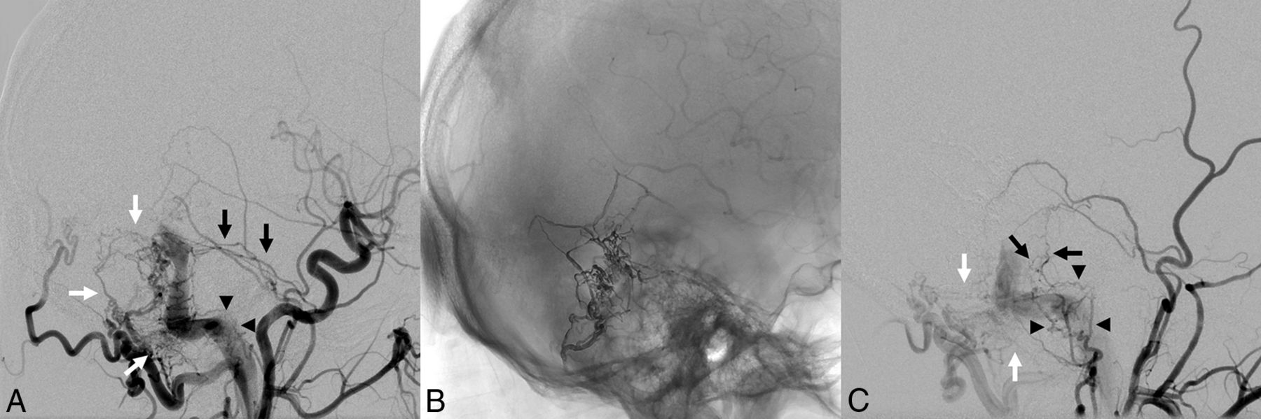
  • Fig 2.
    • Download figure
    • Open in new tab
    • Download powerpoint
    Fig 2.

    Sample case of a DAVF presenting with angioarchitectural features with a negative influence on the treatment success. A DAVF is located at the transverse and sigmoid sinus with antegrade flow in the sinus (Cognard type I) in a 46-year-old man who presented with severe pulsatile tinnitus (A). Multiple feeders supply the DAVF, including the middle meningeal artery (black arrows in A), the occipital artery (white arrows in A), and the ascending pharyngeal artery (black arrowheads in A). The DAVF was treated by transarterial Onyx embolization in combination with transvenous balloon-assisted protection of the venous sinus (B). The DAVF could not be occluded completely, due to small branches of the middle meningeal artery (black arrows in C) and of the occipital artery (white arrows in C), and particularly because of multiple persistent feeders from the ascending pharyngeal artery (black arrowheads in C), which could not be catheterized distal enough because of their small size and their tortuosity. The patient’s symptoms were declining but still persistent at the latest follow-up.

Tables

  • Figures
    • View popup
    Table 1:

    Angioarchitectural features

    FeatureNo. (relative frequency)
    Location
     Transverse and/or sigmoid sinus46 (41.8%)
     Tentorial/petrosal25 (22.7%)
     Superior sagittal sinus20 (18.2%)
     Torcular7 (6.4%)
     Anterior cranial fossa6 (5.5%)
     Sphenoparietal sinus4 (3.6%)
     Others2 (1.8%)
    Cognard and Borden type
     Cognard I12 (10.9%)
     Cognard IIa7 (6.4%)
     Cognard IIb2 (1.8%)
     Cognard IIa+b10 (9.1%)
     Cognard III28 (25.5%)
     Cognard IV51 (46.4%)
     Borden I19 (17.3%)
     Borden II11 (10.9%)
     Borden III79 (71.8%)
    Feeder territories
     Middle meningeal artery96 (87.3%)
     Occipital artery83 (75.5%)
     Internal carotid artery (dural branches)41 (37.3%)
     Verterbral artery (dural branches)41 (37.3%)
     Superficial temporal artery32 (29.1%)
     Ascending pharyngeal artery27 (24.5%)
    Other features
     DAVFs with <10 arterial feeders56 (50.9%)
     DAVFs with ≥10 arterial feeders54 (49.1%)
     Bilateral feeders56 (50.9%)
     Pial artery supply41 (37.3%)
    • View popup
    Table 2:

    Procedural parameters, complications, follow-up, and outcome

    ParameterNo. (relative frequency) / Mean ± SD
    Selected feeder for embolization
     Middle meningeal artery86 (65.2%)
     Occipital artery31 (23.5%)
     Others14 (10.6%)
    No. of embolization positions
     1103 (78.0%)
     224 (18.2%)
     34 (3.0%)
    Embolization technique
     Onyx transarterial76 (57.6%)
     Onyx transarterial combined with venous balloon protection28 (21.2%)
     Onyx transarterial with a dual-lumen balloon catheter19 (14.4%)
     Onyx transvenous combined with coiling3 (2.3%)
     Others7 (5.3%)
    Complications
     Overall complications11 (8.3%)
     Asymptomatic complications6 (4.5%)
     Transient symptomatic complications3 (2.3%)
     Permanent complicationsa2 (1.5%)
    Follow-up
     Follow-up period (mo)23.6 ± 23.5
     Total follow-up time (patient yr)216.3
    Angiographic outcome
     Initial complete angiographic occlusion86 (78.2%)
     Spontaneous occlusion after subtotal endovascular occlusion14 (12.7%)
     Time period from last treatment to diagnosis of spontaneous occlusion (months)6.6 ± 8.1
     Overall complete occlusion at last examination100 (90.9%)
     Recurrence4 (3.6%)
    Clinical outcome
     Preinterventional mRS score0.8 ± 0.9
     Postinterventional mRS score at discharge0.7 ± 1.1
     Postinterventional mRS score at 6 mo after treatment0.4 ± 0.9
     Complete symptom remission after treatment73 (66.4%)
     Symptom relief after treatment32 (29.1%)
     Stable symptoms after treatment3 (2.7%)
     Worsening of symptoms after treatment2 (1.8%)
    Treatment successb90 (81.8%)
    • ↵a Both permanent complications were lethal intracerebral hemorrhages.

    • ↵b Defined as complete symptom remission for low-grade DAVFs (no cortical venous reflux/Cognard I–IIa) and complete angiographic occlusion for high-grade DAVFs (to presence of cortical venous reflux/Cognard IIa+b to IV).

    • View popup
    Table 3:

    Influence of angioarchitectural features on the treatment success—summary and univariate models

    Angioarchitectural Feature, Treatment Success RateP Value (Univariate Analysis)
    LocationTransverse/Sigmoid Sinus, 80.4%Tentorial/Petrosal, 72.0%Superior Sagittal Sinus, 95.0%Torcular, 100%Anterior Cranial Fossa, 83.3%Sphenoparietal Sinus, 75.0%.084
    Cognard typeI, 60.0%IIa, 75.0%IIb, 100%IIa+b 70.0%III, 96.4%IV, 80.4%.047a
    Borden typeI, 66.7%II, 76.9%III, 86.1%.144
    Venous drainageInto dural venous sinus, 71.4%Directly into cortical veins, 85.4%.099
    Presence of cortical venous refluxYes, 85.9%No, 61.1%.013
    No. of feeding arteries<10, 92.6%≥10, 71.4%.004a
    Bilateral feedersYes, 83.3%No, 80.4%.686
    Pial artery supplyYes, 77.8%No, 83.1%.531
    Involvement of the middle meningeal arteryYes, 80.2%No, 92.9%.252
    Involvement of the occipital arteryYes, 80.7%No, 85.2%.602
    Involvement of the internal carotid arterybYes, 70.7%No, 88.4%.020a
    Involvement of the vertebral arterybYes, 75.6%No, 85.5%.193
    Involvement of the superficial temporal arteryYes, 81.3%No, 82.1%.921
    Involvement of the ascending pharyngeal arteryYes, 61.5%No, 88.1%.002a
    • ↵a Statistically significant.

    • ↵b Dural branches.

    • View popup
    Table 4:

    Influence of angioarchitectural features on the treatment success—final multivariable logistic regression model

    Angioarchitectural FeatureP ValueOdds Ratio (95% Confidence Interval)
    No. of feeding arteries.0410.278 (0.081–0.949)
    Involvement of the ascending pharyngeal artery.0390.319 (0.107–0.945)
PreviousNext
Back to top

In this issue

American Journal of Neuroradiology: 40 (12)
American Journal of Neuroradiology
Vol. 40, Issue 12
1 Dec 2019
  • Table of Contents
  • Index by author
  • Complete Issue (PDF)
Advertisement
Print
Download PDF
Email Article

Thank you for your interest in spreading the word on American Journal of Neuroradiology.

NOTE: We only request your email address so that the person you are recommending the page to knows that you wanted them to see it, and that it is not junk mail. We do not capture any email address.

Enter multiple addresses on separate lines or separate them with commas.
The Influence of Angioarchitectural Features on the Success of Endovascular Embolization of Cranial Dural Arteriovenous Fistulas with Onyx
(Your Name) has sent you a message from American Journal of Neuroradiology
(Your Name) thought you would like to see the American Journal of Neuroradiology web site.
CAPTCHA
This question is for testing whether or not you are a human visitor and to prevent automated spam submissions.
Cite this article
D.F. Vollherbst, C. Herweh, S. Schönenberger, F. Seker, S. Nagel, P.A. Ringleb, M. Bendszus, M.A. Möhlenbruch
The Influence of Angioarchitectural Features on the Success of Endovascular Embolization of Cranial Dural Arteriovenous Fistulas with Onyx
American Journal of Neuroradiology Dec 2019, 40 (12) 2130-2136; DOI: 10.3174/ajnr.A6326

Citation Manager Formats

  • BibTeX
  • Bookends
  • EasyBib
  • EndNote (tagged)
  • EndNote 8 (xml)
  • Medlars
  • Mendeley
  • Papers
  • RefWorks Tagged
  • Ref Manager
  • RIS
  • Zotero
0 Responses
Respond to this article
Share
Bookmark this article
The Influence of Angioarchitectural Features on the Success of Endovascular Embolization of Cranial Dural Arteriovenous Fistulas with Onyx
D.F. Vollherbst, C. Herweh, S. Schönenberger, F. Seker, S. Nagel, P.A. Ringleb, M. Bendszus, M.A. Möhlenbruch
American Journal of Neuroradiology Dec 2019, 40 (12) 2130-2136; DOI: 10.3174/ajnr.A6326
del.icio.us logo Twitter logo Facebook logo Mendeley logo
  • Tweet Widget
  • Facebook Like
  • Google Plus One
Purchase

Jump to section

  • Article
    • Abstract
    • ABBREVIATION:
    • MATERIALS AND METHODS
    • RESULTS
    • DISCUSSION
    • CONCLUSIONS
    • Footnotes
    • References
  • Figures & Data
  • Info & Metrics
  • Responses
  • References
  • PDF

Related Articles

  • No related articles found.
  • PubMed
  • Google Scholar

Cited By...

  • Pioneering artificial intelligence-based real time assistance for intracranial liquid embolization in humans: an initial experience
  • Efficacy and safety of the PHIL embolic agent in the treatment of intracranial dural arteriovenous fistulas: results of the PHIL-dAVF study
  • LIQUID - Treatment of high-grade dural arteriovenous fistulas with Squid liquid embolic agent: a prospective, observational multicenter study
  • Crossref
  • Google Scholar

This article has not yet been cited by articles in journals that are participating in Crossref Cited-by Linking.

More in this TOC Section

  • A Retrospective Study in Tentorial DAVFs
  • Proximal Protection Devices for Carotid Stenting
  • Hydrocoils: an Updated Meta-Analysis
Show more NEUROINTERVENTION

Similar Articles

Advertisement

Indexed Content

  • Current Issue
  • Accepted Manuscripts
  • Article Preview
  • Past Issues
  • Editorials
  • Editor's Choice
  • Fellows' Journal Club
  • Letters to the Editor
  • Video Articles

Cases

  • Case Collection
  • Archive - Case of the Week
  • Archive - Case of the Month
  • Archive - Classic Case

More from AJNR

  • Trainee Corner
  • Imaging Protocols
  • MRI Safety Corner
  • Book Reviews

Multimedia

  • AJNR Podcasts
  • AJNR Scantastics

Resources

  • Turnaround Time
  • Submit a Manuscript
  • Submit a Video Article
  • Submit an eLetter to the Editor/Response
  • Manuscript Submission Guidelines
  • Statistical Tips
  • Fast Publishing of Accepted Manuscripts
  • Graphical Abstract Preparation
  • Imaging Protocol Submission
  • Evidence-Based Medicine Level Guide
  • Publishing Checklists
  • Author Policies
  • Become a Reviewer/Academy of Reviewers
  • News and Updates

About Us

  • About AJNR
  • Editorial Board
  • Editorial Board Alumni
  • Alerts
  • Permissions
  • Not an AJNR Subscriber? Join Now
  • Advertise with Us
  • Librarian Resources
  • Feedback
  • Terms and Conditions
  • AJNR Editorial Board Alumni

American Society of Neuroradiology

  • Not an ASNR Member? Join Now

© 2025 by the American Society of Neuroradiology All rights, including for text and data mining, AI training, and similar technologies, are reserved.
Print ISSN: 0195-6108 Online ISSN: 1936-959X

Powered by HighWire