
Article Figures & Data
Figures
- Fig 1.
The FLAIR images of 3 patients acquired at 1 month after CO intoxication. A, A male patient (32 years of age) who had only GP lesions did not develop DNS. B, A female patient (37 years of age) who had only diffuse WMLs developed DNS. C, A male patient (49 years of age) who had both diffuse WMLs and GP lesions developed DNS.
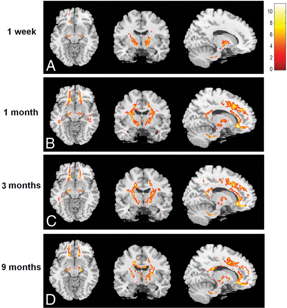
- Fig 2.
The voxelwise comparison of FA values between patients with WMLs and healthy controls. The yellow-to-red areas indicate regions with values that were significantly decreased in the patients, and the color bar on the right-hand side indicates T values.
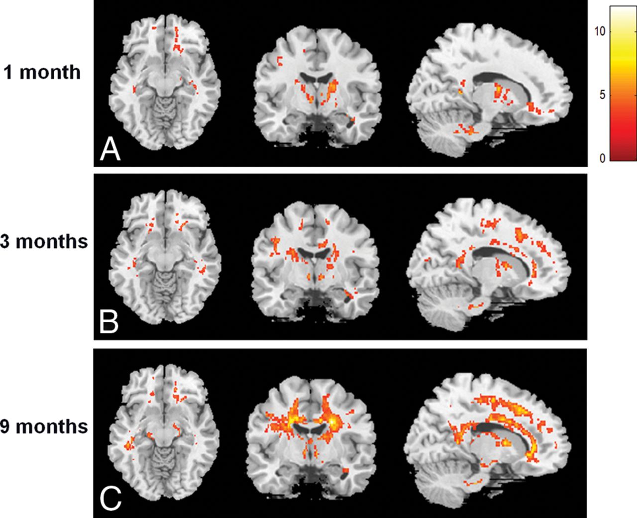
- Fig 3.
The voxelwise comparison of MK values between patients with WMLs and healthy controls. The yellow-to-red areas indicate regions with values that were significantly decreased in the patients, and the color bar on the right-hand side indicates T values.


Tables
Patients with CO Intoxication Controls WML Non-WML Sex (M/F) 6:5 5:8 10:11 Age (mean) (yr) 44.5 ± 15.3 35.7 ± 9.3 41.0 ± 11.9 Duration of coma (mean) (hr) 44.7 ± 48.8a 11.7 ± 17.6a NA Carboxyhemoglobin (mean) (%) 22.3 ± 20.9 31.37 ± 15.1 NA Sessions of HBOT (mean) 11.2 ± 10.1 8.9 ± 2.5 NA DNS (No.) (M/F) 5 (3:2) 0 NA GP involvement (No.) (M/F) 8 (5:3) 4 (2:2) NA Note:—HBOT indicates hyperbaric oxygen therapy; NA, not applicable.
↵a P < .05.
- Table 2:
The ROC analysis of diffusional indices for CP, IC, and FWM in differentiating patients with DNS from those without DNS at 1 week after CO intoxicationa
Brain Regions (MNI Coordinates) (mm) DNS Non-DNS Controls AUC (DNS vs non-DNS) Lt. CP (−14, −14, −12) FA (mean) 0.52 ± 0.03 0.56 ± 0.06 0.60 ± 0.05 .7394 Rt. IC (17, −10, 0) AD (mean) 1.38 ± 0.08 1.49 ± 0.14 1.49 ± 0.08 .7394 AK (mean) 0.87 ± 0.05 0.74 ± 0.08 0.80 ± 0.03 .9156b Lt. IC (−17, −10, 0) AD (mean) 1.38 ± 0.06 1.49 ± 0.13 1.48 ± 0.11 .7762 AK (mean) 0.89 ± 0.06 0.80 ± 0.08 0.81 ± 0.08 .7113 Rt. FWM (14, 31, −12) AD (mean) 1.30 ± 0.03 1.18 ± 0.14 1.19 ± 0.14 .7436 AK (mean) 1.03 ± 0.04 0.86 ± 0.12 0.83 ± 0.09 .9057b Lt. FWM (−14, 31, −12) RD (mean) 0.78 ± 0.04 0.74 ± 0.07 0.62 ± 0.07 .7436 MD (mean) 0.94 ± 0.03 0.94 ± 0.05 0.90 ± 0.06 .7299 MK (mean) 0.98 ± 0.06 0.86 ± 0.07 0.89 ± 0.12 .8627b


Jump to section
Related Articles
Cited By...
- No citing articles found.