Advertisement

Main menu

  • Home
  • Content
    • Current Issue
    • Accepted Manuscripts
    • Article Preview
    • Past Issue Archive
    • AJNR Case Collection
    • Case of the Week Archive
    • Classic Case Archive
    • Case of the Month Archive
  • Special Collections
    • Spinal CSF Leak Articles (Jan 2020-June 2024)
    • 2024 AJNR Journal Awards
    • Most Impactful AJNR Articles
  • Multimedia
    • AJNR Podcast
    • AJNR Scantastics
    • Video Articles
  • For Authors
    • Submit a Manuscript
    • Author Policies
    • Fast publishing of Accepted Manuscripts
    • Graphical Abstract Preparation
    • Manuscript Submission Guidelines
    • Imaging Protocol Submission
    • Submit a Case for the Case Collection
  • About Us
    • About AJNR
    • Editorial Board
  • More
    • Become a Reviewer/Academy of Reviewers
    • Subscribers
    • Permissions
    • Alerts
    • Feedback
    • Advertisers
    • ASNR Home
  • Other Publications
    • ajnr

User menu

  • Alerts
  • Log in
  • Log out

Search

  • Advanced search
American Journal of Neuroradiology

American Journal of Neuroradiology

ASHNR American Society of Functional Neuroradiology ASHNR American Society of Pediatric Neuroradiology ASSR
  • Alerts
  • Log in
  • Log out

Advanced Search

  • Home
  • Content
    • Current Issue
    • Accepted Manuscripts
    • Article Preview
    • Past Issue Archive
    • AJNR Case Collection
    • Case of the Week Archive
    • Classic Case Archive
    • Case of the Month Archive
  • Special Collections
    • Spinal CSF Leak Articles (Jan 2020-June 2024)
    • 2024 AJNR Journal Awards
    • Most Impactful AJNR Articles
  • Multimedia
    • AJNR Podcast
    • AJNR Scantastics
    • Video Articles
  • For Authors
    • Submit a Manuscript
    • Author Policies
    • Fast publishing of Accepted Manuscripts
    • Graphical Abstract Preparation
    • Manuscript Submission Guidelines
    • Imaging Protocol Submission
    • Submit a Case for the Case Collection
  • About Us
    • About AJNR
    • Editorial Board
  • More
    • Become a Reviewer/Academy of Reviewers
    • Subscribers
    • Permissions
    • Alerts
    • Feedback
    • Advertisers
    • ASNR Home
  • Follow AJNR on Twitter
  • Visit AJNR on Facebook
  • Follow AJNR on Instagram
  • Join AJNR on LinkedIn
  • RSS Feeds

Welcome to the new AJNR, Updated Hall of Fame, and more. Read the full announcements.


AJNR is seeking candidates for the position of Associate Section Editor, AJNR Case Collection. Read the full announcement.


Research ArticleInterventional

Aneurysm Remnants after Flow Diversion: Clinical and Angiographic Outcomes

T.P. Madaelil, J.A. Grossberg, B.M. Howard, C.M. Cawley, J. Dion, R.G. Nogueira, D.C. Haussen and F.C. Tong
American Journal of Neuroradiology April 2019, 40 (4) 694-698; DOI: https://doi.org/10.3174/ajnr.A6010
T.P. Madaelil
aFrom the Departments of Neurointerventional Radiology and Neurosurgery (T.P.M., J.A.G., B.M.H., C.M.C., F.C.T.), Emory University School of Medicine, Atlanta, Georgia
  • Find this author on Google Scholar
  • Find this author on PubMed
  • Search for this author on this site
  • ORCID record for T.P. Madaelil
J.A. Grossberg
aFrom the Departments of Neurointerventional Radiology and Neurosurgery (T.P.M., J.A.G., B.M.H., C.M.C., F.C.T.), Emory University School of Medicine, Atlanta, Georgia
  • Find this author on Google Scholar
  • Find this author on PubMed
  • Search for this author on this site
  • ORCID record for J.A. Grossberg
B.M. Howard
aFrom the Departments of Neurointerventional Radiology and Neurosurgery (T.P.M., J.A.G., B.M.H., C.M.C., F.C.T.), Emory University School of Medicine, Atlanta, Georgia
  • Find this author on Google Scholar
  • Find this author on PubMed
  • Search for this author on this site
  • ORCID record for B.M. Howard
C.M. Cawley
aFrom the Departments of Neurointerventional Radiology and Neurosurgery (T.P.M., J.A.G., B.M.H., C.M.C., F.C.T.), Emory University School of Medicine, Atlanta, Georgia
  • Find this author on Google Scholar
  • Find this author on PubMed
  • Search for this author on this site
  • ORCID record for C.M. Cawley
J. Dion
cMicroVention (J.D.), Aliso Viejo, California.
  • Find this author on Google Scholar
  • Find this author on PubMed
  • Search for this author on this site
  • ORCID record for J. Dion
R.G. Nogueira
bMarcus Stroke and Neuroscience Center (R.G.N., D.C.H.), Departments of Neurology and Neurosurgery, Grady Memorial Hospital, Atlanta, Georgia
  • Find this author on Google Scholar
  • Find this author on PubMed
  • Search for this author on this site
  • ORCID record for R.G. Nogueira
D.C. Haussen
bMarcus Stroke and Neuroscience Center (R.G.N., D.C.H.), Departments of Neurology and Neurosurgery, Grady Memorial Hospital, Atlanta, Georgia
  • Find this author on Google Scholar
  • Find this author on PubMed
  • Search for this author on this site
  • ORCID record for D.C. Haussen
F.C. Tong
aFrom the Departments of Neurointerventional Radiology and Neurosurgery (T.P.M., J.A.G., B.M.H., C.M.C., F.C.T.), Emory University School of Medicine, Atlanta, Georgia
  • Find this author on Google Scholar
  • Find this author on PubMed
  • Search for this author on this site
  • ORCID record for F.C. Tong
  • Article
  • Figures & Data
  • Info & Metrics
  • Responses
  • References
  • PDF
Loading

Abstract

BACKGROUND AND PURPOSE: Flow diversion is an established method to treat complex intracranial aneurysms. The natural history of flow-diversion treatment failure resulting in aneurysm remnants is not well-defined. We aimed to delineate the clinical and angiographic features of this entity.

MATERIALS AND METHODS: Review of a prospectively maintained Pipeline Embolization Device data base from inception to October 2017 was performed for aneurysms that demonstrated residual filling on follow-up imaging. Procedural and follow-up clinical details were recorded. Independent, blinded, angiographic assessment of occlusion was performed on the basis of the O'Kelly-Marotta scale. Aggregated outcomes were analyzed using the Fisher exact and Mann-Whitney U tests for categoric and continuous variables, respectively (statistical significance, α = .05).

RESULTS: During the study period, 283 sequential patients were treated; 87% (246/283) were women. The median patient age was 55 years (interquartile range, 47–65 years). Six-month follow-up imaging was available in 83.7% (237/283) of patients, which showed 62.4% (148/237) complete occlusion (class D, O'Kelly-Marotta grading scale). Adjunctive coiling (P = .06), on-label Pipeline Embolization Device use (P = .04), and multiple device constructs (P = .02) had higher rates of complete occlusion at 6 months. Aneurysm remnants were identified in 25 cases on long-term follow-up imaging (median, 16 months; interquartile range, 12–24 months). No patient with an aneurysm remnant after flow diversion presented with delayed rupture or other clinical sequelae, with a median clinical follow-up of 31 months (interquartile range, 23–33 months).

CONCLUSIONS: Aneurysm remnants after flow diversion are infrequent with minimal clinical impact. When appropriate, the presence of overlapping devices and possibly adjunctive coiling may result in higher rates of complete occlusion.

ABBREVIATIONS:

IQR
interquartile range
OKM
O'Kelly-Marotta grading scale

Flow diversion with the Pipeline Embolization Device (PED; Covidien, Irvine, California) was first reported in 2008.1 Since that time, multiple trials2,3 and retrospective case series4,5 have supported the role of the PED in the treatment of complex intracranial aneurysms. Current follow-up data show high rates of angiographic occlusion at 5 years with only a 4.8% rate of aneurysm persistence and no evidence of recanalization of previously occluded aneurysms.6 In contrast, coil embolization of intracranial aneurysms results in higher rates of aneurysm persistence.7 Increasingly, natural history data describing the angiographic8 and clinical outcomes9 of aneurysm remnants after endovascular therapy are now available. Preliminary evidence suggests that the type of aneurysm remnant (neck versus body filling) dictates the rate of recanalization after coil embolization (with neck filling having lower rates of recanalization compared with body filling)8 and remnants of previously ruptured aneurysms are at a higher risk of rerupture.9 However, long-term imaging and clinical data with respect to aneurysm remnants after flow diversion remain sparse. Our goal was to evaluate the longitudinal angiographic and clinical outcomes of aneurysm remnants after flow diversion with the PED.

Materials and Methods

A prospectively maintained institutional (Emory University) data base of patients treated with the PED from 2011 through October 2017 was searched for patients who demonstrated residual filling on follow-up imaging. Patient-level information regarding aneurysm characteristics, procedural details, and clinical follow-up was collected into an electronic data base. The main exclusion criterion was ruptured aneurysms treated in the acute or subacute period (0–14 days). Collected data points included patient characteristics of age, sex, family history of aneurysms, tobacco use; aneurysm characteristics, including type, size, and location; and procedural details such as the number of devices used, adjunctive coiling, and clinical outcome along with the duration of both clinical and radiographic follow-up. If data were available at 6 months, changes to dual antiplatelet therapy after aneurysm occlusion status were noted. Incomplete occlusion status was defined as classes A–C based on the O'Kelly-Marotta (OKM) grading scale10 for assessment of aneurysms treated by flow diversion. Independent, retrospective review of follow-up imaging was completed by a fellowship-trained neuroradiologist (3 years of dedicated cerebrovascular experience) who did not participate in the initial procedure. Complete occlusion was defined as class D based on the OKM grading scale.

The association between demographic and clinical risk factors with incomplete occlusion and long-term clinical outcome for patients with incomplete occlusion versus patients with complete occlusion was evaluated using the Fisher exact and Mann-Whitney U test for categoric and continuous variables, respectively. The threshold of statistical significance was α = .05. All statistical analyses were performed using SPSS, Version 22 (IBM, Armonk, New York) and Excel 2007 (Microsoft, Redmond, Washington).

Results

During the study period, 296 interventions were performed in 283 patients to treat 294 aneurysms. Overwhelmingly, the patients treated were women (87%). The median patient age was 55 years (interquartile range [IQR], 47–65 years). Minimum 6-month follow-up imaging was available in 83.7% (237/283) of patients, which included either DSA (50.2%; 119/237), MRA and DSA (36.3%; 86/237), or MRA (13.5%; 32/237). Table 1 provides summary demographic data.

View this table:
  • View inline
  • View popup
Table 1:

Basic clinical and imaging demographics

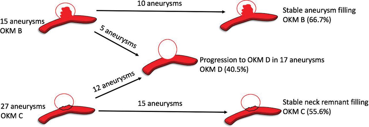
At 6 months, 62.4% (148/237) of aneurysms demonstrated complete occlusion (class D, OKM scale), 24.1% (57/237) of aneurysms had an entry remnant (class C, OKM scale), and 13.5% (32/237) of aneurysms had subtotal filling (class B, OKM scale). In patients with available late surveillance imaging at a median duration of 16 months (IQR, 12–24 months), progressive complete occlusion was observed in 40.5% (17/42) of aneurysms (class D, OKM scale). The most common dual antiplatelet regimen (73.4%; 174/237) at midterm follow-up imaging was 75 mg of clopidogrel and 325 mg of aspirin. Tapering of antiplatelet therapy was initiated by the treating physician only if progressive aneurysm occlusion was noted on midterm imaging and in the absence of in-stent intimal hyperplasia. The precise regimen was both patient- and operator-specific. Higher rates of progressive complete occlusion were observed in aneurysms with remnant necks (44.4%, 12/27; class C, OKM scale) as opposed to aneurysms with subtotal filling (33.3%, 5/15; class B, OKM scale), though this trend was not statistically significant (P = .53). Figure 1 demonstrates progressive occlusion rates of aneurysms with residual filling on late surveillance imaging in a pictorial format.

Fig 1.
  • Download figure
  • Open in new tab
  • Download powerpoint
Fig 1.

Occlusion status on late-surveillance follow-up imaging.

Procedural characteristics and aneurysm morphology played an important role in aneurysm occlusion on midterm (6 month) imaging follow-up. Adjunctive coiling during the initial PED placement resulted in higher rates of complete occlusion at 6 months (73.9%, 34/46 versus 58.6%, 99/169); this result almost reached statistical significance (P = .06). On-label use of the PED was associated with a higher rate of occlusion (73.0%, 54/74) than off-label use (58.5%, 93/159) (P = .04). In addition, deployment of >1 device resulted in higher rates of complete occlusion at 6 months (79.5%, 31/39) versus a single device (58.9%, 116/197) (P = .02). No correlation between wide-neck aneurysms (>4 mm; P = .29) or diameter (>10 mm; P = .52) and the rate of occlusion was found.

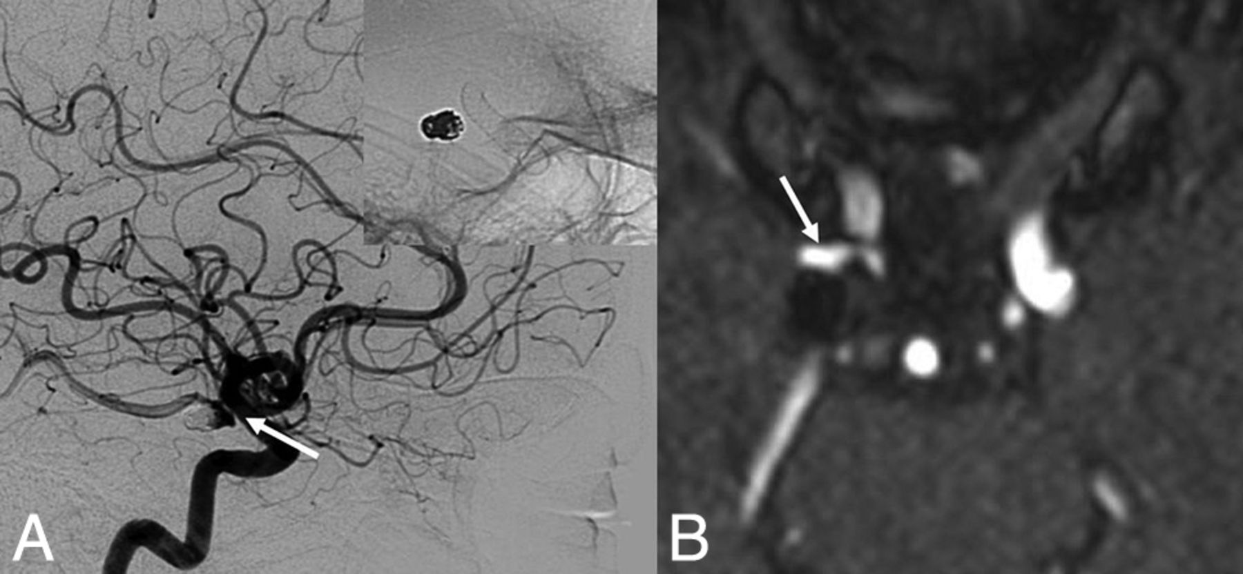
A total of 25 aneurysm remnants were available for clinical analysis based on last known follow-up imaging. No patient with an aneurysm remnant after flow diversion presented with delayed rupture or other clinical sequelae (median clinical follow-up, 31 months; IQR, 23–33 months). Details related to aneurysm remnants are noted in Table 2. In addition, angiographic follow-up demonstrated no progression of neck remnants. Figure 2 is one such representative case.

View this table:
  • View inline
  • View popup
Table 2:

Characteristics of aneurysm remnants after flow diversion

Fig 2.
  • Download figure
  • Open in new tab
  • Download powerpoint
Fig 2.

Aneurysm persistence after flow diversion. A 67-year-old woman who had a previously ruptured right posterior communicating artery aneurysm with evidence of recanalization on the 6-month follow-up angiogram. She was treated with flow diversion for the neck recurrence. A 6-month follow-up DSA (lateral x-ray) after flow diversion demonstrates class C OKM grade (continued filling at the neck of the aneurysm) (white arrow). At last clinical and angiographic follow-ups at 3 years, there is no evidence of aneurysm rerupture or progression of the neck remnant on MRA (white arrow).

Discussion

Flow diversion has emerged as a paradigm shift in the treatment of intracranial aneurysms. As more data define characteristics associated with aneurysm persistence after flow diversion,11⇓–13 improved patient selection and procedural technique may increase long-term occlusion rates. Parent vessel remodeling resulting in aneurysm occlusion after flow diversion is a remarkably different mechanism compared with coil embolized aneurysms. In addition, high rates of aneurysm occlusion and durability of treatment6 make studying aneurysm remnants after flow diversion difficult. Therefore, the natural history of this entity is ill-defined. Our study emphasizes the importance of adjunctive coiling and overlapping devices when appropriate to achieve higher rates of occlusion on midterm imaging follow-up. Moreover, we find that the clinical impact of aneurysm remnants after flow diversion is benign, with no evidence of rerupture in our cohort.

Predictors of Occlusion after Flow Diversion

Adjunctive coil embolization during flow diversion has been previously described by many reports.14,15 The placement of loosely packed coils inside the aneurysm at the time of flow diversion provides an additional element of flow disruption to aid aneurysm thrombosis. This observation is confirmed in our study and falls in line with the literature: The presence of adjunctive coiling leads to greater rates of aneurysm occlusion on midterm follow-up imaging. Although our result is slightly underpowered to reach statistical significance (P = .06), Lin et al14 noted a higher proportion of complete occlusion with adjunctive coiling at the time of flow diversion in 29 patients compared with flow diversion alone in 75 patients (93.1% versus 74.7%; P = .03). High rates of occlusion on follow-up imaging were similarly replicated by Nossek et al15 because all aneurysms that underwent flow diversion and adjunctive coiling were occluded. Enthusiasm for adjunctive coiling has been tempered by a few studies16 reporting that robust coil packing during flow diversion can cause device occlusion due to mass effect and increased thrombogenicity from the large coil mass. However, we did not experience device occlusion or an increased rate of ischemic complications with adjunctive coiling. This outcome has been confirmed by multiple case series that show the equivalent risk profile of flow diversion with or without adjunctive coiling.14,15,17

Aneurysm occlusion after flow diversion is dependent on metal surface coverage.18 Constructs with multiple overlapping flow diverters placed across the aneurysm neck favorably increase surface coverage both in ex vivo19 and computational fluid dynamics models.20 Multiple studies corroborate these basic science observations, with both the Pipeline embolization device for the Intracranial Treatment of Aneurysms trial (PITA)3 and Pipeline for Uncoilable or Failed Aneurysms (PUFS)2 trials supporting the efficacy of using overlapping flow diverters to treat aneurysms. Similarly, we show that the use of multiple devices resulted in higher rates of occlusion compared with 1 device (P = .02). Understandably, increased metal surface coverage and number of devices may increase thrombogenicity and, as a result, thromboembolic complications.21 Chalouhi et al22 proposed a single-device rationale for treatment of aneurysms on the basis of decreased complication rates and equivalent rates of occlusion between single- and multiple-device constructs. On the contrary, a larger study by Brinjikji et al23 showed that in 906 treated aneurysms, only fusiform aneurysm morphology was independently associated with ischemic complications after multivariate analysis (P < .001). Given the conflicting data, larger trials will be required to determine the safety profile of overlapping devices.

Aneurysm Remnants after Flow Diversion

The natural history of remnants after coil embolization of a ruptured aneurysm is well-defined: The more complete occlusion on postprocedural DSA, the lower is the risk of rerupture.24 Increasingly, studies show that unruptured neck remnants after endovascular treatment (stent-assisted coiling/coiling) are both angiographically8 and clinically9 benign on long-term follow-up. Mascitelli et al8 showed that in 99 completely occluded aneurysms and 110 neck remnants after coil embolization, angiographic outcome was similar with very few recanalizations. Munich et al9 analyzed the rerupture rate in 626 aneurysms with residual filling on immediate posttreatment angiography. Ruptured aneurysms with neck remnants pose a high risk of rerupture (3.4%), while unruptured aneurysms with residual necks confer a very low risk of rupture (0.6%). Although these results may not directly correlate with aneurysm remnants after flow diversion, the benign nature of unruptured flow-diverted aneurysm neck remnants should be considered. In addition, the progressive occlusion, parent vessel remodeling, and stasis of flow within the aneurysm sac after flow diversion may be protective against progression of aneurysm neck remnants to definite recurrence. In 25 aneurysm remnants with a median clinical follow-up of 31 months, we did not observe any evidence of rerupture or neck progression. Moreover, in a recent report by Kan et al,25 none of 16 cerebral aneurysms that failed to occlude after flow diversion ruptured during the follow-up period (mean follow-up duration, 24 months). Like the results of Mascitelli et al and Munich et al, the results of Kan et al suggest that remnants of unruptured aneurysms, despite being treated by different devices, are benign.

Although these results are preliminary, the clinical implications of our findings can be considered. Treatment failures after flow diversion resulting in neck remnants or persistent aneurysm filling did not progress in our series. Therefore, these cases could be monitored with noninvasive temporal MRA imaging.26 If progression of an aneurysm is confirmed on DSA, then a treatment decision about a second device can be considered. In addition, antiplatelet therapy can be de-escalated in cases in which remnants have persisted past 12 months because endothelization of the stent construct has likely already occurred.

Limitations

The retrospective nature of this study and analysis from a single academic center introduces sampling bias and possibly limits external validity. In addition, other angiographic findings that may be associated with aneurysm persistence that were not measured include inflow angle to aneurysm ostium, vessel size arising from aneurysm neck, and degree of malapposition as measured by VasoCT (Philips Healthcare, Best, the Netherlands).27 Angiographic follow-up at the 6-month time point was relatively consistent for the entire cohort; however, long-term imaging follow-up (MRA/DSA) was variable and at the discretion of the treating neurointerventionalist. The variable regiment of long-term imaging follow-up of the treating physician introduces heterogeneity with respect to our results of occlusion rates on long-term follow-up. In addition, long-term imaging follow-up was not available for all patients (52.8%; 47/89) with aneurysm persistence at 6 months. Some patients were lost to follow-up (72.3%; 34/47), or no follow-up was available for miniscule aneurysm remnants (27.7%; 13/47) that were initially interpreted as complete thrombosis or a tiny remnant that would eventually thrombose.

Conclusions

Preliminary results suggest that aneurysm remnants after flow diversion are infrequent with minimal clinical impact. When appropriate, the presence of overlapping devices and possibly adjunctive coiling may result in higher rates of complete occlusion. Larger studies with long-term clinical follow-up will be needed to confirm these findings.

Acknowledgments

We thank Duk Soo Han, MPH, for statistical review.

Footnotes

  • Drs Thomas P. Madaelil and Jonathan A. Grossberg are co-first authors.

  • Disclosures: Jonathan A. Grossberg—UNRELATED: Consultancy: Cognition Medical, Comments: no fees, only stock options; Grants/Grants Pending: Georgia Research Alliance; Stock/Stock Options: equity in Neurotechnology Investors (NTI). Jacques Dion—UNRELATED: Employment: MicroVention/Terumo, Comments: employee, Vice President of Scientific Affairs; Travel/Accommodations/Meeting Expenses Unrelated to Activities Listed: MicroVention/Terumo, Comments: as part of my employment duties. Raul G. Nogueira—OTHER RELATIONSHIPS: Stryker Neurovascular (DAWN Trial Principal Investigator, no compensation; TREVO Registry Steering Committee, no compensation; TREVO 2 trial Principal Investigator, modest; consultant, modest), Medtronic (SWIFT Trial Steering Committee, modest; SWIFT PRIME Trial Steering Committee, no compensation; STAR Trial Angiographic Core Lab, modest compensation), Penumbra (3D Separator Trial Executive Committee, no compensation), Cerenovus/Neuravi (ENDOLOW Trial Principal Investigator; EXCELLENT Registry Principal Investigator; ARISE 2 trial Steering Committee, no compensation; Physician Advisory Board, modest compensation), phenox (Physician Advisory Board, modest compensation), Anaconda (Physician Advisory Board, modest compensation), Genentech (Physician Advisory Board, modest compensation), Biogen (Physician Advisory Board, modest compensation), Prolong Pharmaceuticals (Physician Advisory Board, modest compensation), Allm Inc (Physician Advisory Board, no compensation), iSchemaView (speaker, modest compensation), Brainomix (research software use, no compensation), Sensome (research device use, no compensation), Viz.ai (Physician Advisory Board, stock options), Philips Healthcare (research software use, no compensation; speaker, modest), Corindus Vascular Robotics (Physician Advisory Board, stock options).

  • Paper previously presented at: Annual Meeting of the American Society of Neuroradiology and the Foundation of the ASNR Symposium, June 2–7, 2018; Vancouver, British Columbia, Canada.

REFERENCES

  1. 1.↵
    1. Fiorella D,
    2. Woo HH,
    3. Albuquerque FC, et al
    . Definitive reconstruction of circumferential, fusiform intracranial aneurysms with the Pipeline embolization device. Neurosurgery 2008;62:1115–20; discussion 1120–21 doi:10.1227/01.neu.0000325873.44881.6e pmid:18580809
    CrossRefPubMed
  2. 2.↵
    1. Becske T,
    2. Kallmes DF,
    3. Saatci I, et al
    . Pipeline for Uncoilable or Failed Aneurysms: results from a multicenter clinical trial. Radiology 2013;267:858–68 doi:10.1148/radiol.13120099 pmid:23418004
    CrossRefPubMed
  3. 3.↵
    1. Nelson PK,
    2. Lylyk P,
    3. Szikora I, et al
    . The Pipeline embolization device for the intracranial treatment of aneurysms trial. AJNR Am J Neuroradiology 2011;32:34–40 doi:10.3174/ajnr.A2421 pmid:21148256
    Abstract/FREE Full Text
  4. 4.↵
    1. Lylyk P,
    2. Miranda C,
    3. Ceratto R, et al
    . Curative endovascular reconstruction of cerebral aneurysms with the Pipeline Embolization Device: the Buenos Aires experience. Neurosurgery 2009;64:632–42; discussion 642–43; quiz N6 doi:10.1227/01.NEU.0000339109.98070.65 pmid:19349825
    CrossRefPubMed
  5. 5.↵
    1. Kallmes DF,
    2. Brinjikji W,
    3. Cekirge S, et al
    . Safety and efficacy of the Pipeline Embolization Device for treatment of intracranial aneurysms: a pooled analysis of 3 large studies. J Neurosurg 2017;127:775–80 doi:10.3171/2016.8.JNS16467 pmid:27791519
    CrossRefPubMed
  6. 6.↵
    1. Becske T,
    2. Brinjikji W,
    3. Potts MB, et al
    . Long-term clinical and angiographic outcomes following Pipeline Embolization Device treatment of complex internal carotid artery aneurysms: five-year results of the Pipeline for Uncoilable or Failed Aneurysms trial. Neurosurgery 2017;80:40–48 doi:10.1093/neuros/nyw014 pmid:28362885
    CrossRefPubMed
  7. 7.↵
    1. Lecler A,
    2. Raymond J,
    3. Rodriguez-Régent C, et al
    . Intracranial aneurysms: recurrences more than 10 years after endovascular treatment—a prospective cohort study, systematic review, and meta-analysis. Radiology 2015;277:173–80 doi:10.1148/radiol.2015142496 pmid:26057784
    CrossRefPubMed
  8. 8.↵
    1. Mascitelli JR,
    2. Oermann EK,
    3. De Leacy RA, et al
    . Angiographic outcome of intracranial aneurysms with neck remnant following coil embolization. J Neurointerv Surg 2015;7:484–89 doi:10.1136/neurintsurg-2014-011226 pmid:24792578
    Abstract/FREE Full Text
  9. 9.↵
    1. Munich SA,
    2. Cress MC,
    3. Rangel-Castilla L, et al
    . Neck remnants and the risk of aneurysm rupture after endovascular treatment with coiling or stent-assisted coiling: much ado about nothing? Neurosurgery 2019;84:421–27 doi:10.1093/neuros/nyy056 pmid:29528449
    CrossRefPubMed
  10. 10.↵
    1. O'Kelly C,
    2. Krings T,
    3. Fiorella D, et al
    . A novel grading scale for the angiographic assessment of intracranial aneurysms treated using flow diverting stents. Interv Neuroradiol 2010;16:133–37 doi:10.1177/159101991001600204 pmid:20642887
    CrossRefPubMed
  11. 11.↵
    1. Adeeb N,
    2. Moore J,
    3. Wirtz M, et al
    . Predictors of incomplete occlusion following Pipeline embolization of intracranial aneurysms: is it less effective in older patients? AJNR Am J Neuroradiol 2017;38:2295–300 doi:10.3174/ajnr.A5375 pmid:28912285
    Abstract/FREE Full Text
  12. 12.↵
    1. Trivelato FP,
    2. Salles Rezende MT,
    3. Ulhôa AC, et al
    . Occlusion rates of intracranial aneurysms treated with the Pipeline Embolization Device: the role of branches arising from the sac. J Neurosurg 18 Apr 1:1–7. [Epub ahead of print] doi:10.3171/2017.10.JNS172175 pmid:29624153
    CrossRefPubMed
  13. 13.↵
    1. Bender MT,
    2. Colby GP,
    3. Lin LM, et al
    . Predictors of cerebral aneurysm persistence and occlusion after flow diversion: a single-institution series of 445 cases with angiographic follow-up. J Neurosurg 2018 Mar 1:1–9. [Epub ahead of print] doi:10.3171/2017.11.JNS171738 pmid:29600915
    CrossRefPubMed
  14. 14.↵
    1. Lin N,
    2. Brouillard AM,
    3. Krishna C, et al
    . Use of coils in conjunction with the Pipeline embolization device for treatment of intracranial aneurysms. Neurosurgery 2015;76:142–49 doi:10.1227/NEU.0000000000000579 pmid:25255261
    CrossRefPubMed
  15. 15.↵
    1. Nossek E,
    2. Chalif DJ,
    3. Chakraborty S, et al
    . Concurrent use of the Pipeline Embolization Device and coils for intracranial aneurysms: technique, safety, and efficacy. J Neurosurg 2015;122:904–11 doi:10.3171/2014.12.JNS141259 pmid:25658781
    CrossRefPubMed
  16. 16.↵
    1. Siddiqui AH,
    2. Kan P,
    3. Abla AA, et al
    . Complications after treatment with Pipeline embolization for giant distal intracranial aneurysms with or without coil embolization. Neurosurgery 2012;71:E509–13 doi:10.1227/NEU.0b013e318258e1f8 pmid:22710418
    CrossRefPubMed
  17. 17.↵
    1. Park M,
    2. Kilburg C,
    3. Taussky P, et al
    . Pipeline embolization device with or without adjunctive coil embolization: analysis of complications from the IntrePED Registry. AJNR Am J Neuroradiol 2016;37:1127–31 doi:10.3174/ajnr.A4678 pmid:26767709
    Abstract/FREE Full Text
  18. 18.↵
    1. Wang K,
    2. Huang Q,
    3. Hong B, et al
    . Correlation of aneurysm occlusion with actual metal coverage at neck after implantation of flow-diverting stent in rabbit models. Neuroradiology 2012;54:607–13 doi:10.1007/s00234-011-0922-7 pmid:21874284
    CrossRefPubMed
  19. 19.↵
    1. Shapiro M,
    2. Raz E,
    3. Becske T, et al
    . Building multidevice Pipeline constructs of favorable metal coverage: a practical guide. AJNR Am J Neuroradiol 2014;35:1556–61 doi:10.3174/ajnr.A3902 pmid:24676003
    Abstract/FREE Full Text
  20. 20.↵
    1. Kim M1,
    2. Levy EI,
    3. Meng H, et al
    . Quantification of hemodynamic changes induced by virtual placement of multiple stents across a wide-necked basilar trunk aneurysm. Neurosurgery 2007;61:1305–12; discussion 1312–13 doi:10.1227/01.neu.0000306110.55174.30 pmid:18162911
    CrossRefPubMed
  21. 21.↵
    1. Tan LA,
    2. Keigher KM,
    3. Munich SA, et al
    . Thromboembolic complications with Pipeline Embolization Device placement: impact of procedure time, number of stents and pre-procedure P2Y12 reaction unit (PRU) value. J Neurointerv Surg 2015;7:217–21 doi:10.1136/neurintsurg-2014-011111 pmid:24553344
    Abstract/FREE Full Text
  22. 22.↵
    1. Chalouhi N,
    2. Tjoumakaris S,
    3. Phillips J, et al
    . A single Pipeline embolization device is sufficient for treatment of intracranial aneurysms. AJNR Am J Neuroradiol 2014;35:1562–66 doi:10.3174/ajnr.A3957 pmid:24788125
    Abstract/FREE Full Text
  23. 23.↵
    1. Brinjikji W,
    2. Lanzino G,
    3. Cloft H, et al
    . Risk factors for ischemic complications following Pipeline Embolization Device treatment of intracranial aneurysms: results from the IntrePED study. AJNR Am J Neuroradiol 2016;37:1673–78 doi:10.3174/ajnr.A4807 pmid:27102308
    Abstract/FREE Full Text
  24. 24.↵
    1. Johnston SC,
    2. Dowd CF,
    3. Higashida RT, et al
    ; CARAT Investigators. Predictors of rehemorrhage after treatment of ruptured intracranial aneurysms: the Cerebral Aneurysm Rerupture After Treatment (CARAT) study. Stroke 2008;39:120–25 doi:10.1161/STROKEAHA.107.495747 pmid:18048860
    Abstract/FREE Full Text
  25. 25.↵
    1. Kan P,
    2. Srinivasan VM,
    3. Mbabuike N, et al
    . Aneurysms with persistent patency after treatment with the Pipeline Embolization Device. J Neurosurg 2017;126:1894–98 doi:10.3171/2016.6.JNS16402 pmid:27636182
    CrossRefPubMed
  26. 26.↵
    1. Boddu S,
    2. Tong F,
    3. Dehkharghani S, et al
    . Contrast-enhanced time-resolved MRA for follow-up of intracranial aneurysms treated with the Pipeline Embolization Device. AJNR Am J Neuroradiol 2014;35:2112–18 doi:10.3174/ajnr.A4008 pmid:24994828
    Abstract/FREE Full Text
  27. 27.↵
    1. van der Marel K,
    2. Gounis MJ,
    3. Weaver JP, et al
    . Grading of Regional Apposition after Flow-Diverter Treatment (GRAFT): a comparative evaluation of VasoCT and intravascular OCT. J Neurointerv Surg 2016;8:847–52 doi:10.1136/neurintsurg-2015-011843 pmid:26220411
    Abstract/FREE Full Text
  • Received September 3, 2018.
  • Accepted after revision February 4, 2019.
  • © 2019 by American Journal of Neuroradiology
View Abstract
PreviousNext
Back to top

In this issue

American Journal of Neuroradiology: 40 (4)
American Journal of Neuroradiology
Vol. 40, Issue 4
1 Apr 2019
  • Table of Contents
  • Index by author
  • Complete Issue (PDF)
Advertisement
Print
Download PDF
Email Article

Thank you for your interest in spreading the word on American Journal of Neuroradiology.

NOTE: We only request your email address so that the person you are recommending the page to knows that you wanted them to see it, and that it is not junk mail. We do not capture any email address.

Enter multiple addresses on separate lines or separate them with commas.
Aneurysm Remnants after Flow Diversion: Clinical and Angiographic Outcomes
(Your Name) has sent you a message from American Journal of Neuroradiology
(Your Name) thought you would like to see the American Journal of Neuroradiology web site.
CAPTCHA
This question is for testing whether or not you are a human visitor and to prevent automated spam submissions.
Cite this article
T.P. Madaelil, J.A. Grossberg, B.M. Howard, C.M. Cawley, J. Dion, R.G. Nogueira, D.C. Haussen, F.C. Tong
Aneurysm Remnants after Flow Diversion: Clinical and Angiographic Outcomes
American Journal of Neuroradiology Apr 2019, 40 (4) 694-698; DOI: 10.3174/ajnr.A6010

Citation Manager Formats

  • BibTeX
  • Bookends
  • EasyBib
  • EndNote (tagged)
  • EndNote 8 (xml)
  • Medlars
  • Mendeley
  • Papers
  • RefWorks Tagged
  • Ref Manager
  • RIS
  • Zotero
0 Responses
Respond to this article
Share
Bookmark this article
Aneurysm Remnants after Flow Diversion: Clinical and Angiographic Outcomes
T.P. Madaelil, J.A. Grossberg, B.M. Howard, C.M. Cawley, J. Dion, R.G. Nogueira, D.C. Haussen, F.C. Tong
American Journal of Neuroradiology Apr 2019, 40 (4) 694-698; DOI: 10.3174/ajnr.A6010
del.icio.us logo Twitter logo Facebook logo Mendeley logo
  • Tweet Widget
  • Facebook Like
  • Google Plus One
Purchase

Jump to section

  • Article
    • Abstract
    • ABBREVIATIONS:
    • Materials and Methods
    • Results
    • Discussion
    • Conclusions
    • Acknowledgments
    • Footnotes
    • REFERENCES
  • Figures & Data
  • Info & Metrics
  • Responses
  • References
  • PDF

Related Articles

  • No related articles found.
  • PubMed
  • Google Scholar

Cited By...

  • Brain aneurysm and parent vessel remodeling after flow diversion treatment: a proposed modification for Cekirge-Saatci classification (mCSC)
  • Endoluminal flow diverting stents for middle cerebral artery bifurcation aneurysms: multicenter cohort
  • Neck Location on the Outer Convexity is a Predictor of Incomplete Occlusion in Treatment with the Pipeline Embolization Device: Clinical and Angiographic Outcomes
  • Large Neck and Strong Ostium Inflow as the Potential Causes for Delayed Occlusion of Unruptured Sidewall Intracranial Aneurysms Treated by Flow Diverter
  • Crossref (14)
  • Google Scholar

This article has been cited by the following articles in journals that are participating in Crossref Cited-by Linking.

  • ‘Plug and pipe’ strategy for treatment of ruptured intracranial aneurysms
    Brian M Howard, Jason M Frerich, Thomas P Madaelil, Jacques E Dion, Frank C Tong, C Michael Cawley, Jonathan A Grossberg
    Journal of NeuroInterventional Surgery 2019 11 1
  • Predictors of aneurysm occlusion following treatment with the WEB device: systematic review and case series
    Fadi Al Saiegh, Lohit Velagapudi, Omaditya Khanna, Ahmad Sweid, Nikolaos Mouchtouris, Michael P. Baldassari, Thana Theofanis, Rizwan Tahir, Victoria Schunemann, Carrie Andrews, Lucas Philipp, Nohra Chalouhi, Stavropoula I. Tjoumakaris, David Hasan, M. Reid Gooch, Nabeel A. Herial, Robert H. Rosenwasser, Pascal Jabbour
    Neurosurgical Review 2022 45 2
  • Endoluminal flow diverting stents for middle cerebral artery bifurcation aneurysms: multicenter cohort
    Mohamed M Salem, Mirhojjat Khorasanizadeh, Sovann V Lay, Leonardo Renieri, Anna L Kuhn, Ahmad Sweid, Francesco Massari, Justin M Moore, Stavropoula I. Tjoumakaris, Pascal Jabbour, Ajit S Puri, Christopher S Ogilvy, Brian T Jankowitz, Jan-Karl Burkhardt, Peter Kan, Nicola Limbucci, Christophe Cognard, Ajith J Thomas
    Journal of NeuroInterventional Surgery 2022 14 11
  • Large Neck and Strong Ostium Inflow as the Potential Causes for Delayed Occlusion of Unruptured Sidewall Intracranial Aneurysms Treated by Flow Diverter
    T. Su, P. Reymond, O. Brina, P. Bouillot, P. Machi, B.M.A. Delattre, L. Jin, K.O. Lövblad, M.I. Vargas
    American Journal of Neuroradiology 2020 41 3
  • Neck Location on the Outer Convexity is a Predictor of Incomplete Occlusion in Treatment with the Pipeline Embolization Device: Clinical and Angiographic Outcomes
    T. Sunohara, H. Imamura, M. Goto, R. Fukumitsu, S. Matsumoto, N. Fukui, Y. Oomura, T. Akiyama, T. Fukuda, K. Go, S. Kajiura, M. Shigeyasu, K. Asakura, R. Horii, C. Sakai, N. Sakai
    American Journal of Neuroradiology 2021 42 1
  • Pipeline Embolization Device for the Treatment of Ruptured Intracerebral Aneurysms: A Multicenter Retrospective Study
    Weiying Zhong, Hong Kuang, Ping Zhang, Xinjian Yang, Bin Luo, Aisha Maimaitili, Yuanli Zhao, Donglei Song, Sheng Guan, Hongqi Zhang, Yang Wang, Donghai Wang, Wandong Su, Yunyan Wang
    Frontiers in Neurology 2021 12
  • Usefulness of 3D T1-SPACE in Combination With 3D-TOF MRA for Follow-Up Evaluation of Intracranial Aneurysms Treated With Pipeline Embolization Devices
    Qiuji Shao, Qiaowei Wu, Qiang Li, Tianxiao Li, Li Li, Kaitao Chang, Meiyun Wang
    Frontiers in Neurology 2020 11
  • Pipeline flow diversion with adjunctive coil embolization for internal carotid artery aneurysms following an intradural component: results in 46 consecutive aneurysms from a Japanese single-center experience
    Tomoaki Akiyama, Hirotoshi Imamura, Masanori Goto, Ryu Fukumitsu, Tadashi Sunohara, Shirabe Matsumoto, Nobuyuki Fukui, Yoshihiro Omura, Tatsumaru Fukuda, Koichi Go, Shinji Kajiura, Masashi Shigeyasu, Kento Asakura, Ryo Horii, Yuji Naramoto, Rikuo Nishii, Yasuhiro Yamamoto, Chiaki Sakai, Nobuyuki Sakai
    Neurosurgical Review 2022 45 3
  • A Review of Current Flow Diverters
    Kiyofumi Yamada, Hirotoshi Imamura, Saya Ozaki, Akihiro Niwa, Yuji Kushi, Naoto Yamada, Taichi Ikedo, Eika Hamano, Hisae Mori, Koji Iihara, Shinichi Yoshimura, Hiroharu Kataoka
    Journal of Neuroendovascular Therapy 2024 18 3
  • Brain aneurysm and parent vessel remodeling after flow diversion treatment: a proposed modification for Cekirge-Saatci classification (mCSC)
    Ricardo A Hanel, Gustavo M Cortez, Demetrius Klee Lopes, Isil Saatci, H Saruhan Cekirge
    Journal of NeuroInterventional Surgery 2023 15 2

More in this TOC Section

  • SAVE vs. Solumbra Techniques for Thrombectomy
  • CT Perfusion&Reperfusion in Acute Ischemic Stroke
  • Delayed Reperfusion Post-Thrombectomy&Thrombolysis
Show more Interventional

Similar Articles

Advertisement

Indexed Content

  • Current Issue
  • Accepted Manuscripts
  • Article Preview
  • Past Issues
  • Editorials
  • Editors Choice
  • Fellow Journal Club
  • Letters to the Editor

Cases

  • Case Collection
  • Archive - Case of the Week
  • Archive - Case of the Month
  • Archive - Classic Case

Special Collections

  • Special Collections

Resources

  • News and Updates
  • Turn around Times
  • Submit a Manuscript
  • Author Policies
  • Manuscript Submission Guidelines
  • Evidence-Based Medicine Level Guide
  • Publishing Checklists
  • Graphical Abstract Preparation
  • Imaging Protocol Submission
  • Submit a Case
  • Become a Reviewer/Academy of Reviewers
  • Get Peer Review Credit from Publons

Multimedia

  • AJNR Podcast
  • AJNR SCANtastic
  • Video Articles

About Us

  • About AJNR
  • Editorial Board
  • Not an AJNR Subscriber? Join Now
  • Alerts
  • Feedback
  • Advertise with us
  • Librarian Resources
  • Permissions
  • Terms and Conditions

American Society of Neuroradiology

  • Not an ASNR Member? Join Now

© 2025 by the American Society of Neuroradiology All rights, including for text and data mining, AI training, and similar technologies, are reserved.
Print ISSN: 0195-6108 Online ISSN: 1936-959X

Powered by HighWire