
Article Figures & Data
Figures
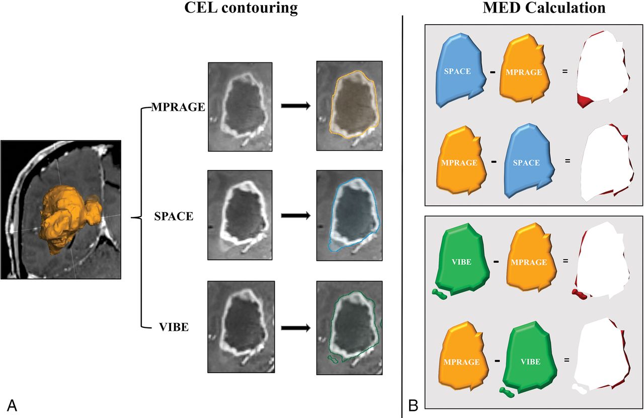
- Fig 1.
Diagram showing the contrast-enhancing lesion margin extent discrepancy (MED) estimation procedure. This approach is aimed at highlighting the spatial mismatch of the tumor border segmentation obtained from MPRAGE, with respect to SPACE and VIBE, and vice versa. A, First, for each CEL and sequence type, volume segmentation is performed using a validated computer-assisted tool dedicated to pretreatment planning and neuronavigation (SmartBrush 2.5; Brainlab). Segmentations obtained on MPRAGE, SPACE, and VIBE images are, respectively, represented in orange, blue, and green. B, The segmented volumes are reciprocally subtracted, generating maps of the areas where SPACE and VIBE volumes, respectively, exceed MPRAGE, and vice versa. Finally, the resulting MED areas are represented in red.
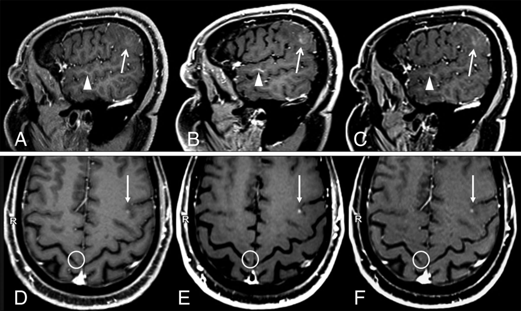
- Fig 2.
Two sample cases showing differences in contrast-enhancing lesion conspicuity between sequences. A and D, MPRAGE. B and E, SPACE. C and F, VIBE. A–C, Case 1: a patient with a faintly enhancing glioblastoma. Compared with MPRAGE (A), the lesion enhancement (arrows) and its boundary demarcations are much better appreciated on SPACE and VIBE images. The corresponding contrast rate/contrast-to-noise ratio values are 24.75/2.45, 51.32/8.96, and 41.23/6.25, and the rankings are worst, best, and intermediate, respectively, for MPRAGE, SPACE, and VIBE. Also incidentally noted is a developmental venous anomaly (arrowheads), which shows a strong contrast enhancement on black-blood SPACE images. This is probably related to the extremely slow flow seen in such small venous malformations. Images were acquired at 5 minutes after contrast injection in the following order: VIBE, SPACE, MPRAGE. D–F, Case 2: a patient with metastases from renal carcinoma (D, MPRAGE. E, SPACE. F, VIBE). A small CEL is seen in the left frontal lobe (arrows) whose conspicuity with respect to the background parenchyma was ranked worst on MPRAGE, intermediate on VIBE, and best on SPACE images. The corresponding contrast rate/contrast-to-noise ratio values are 8.85/3.08, 18.96/9.15, and 16.63/6.89, respectively. An example of a very tiny metastasis in the right precentral gyrus cortex, which was missed when inspecting MPRAGE images alone but was visible on SPACE and VIBE, is highlighted by circles. This lesion was not included in the analyses. Images were acquired after 5 minutes from contrast injection in the following order: SPACE, VIBE, and MPRAGE.
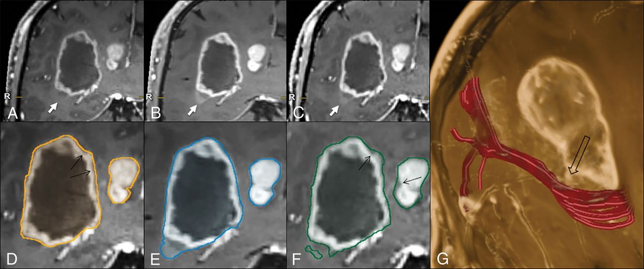
- Fig 3.
Illustrative case comparing the 3D target-object-creation results in a glioblastoma, obtained on MPRAGE, SPACE, or VIBE images (see the text and Fig 1 for method explanation). There is a clear difference among the MPRAGE (A), SPACE (B), and VIBE (C) conspicuities at the level of the faintly enhancing inferolateral border of the lesion (arrows), which is better represented on the SPACE and VIBE images, compared with MPRAGE. This part of the tumor is not included in the MPRAGE lesion segmentation (D); however, it is captured completely on SPACE (E) and partially on VIBE (F) images. The tractographic reconstruction of the optic radiation trajectory (G, in red) demonstrates the close proximity of the tumor to this tract (empty arrow). The black arrows in D and F indicate some thin areas of tumor margin overestimation on MPRAGE and VIBE, respectively, which are not seen on SPACE images. Images were acquired after 5 minutes from contrast injection in the following order: MPRAGE, VIBE, and SPACE.


Tables
CR Median (IQR) CNR Median (IQR) Visual Score Besta Intermediate Worst All CELs (n = 54) 3D-IR GRE MPRAGE 80.88 (43.71–125.65) 10.02 (5.71–16.06) 15 (27.8%) 26 (48.1%) 13 (24.1%) 3D-TSE SPACE 100.92b,c (73–191.59) 19.17b,c (13.21–36.01) 54 (100%) 0 0 3D-GRE VIBE 85.86b,c (43.71–135.22) 16.76b,c (11–37.22) 24 (44.4%)b 25 (46.3%) 5 (9.3%)b Gliomas (n = 38) 3D-IR GRE MPRAGE 90.01 (48.29–125.21) 10.51 (5.68–17.33) 9 (23.7%) 19 (50%) 10 (26.3%) 3D-TSE SPACE 125.64b,c (88.41–187.45) 24.46b,c (12.55–41.23) 38 (100%) 0 0 3D-GRE VIBE 94.19b,c (58.34–88.95) 16.22d,c (9.89–27.25) 16 (42.1%)d 18 (47.4%) 4 (10.5%)d Metastases (n = 16) 3D-IR GRE MPRAGE 54.71 (35.24–130.01) 9.30 (6.77–12.29) 6 (37.5%) 7 (43.8%) 3 (18.8%) 3D-TSE SPACE 80.04b,c (63.13–200) 17.15b,c (13.63–21.26) 16 (100%) 0 0 3D-GRE VIBE 72.95b,c (54.19–154.43) 17.26b,c (12.14–31.18) 8 (50%) 7 (43.8%) 1 (6.3%) Note:—IR indicates inversion recovery; GRE, gradient recalled-echo; CR, contrast rate; CNR, contrast-to-noise ratio; IQR, interquartile range.
↵a Because SPACE in the visual assessment was constantly rated as best in all cases, no statistical comparison tests were performed in this analysis.
↵b P < .001.
↵c P ≤ .01 (adjusted for the order of sequence acquisition; FSL General Linear Model).
↵d P = .001 (compared with MPRAGE, Wilcoxon test).
All CELs (n = 54) Gliomas (n = 38) Metastases (n = 16) TV (median/IQR) (mL) MPRAGE 1.36/0.18–10.93 3.02/0.44–28.93 0.33/0.006–2.81 SPACE 1.78/0.20–11.00 3.5/0.49–29.73 0.39/0.006–3.2 VIBE 1.62/0.16–10.35 3.39/0.43–27.48 0.40/0.007–2.84 TV, SPACE vs MPRAGE Pa .001b .007b .003b P (adjusted for sequence acquisition order)c .034b .033b .075 TV, VIBE vs MPRAGE Pa .259 .201 .343 P (adjusted for sequence acquisition order)c .521 .538 .706 MED (median/% of TV) |CELMPRAGE|–|CELSPACE| 0.10 mL/7.4% 0.18 mL/6% 0.02 mL/6.1% |CELSPACE|–|CELMPRAGE| 0.27 mL/19.9% 0.43 mL/14.2% 0.05 mL/15.2% |CELMPRAGE|–|CELVIBE| 0.15 mL/11% 0.21 mL/7% 0.01 mL/3% |CELVIBE|–|CELMPRAGE| 0.15 mL/11% 0.23 mL/7.6% 0.05 mL/15.2%


Jump to section
Related Articles
Cited By...
- Radiomics-Based Differentiation of Glioblastoma and Metastatic Disease: Impact of Different T1-Contrast-Enhanced Sequences on Radiomics Features and Model Performance
- 7T MRI for Cushing Disease: A Single-Institution Experience and Literature Review
- Nonlesional Sources of Contrast Enhancement on Postgadolinium "Black-Blood" 3D T1-SPACE Images in Patients with Multiple Sclerosis
- Twofold improved tumor-to-brain contrast using a novel T1 relaxation-enhanced steady-state (T1RESS) MRI technique