
Article Figures & Data
Figures
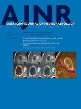
- Fig 1.
MR imaging appearances of true-positive AVM recurrence. A, Recurrent nidus in a 7-year-old girl with a right choroidal AVM that was embolized with n-BCA through a posterolateral choroidal feeder, with postembolization digital subtraction angiography confirming exclusion. B, Axial section from TOF-MRA performed 6 months posttreatment showing a tuft of vessels (arrow) posterior to the right lateral ventricular choroid plexus. This was also seen on contrast-enhanced MR imaging (not shown here) as exuberant and asymmetric choroid enhancement. C, Conventional angiogram, obtained 3 weeks after MR imaging, confirms an AVM nidus. Left vertebral artery injection confirms a right choroidal nidus (arrow) with deep venous drainage. The patient subsequently underwent radiosurgery. D, Recurrent cerebellar AVM in a 11-year-old girl with hereditary hemorrhagic telengiectasia lateral angiogram with negative findings immediately following endovascular embolization through a cerebellar branch of the right posterior inferior cerebellar artery. E, Sagittal section from postgadolinium MR imaging 3 months after treatment shows a nodular juxtamural focus of enhancement (arrow), with a vein traceable to the torcular. F, Conventional angiogram performed 1 month after MR imaging confirms this to be a recurrent nidus with early venous drainage. The recurrent AVM was surgically removed.
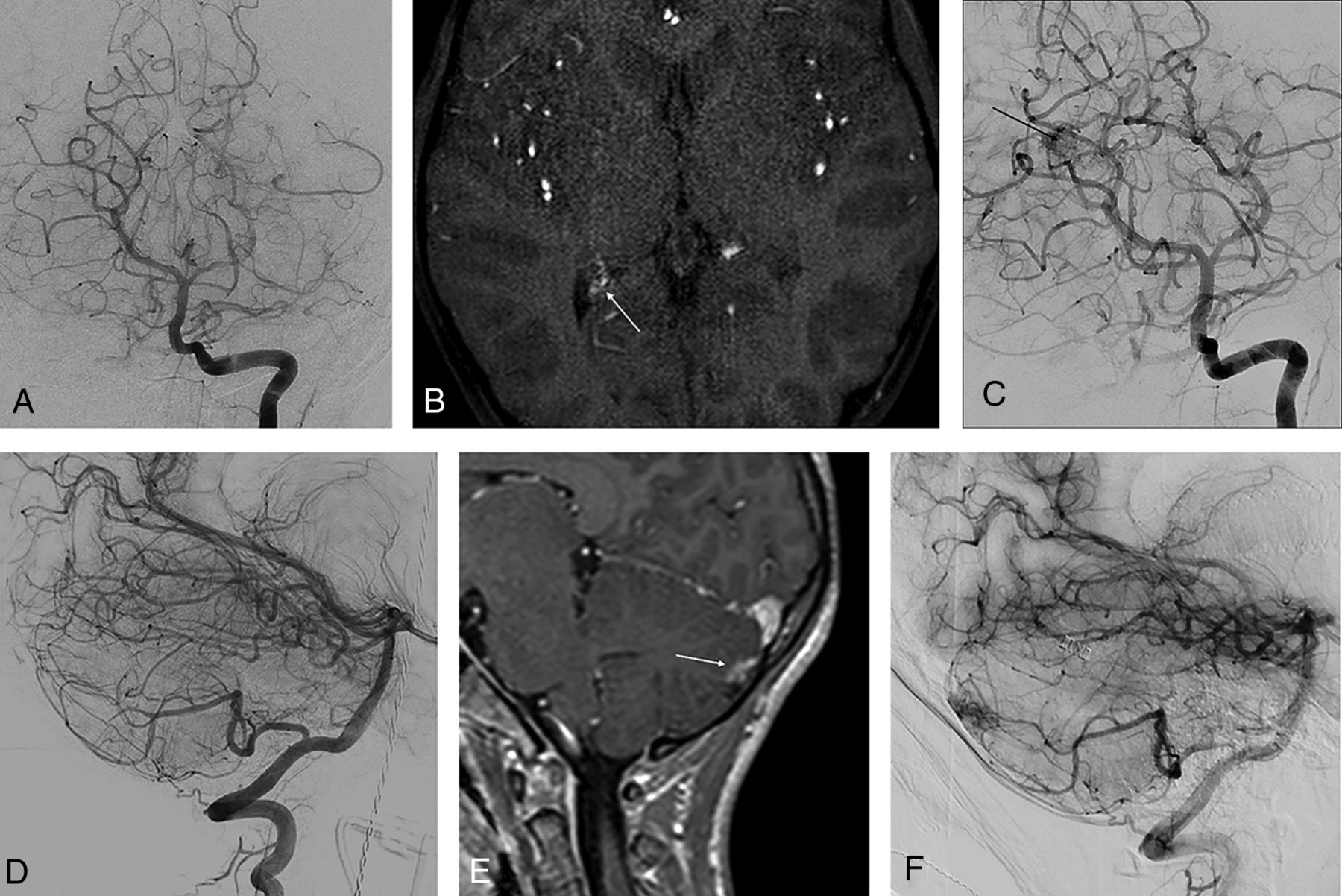
- Fig 2.
False-negative MR imaging findings for brain AVM recurrence, confirmed by digital subtraction and 3D rotational angiography. A 16-year-old boy with a left occipital AVM treated by embolization with Onyx. A, Frontal projection, left internal carotid angiography, shows a hypertrophied temporo-occipital middle cerebral artery branch suppling a compact nidus, with superficial venous drainage. B, Postembolization frontal angiogram shows complete exclusion of the shunt. C, Axial postgadolinium MR imaging section obtained 1 year after angiographically documented cure shows the hematoma cavity with artifacts from the Onyx cast within, but no suggestion of recurrence. D, Volume-rendered reformat obtained from 3D rotational angiography in the left vertebral artery shows a nidus behind the Onyx cast, with a prominent feeder (thin arrow) and early draining vein (thick arrow), which was also confirmed on digital subtraction angiography (E).
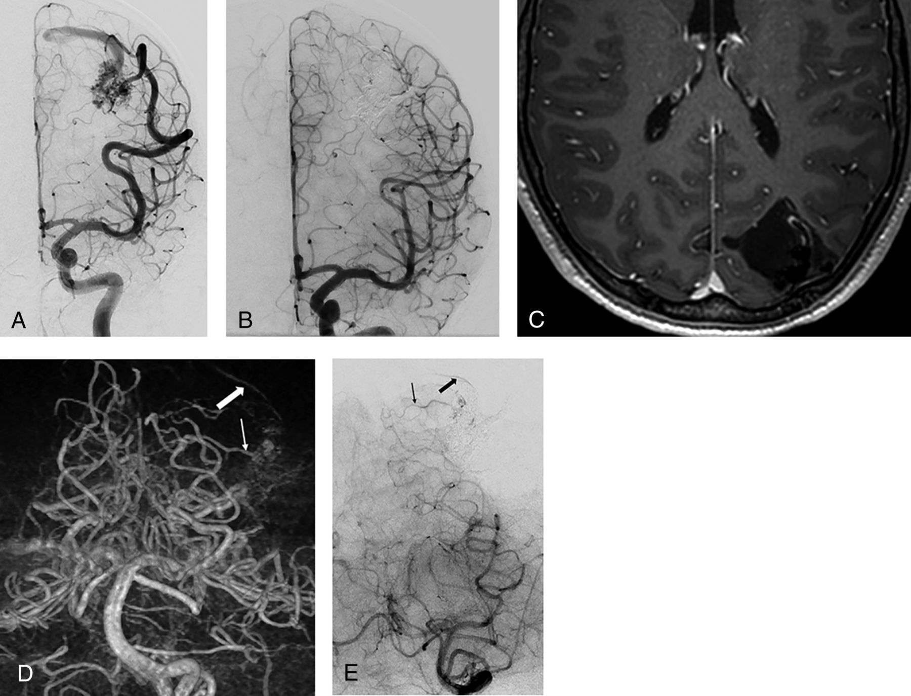
- Fig 3.
False-positive MR imaging for brain AVM recurrence, confirmed by conventional angiography and 3DRA-MR imaging fusion. A 5-year-old boy with a left parietal AVM. A, Left internal carotid injection in the lateral projection during conventional angiography shows a postcentral diffuse AVM nidus with a deep white matter component, hypertrophied anterior and middle cerebral arterial feeders, and venous drainage into the superior sagittal sinus. B, Complete surgical resection was performed after partial embolization, with cure confirmed with postresection angiography. C, Axial postgadolinium section from MR imaging performed 1 year after cure shows central linear and nodular enhancement suspicious for recurrence. D, Digital subtraction angiographic image, left internal carotid injection, shows no AVM recurrence. This was also confirmed from injections into the posterior circulation. E, 3DRA-MR imaging fusion shows no vascularity within the encephalomalacic cavity, excluding recurrence. MR imaging enhancement is believed to be related to scar tissue or dural folds.
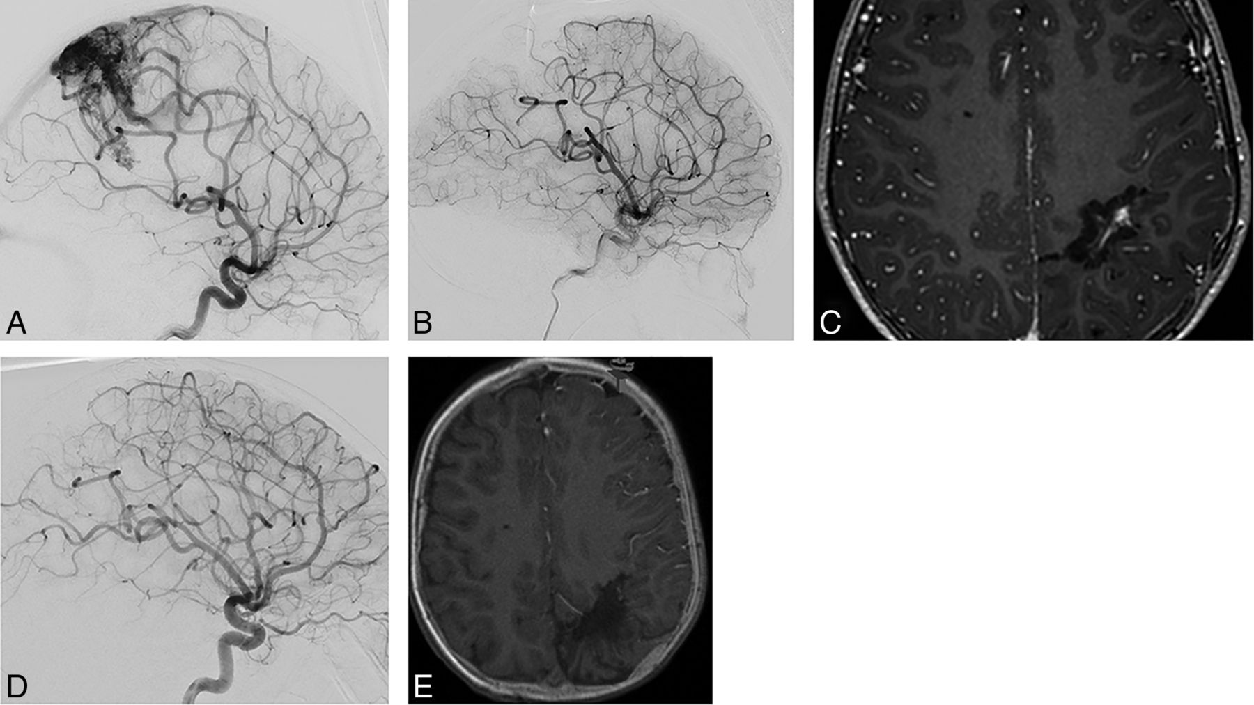
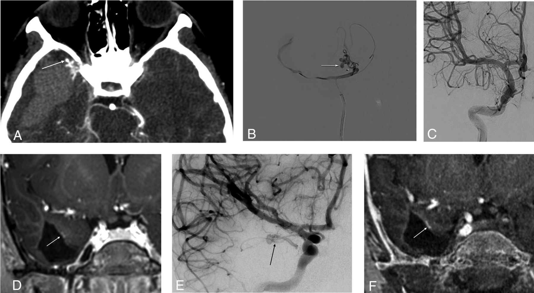
- Fig 4.
3DRA-MR imaging fusion for accurate depiction of a small AVM recurrence. A, Axial CTA section in an 8-year-old child who presented with sudden reduced level of consciousness, showing a right temporal hematoma and an anterior temporal AVM with an anterolaterally directed pseudoaneurysm (arrow). B, Microcatheter injection into the temporopolar branch of the right middle cerebral artery shows the AVM and rupture point (arrow). C, This was embolized with n-BCA in Lipiodol, achieving a complete angiographic cure. D, MR imaging performed 1 year after treatment shows a small juxtamural nodular enhancement (arrow) on this postgadolinium coronal section. E, Basal view from a right internal carotid injection on subsequent conventional angiography confirms recurrent arteriovenous shunting (arrow). F, 3DR-MR imaging coronal fused image confirms CEMRI findings, as well as providing exquisite delineation of feeding arteries and draining vein (not shown here). This sequence, providing all requisite information for surgical resection, was used for intraoperative neuronavigation.


Tables
Demographics Age (mean) (range) (yr) 10.8 ± 3.9, 2–17 Male/female ratio 19/20 Presentation Hemorrhage 28 (71.2) Seizures 5 (12.8) HHT screening 3 (7.7) Incidental (screening for headache) 2 (5.1) Partially treated at outside institution 1 (2.6) SM grade I 10 (26) II 24 (61.5) III 4 (10.3) IV 1 (2.6) Location Frontal 5 (12.8) Parietal 9 (23.1) Temporal 11 (28.2) Occipital 9 (23.1) Brain stem 1 (2.6) Cerebellar 3 (7.7) Choroidal 1 (2.6) Arterial feeder type Terminal 19 (48.7) En passant 20 (51.3) Venous drainage Superficial only 31 (79.5) Deep (±superficial drainage) 8 (20.5) Venous stenosis Present 3 (7.7) Absent 36 (92.3) Compactness Compact 30 (76.9) Diffuse 9 (23.1) Treatment Surgery 20 (51.3) Embolization 11 (28.2) Radiosurgery 3 (7.7) Multimodality 5 (12.8) Note:—HHT indicates hereditary hemorrhagic telengiectasia.
↵a Data are numbers (%) unless otherwise indicated.
Odds Ratio (95% CI) P Value Age at diagnosis (yr) 0.9 (0.7–1.3) .914 Sex (M) 3.3 (0.4–28.7) .287 SM grade 0.2 (0.0–1.2) .079 Diffuse nidus (compact vs diffuse) 0.2 (0.0–2.8) .256 Draining vein stenosis (yes vs no) 1.6 (0.0–130.3) .831 Embolization only treatment (yes vs no) 32.4 (2.7–386.3) .006 - Table 3:
Sensitivity, specificity, PPV, and NPV for CEMRI, TOF-MRA, and a combination of both to diagnose recurrent brain AVM after treatment in children
CEMRI TOF-MRA CEMRI + TOF-MRA Sensitivity 84.6% 50.0% 75.0% Specificity 38.5% 96.1% 90.9% PPV 40.7% 85.7% 85.7% NPV 81.8% 79.3% 83.3%


